. 2015 Sep 8:6:894.
doi: 10.3389/fmicb.2015.00894. eCollection 2015.

HIV-1 Tat and cocaine mediated synaptopathy in cortical and midbrain neurons is prevented by the isoflavone Equol

Affiliations

HIV-1 Tat and cocaine mediated synaptopathy in cortical and midbrain neurons is prevented by the isoflavone Equol

Sarah J Bertrand et al. Front Microbiol. .

Abstract

Illicit drugs, such as cocaine, are known to increase the likelihood and severity of HIV-1 associated neurocognitive disorders (HAND). In the current studies synaptic integrity was assessed following exposure to low concentrations of the HIV-1 viral protein Tat 1-86B, with or without cocaine, by quantifying filamentous actin (F-actin) rich structures (i.e., puncta and dendritic spines) on neuronal dendrites in vitro. In addition, the synapse-protective effects of either R-Equol (RE) or S-Equol (SE; derivatives of the soy isoflavone, daidzein) were determined. Individually, neither low concentrations of HIV-1 Tat (10 nM) nor low concentrations of cocaine (1.6 μM) had any significant effect on F-actin puncta number; however, the same low concentrations of HIV-1 Tat + cocaine in combination significantly reduced dendritic synapses. This synaptic reduction was prevented by pre-treatment with either RE or SE, in an estrogen receptor beta dependent manner. In sum, targeted therapeutic intervention with SE may prevent HIV-1 + drug abuse synaptopathy, and thereby potentially influence the development of HAND.

Keywords: F-actin; HAND; S-Equol; estrogen receptor; soy isoflavone.


Figures

FIGURE 1
FIGURE 1
R-Equol (RE) and S-Equol (SE) prevented synaptic loss induced by HIV-1 Tat (50 nM) via an estrogen receptor mediated mechanism. (A) Conversion of the soy isoflavone daidzein to SE. The dotted circle identifies a chiral center on carbon 3 wherein a conformational change produces RE. However, only SE may be produced by mammalian gut bacteria. (B) A moderate concentration of RE (50 nM), but not a low concentration (33 nM), prevented significant F-actin puncta loss induced by HIV-1 Tat (50 nM; p ≤ 0.01). (C) Both the low (33 nM) and moderate (50 nM) concentrations of SE prevented HIV-1 Tat induced F-actin puncta loss (p ≤ 0.05 and p ≤ 0.001, respectively). (D) 1 h pre-treatment with TMX (100 nM) blocked the protective effects of RE and SE (50 nM) against HIV-1 Tat (50 nM) in midbrain neurons. Mean ± SEM, p ≤ 0.05 compared to HIV-1 Tat treatment.
FIGURE 2
FIGURE 2
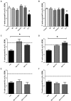
HIV-1 Tat (10 nM) + cocaine (1.6 μM) treatment produced significant synaptic loss in midbrain and cortical neurons, which was prevented by pre-treatment with either RE or SE. (A) Treatment with either SE, RE, HIV-1 Tat, or cocaine does not significantly alter the density of dendritic F-actin puncta compared to controls in midbrain neurons. Treatment with HIV-1 Tat + cocaine produced significant loss of F-actin puncta (p ≤ 0.05). (B) Treatment with either SE, RE, HIV-1 Tat, or cocaine does not significantly alter the density of dendritic F-actin puncta compared to controls in cortical neurons. Treatment with HIV-1 Tat + cocaine produced significant loss of F-actin puncta (p ≤ 0.05). (C) Pre-treatment with either RE or SE (50 nM) prevented dendritic F-actin puncta loss caused by HIV-1 Tat + cocaine treatments in midbrain neurons. Dendrites from pre-treated neurons are not significantly different from vehicle-treated controls (mean values, dotted line). (D) Pre-treatment with either RE or SE (50 nM) prevents HIV-1Tat + cocaine induced loss of dendritic F-actin puncta (p ≤ 0.001) in cortical neurons. Dendrites from pre-treated neurons are not significantly different from vehicle-treated controls (mean values, dotted line). (E) Pre-treatment of midbrain neurons with TMX (100 nM) for 1 h prior to either RE or SE prevented the protective effects of RE and SE, suggesting an estrogen receptor mediated mechanism. Vehicle-treated control mean values are represented by dotted line. (F) Pre-treatment of cortical neurons with TMX (100 nM) for 1 h prior to either RE or SE prevented the protective effects of RE and SE, suggesting an estrogen receptor mediated mechanism. Control mean value represented by dotted line. Mean ± SEM, p ≤ 0.05 compared either to vehicle-treated controls (A,B) or HIV-1 Tat + cocaine (C,D).
FIGURE 3
FIGURE 3
R-Equol and SE prevented HIV-1 Tat + cocaine synaptic loss via a selective estrogen receptor (ERβ) dependent mechanism. Cortical neurons were pre-treated for 1 h with either MPP (ERα), PHTPP (ERβ), or G15 (GPR30) prior to treatment with either RE (A) or SE (B) to identify which specific ER subtype was necessary for equol-mediated synaptic protection from HIV-1 Tat + cocaine. We found that pre-treatment with PHTPP (ERβ) inhibited both the RE (panel A) and SE (panel B) induced synaptic protection in HIV-1 Tat + cocaine treated cortical neurons. (A) HIV-1 Tat + cocaine exposed neurons pre-treated with either MPP or G15, and subsequently RE, had normal levels of dendritic F-actin puncta (i.e., not significantly different from vehicle-treated controls, p > 0.05). In contrast, PHTPP-treated neurons had a significant loss of dendritic F-actin puncta compared to controls (p ≤ 0.05) and were not significantly different from HIV-1 Tat + cocaine treated dendrites (p > 0.05), indicating that PHTPP inhibited RE-induced synaptic protection. Mean ± SEM, p ≤ 0.05. (B) HIV-1 Tat + cocaine exposed neurons pre-treated with either MPP or G15, and subsequently SE, had normal levels of dendritic F-actin puncta (i.e., not significantly different from vehicle-treated controls, p > 0.05). In contrast, PHTPP-treated neurons had a significant loss of dendritic F-actin puncta compared to controls (p ≤ 0.05) and were not significantly different from HIV-1 Tat + cocaine treated dendrites (p > 0.05), indicating that PHTPP inhibited SE-induced synaptic protection. Mean ± SEM, p ≤ 0.05. (C–J) The ERβ antagonist PHTPP significantly blocked equol-mediated protection from HIV-1 Tat + cocaine in cortical neurons. PHTPP pre-treated, SE(I)/RE(J) treated, neurons lacked a fine dendritic network and had reduced dendritic branching, similar characteristics to those in non pre-treated HIV-1Tat + cocaine neurons (D). In contrast, vehicle (C), HIV-1 Tat (10 nM; G), cocaine (H), and SE(E)/RE(F) treated neurons had robust dendrites with complex branching patterns and a lack of aberrant morphology such as bundling or beading. Phalloidin/F-actin(green) and MAP-2 (blue); 20× magnification.

References

    1. Adams S. M., Aksenova M. V., Aksenov M. Y., Mactutus C. F., Booze R. M. (2010). ER-beta mediates 17beta-estradiol attenuation of HIV-1 Tat-induced apoptotic signaling. Synapse 64 829–838. 10.1002/syn.20793 - DOI - PMC - PubMed
    1. Adams S. M., Aksenova M. V., Aksenov M. Y., Mactutus C. F., Booze R. M. (2012). Soy isoflavones genistein and daidzein exert anti-apoptotic actions via a selective er-mediated mechanism in neurons following hiv-1 tat(1-86) exposure. PLoS ONE 7:e37540 10.1371/journal.pone.0037540 - DOI - PMC - PubMed
    1. Aksenov M. Y., Aksenova M. V., Nath A., Ray P. D., Mactutus C. F., Booze R. M. (2006). Cocaine-mediated enhancement of Tat toxicity in rat hippocampal cell cultures: the role of oxidative stress and D1 dopamine receptor. Neurotoxicology 27 217–228. 10.1016/j.neuro.2005.10.003 - DOI - PubMed
    1. Aksenov M. Y., Aksenova M. V., Silvers J. M., Mactutus C. F., Booze R. M. (2008). Different effects of selective dopamine uptake inhibitors, GBR 12909 and WIN 35428 on HIV-1 Tat toxicity in rat fetal midbrain neurons. Neurotoxicolgy 29 971–977. 10.1016/j.neuro.2008.06.003 - DOI - PMC - PubMed
    1. Aksenova M. V., Aksenov M. Y., Adams S. M., Mactutus C. F., Booze R. M. (2009). Neuronal survival and resistance to HIV-1 Tat toxicity in the primary culture of rat fetal neurons. Exp. Neurol. 215 253–263. 10.1016/j.expneurol.2008.10.006 - DOI - PMC - PubMed

LinkOut - more resources