Review
. 2015 Sep 22:6:459.
doi: 10.3389/fimmu.2015.00459. eCollection 2015.

Macroautophagy in Endogenous Processing of Self- and Pathogen-Derived Antigens for MHC Class II Presentation

Affiliations
Review

Macroautophagy in Endogenous Processing of Self- and Pathogen-Derived Antigens for MHC Class II Presentation

Fernanda V Duraes et al. Front Immunol. .

Abstract

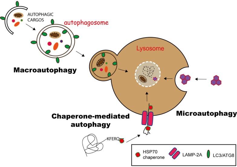
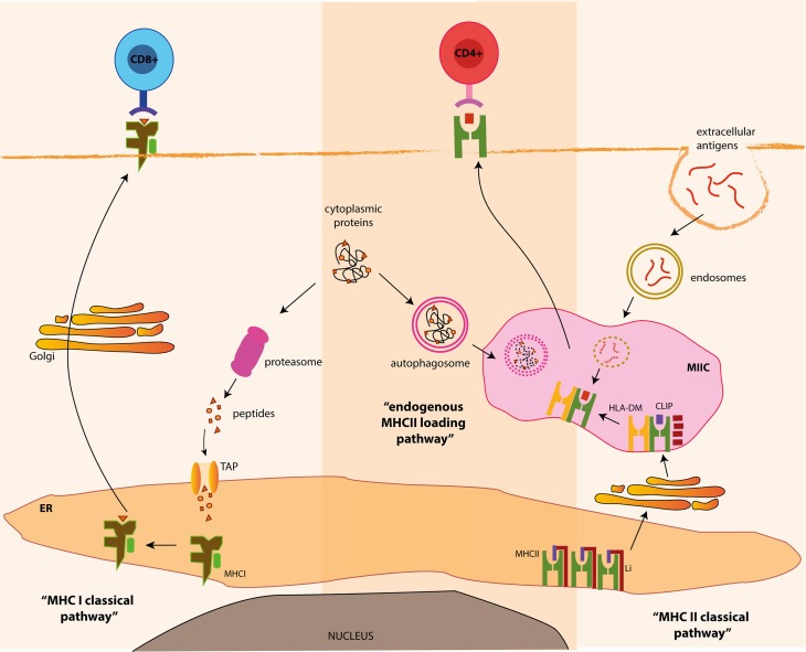
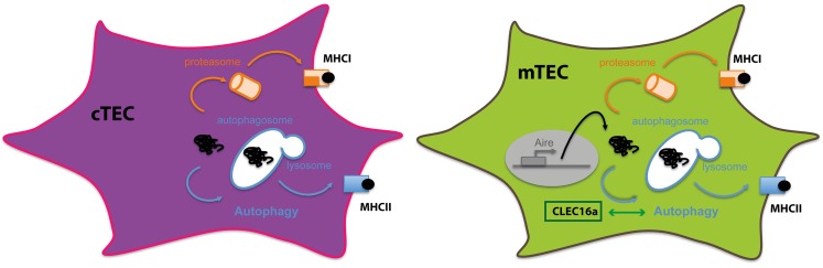
Although autophagy is a process that has been studied for several years its link with antigen presentation and T cell immunity has only recently emerged. Autophagy, which means "self-eating," is important to maintain cell homeostasis and refers to a collection of mechanisms that delivers intracellular material for degradation into lysosomes. Among them, macroautophagy pathway has many implications in different biological processes, including innate and adaptive immunity. In particular, macroautophagy can provide a substantial source of intracellular antigens for loading onto MHC class II molecules using the alternative MHC class II pathway. Through autophagosomes, endogenous self-antigens as well as antigens derived from intracellular pathogens can be delivered to MHC class II compartment and presented to CD4(+) T cells. The pathway will, therefore, impact both peripheral T cell tolerance and the pathogen specific immune response. This review will describe the contribution of autophagy to intracellular presentation of endogenous self- or pathogen-derived antigens via MHC class II and its consequences on CD4(+) T cell responses.

Keywords: CD4-positive T-lymphocytes; MHC class II; antigen presentation/processing; macroautophagy; tolerance mechanisms.


Figures

Figure 1
Figure 1
Pathways of autophagy. Autophagy can deliver cytosolic components to lysosomes for degradation via three different pathways. In chaperone-mediated autophagy (CMA), proteins having a KFERQ-like motif are translocated into the lysosome via the LAMP-2A transporter, with the help of Hsp70 chaperones. Microautophagy involves the sequestration of substrates via the invagination of the lysosomal membrane, while in macroautophagy, the substrates are engulfed in a double membrane vesicle, called autophagosome, which subsequently fuses with the lysosome to deliver its content for degradation.
Figure 2
Figure 2
MHC class I and class II processing pathways and autophagy. Classically, MHC class I bound antigens are originated from intracellular proteins through proteasomal proteolysis and are transferred to the outer membrane, where the resulting peptides are presented to CD8+ T cells. On the other hand, MHC class II products originate from extracellular antigens, which are endocytosed and delivered to MHC class II containing compartments (MIIC), where they meet newly generated MHC class II molecules. Alternatively, autophagy can deliver cytosolic antigens for MHC class II presentation, via the fusion of autophagosomes and MIIC, for the presentation of antigens to CD4+ T cells.
Figure 3
Figure 3
Autophagy in thymic epithelial cells: thymic epithelial cells are specialized in inducing tolerance. To sample intracellular- and extracellular-derived antigens, TECs rely on different mechanisms to present antigens via MHC class I or class II molecules. Autophagy in both cTECS and mTECS plays an important role in unconventional cytosolic peripheral self-antigens presentation via MHC class II molecules to establish CD4+ T cell tolerance.

References

    1. Ciechanover A. Proteolysis: from the lysosome to ubiquitin and the proteasome. Nat Rev Mol Cell Biol (2005) 6:79–87.10.1038/nrm1552 - DOI - PubMed
    1. Ashford TP, Porter KR. Cytoplasmic components in hepatic cell lysosomes. J Cell Biol (1962) 12:198–202.10.1083/jcb.12.1.198 - DOI - PMC - PubMed
    1. Novikoff AB, Essner E. Cytolysomes and mitochondrial degeneration. J Cell Biol (1962) 15:140–6.10.1083/jcb.15.1.140 - DOI - PMC - PubMed
    1. De Duve C, Pressman BC, Gianetto R, Wattiaux R, Appelmans F. Tissue fractionation studies. 6. Intracellular distribution patterns of enzymes in rat-liver tissue. Biochem J (1955) 60:604–17.10.1042/bj0600604 - DOI - PMC - PubMed
    1. Levine B, Klionsky DJ. Development by self-digestion: molecular mechanisms and biological functions of autophagy. Dev Cell (2004) 6:463–77.10.1016/S1534-5807(04)00099-1 - DOI - PubMed