. 2015 Sep 7:6:702.
doi: 10.3389/fpls.2015.00702. eCollection 2015.

Nuclear localization of the dehydrin OpsDHN1 is determined by histidine-rich motif

Affiliations

Nuclear localization of the dehydrin OpsDHN1 is determined by histidine-rich motif

Itzell E Hernández-Sánchez et al. Front Plant Sci. .

Abstract

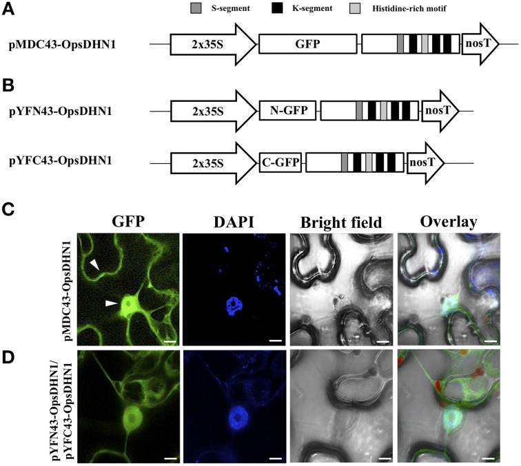
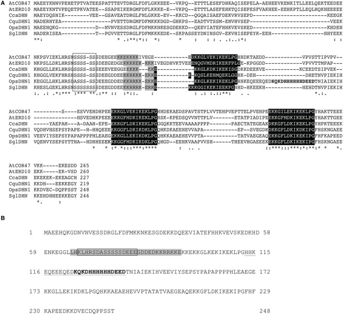
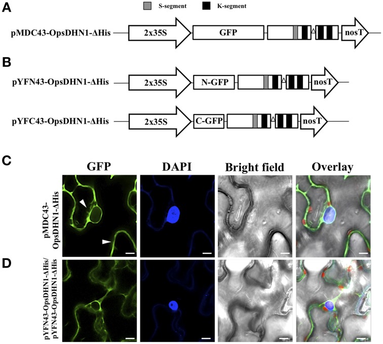
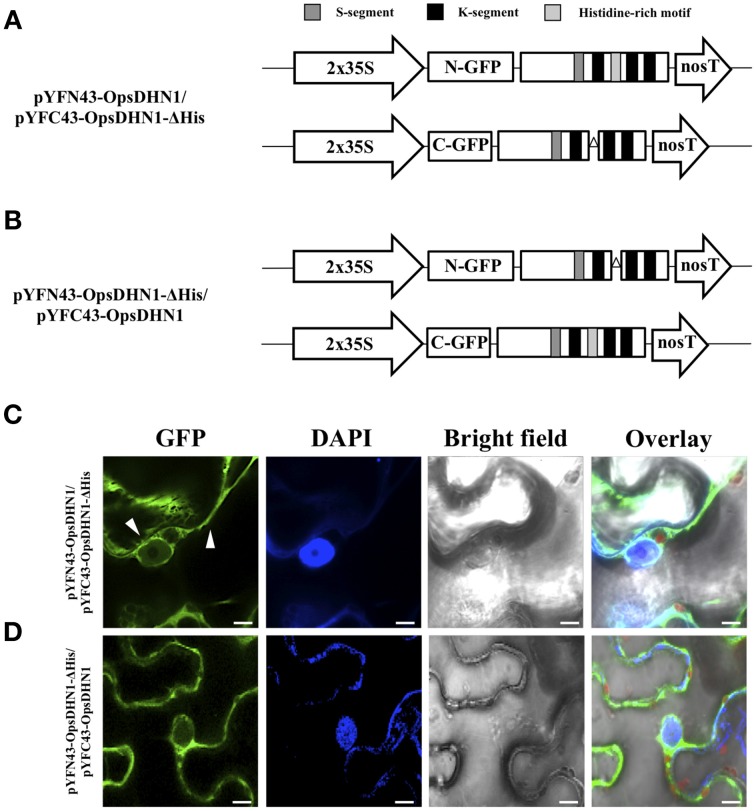
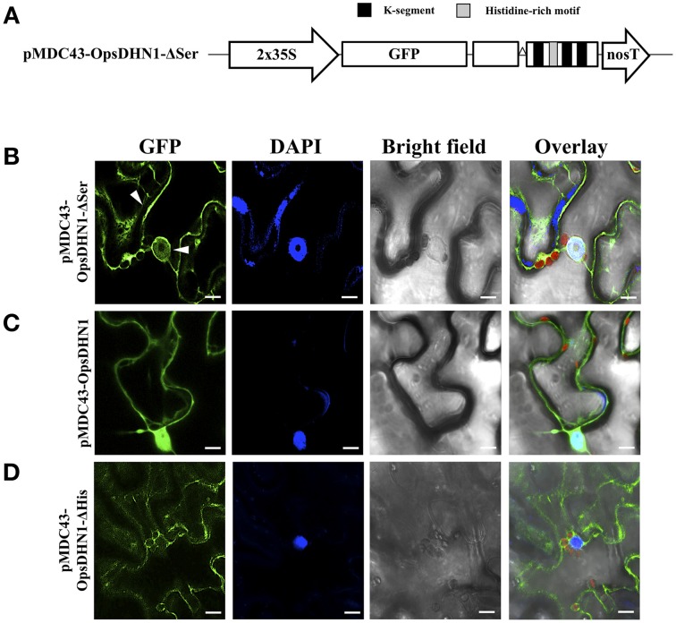
The cactus OpsDHN1 dehydrin belongs to a large family of disordered and highly hydrophilic proteins known as Late Embryogenesis Abundant (LEA) proteins, which accumulate during the late stages of embryogenesis and in response to abiotic stresses. Herein, we present the in vivo OpsDHN1 subcellular localization by N-terminal GFP translational fusion; our results revealed a cytoplasmic and nuclear localization of the GFP::OpsDHN1 protein in Nicotiana benthamiana epidermal cells. In addition, dimer assembly of OpsDHN1 in planta using a Bimolecular Fluorescence Complementation (BiFC) approach was demonstrated. In order to understand the in vivo role of the histidine-rich motif, the OpsDHN1-ΔHis version was produced and assayed for its subcellular localization and dimer capability by GFP fusion and BiFC assays, respectively. We found that deletion of the OpsDHN1 histidine-rich motif restricted its localization to cytoplasm, but did not affect dimer formation. In addition, the deletion of the S-segment in the OpsDHN1 protein affected its nuclear localization. Our data suggest that the deletion of histidine-rich motif and S-segment show similar effects, preventing OpsDHN1 from getting into the nucleus. Based on these results, the histidine-rich motif is proposed as a targeting element for OpsDHN1 nuclear localization.

Keywords: BiFC; dehydrin; histidine-rich motif; homodimer; nuclear/cytoplasmic localization.


Figures

Figure 1
Figure 1
In vivo OpsDHN1 subcellular localization and dimer formation. (A) Schematic representation of the pMDC43-OpsDHN1 construct. (B) Schematic representation of BiFC pYFN43- and pYFC43-OpsDHN1 vectors. (C) Cytoplasm and nuclear localization of the GFP::OpsDHN1 translational fusion in N. benthamiana epidermal cells. White arrowheads indicate cytosolic and nuclear signals. (D) Detection of the OpsDHN1 dimer formation by BiFC approach. The fluorescence was assessed by laser-scanning confocal microscopy. From left to right: the GFP and DAPI fluorescence spectrum, bright field, chlorophyll fluorescence and overlay signals. The S-segment, K-segments, and histidine-rich motif are depicted as dark-gray, black, and light-gray boxes, respectively. The scale bar corresponds to 10 μm.
Figure 2
Figure 2
Multiple sequence alignment of SK3 DHN proteins and the OpsDHN1. (A) Identical residues (asterisk) in the six DHN sequences and conserved amino acid substitutions (dots) are indicated, dashes show gaps in the amino acid sequences introduced to optimize alignment, conserved regions, and distinctive motifs of the group: S-segment (open box), poly-lysine rich sequence (gray shadow), K-segments (black shadow), and particular histidine-rich motif of OpsDHN1 (bold letters). (B) Schematic representation of bipartite NLS (gray shadow) and the metal-binding site (bold letters) in OpsDHN1 protein sequence, deleted regions: in OpsDHN1-ΔSer version the open box and for OpsDHN1-ΔHis version the underlined sequence.
Figure 3
Figure 3
The OpsDHN1 histidine-rich motif is required for its nuclear subcellular localization. (A) Schematic representation of the pMDC43-OpsDHN1-ΔHis construct. (B) Schematic representation of BiFC pYFN43- and pYFC43- OpsDHN1-ΔHis vectors. (C) Cytoplasm localization of GFP::OpsDHN1-ΔHis translational fusion in N. benthamiana leaves. White arrowheads indicate cytosolic and nuclear compartments. (D) BiFC assay of OpsDHN1-ΔHis version. Transient fluorescent expression was analyzed by laser-scanning confocal microscopy. From left to right: the GFP and DAPI fluorescence spectrum, bright field, chlorophyll fluorescence and overlay signals. The S-segment and K-segments are depicted as dark-gray box and black boxes, respectively. The deleted histidine-rich motif is represented by an open triangle. The scale bar corresponds to 10 μm.
Figure 4
Figure 4
Visualization of OpsDHN1/OpsDHN1-ΔHis dimer formation using BiFC approach. (A,B) Schematic representation of OpsDHN1 derived versions cloned into pYFN43 and pYFC43 vectors. (C,D) BiFC analysis of OpsDHN1/OpsDHN1-ΔHis and swapped constructs. White arrowheads indicate cytosolic and nuclear compartments. The fluorescence was assessed by laser-scanning confocal microscopy. From left to right: the GFP and DAPI fluorescence spectrum, bright field, chlorophyll fluorescence and overlay signals. The S-segment, K-segments, and histidine-rich motif are depicted as dark-gray, black, and light-gray boxes, respectively. The deleted histidine-rich motif is represented by open triangle. The scale bar corresponds to 10 μm.
Figure 5
Figure 5
The OpsDHN1 S-segment is involved in its nuclear location. (A) Schematic representation of the pMDC43-OpsDHN1-ΔSer construct. Fluorescent visualization of (B) GFP::OpsDHN1-ΔSer, (C) GFP::OpsDHN1, (D) GFP::OpsDHN1-ΔHis translational fusions in N. benthamiana leaves. White arrowheads indicate cytosol and nuclear signals. The fluorescence was examined by laser-scanning confocal microscopy. From left to right: the GFP and DAPI fluorescence spectrum, bright field, chlorophyll fluorescence and overlay signals. The K-segments and histidine-rich motif are depicted as black and light-gray boxes, respectively. The deleted S-segment is represented by open triangle. The scale bar corresponds to 10 μm.

References

    1. Alsheikh M. K., Heyen B. J., Randall S. K. (2003). Ion binding properties of the dehydrin ERD14 are dependent upon phosphorylation. J. Biol. Chem. 278, 40882–40889. 10.1074/jbc.M307151200 - DOI - PubMed
    1. Belda-Palazón B., Ruiz L., Martí E., Tárraga S., Tiburcio A. F., Culiáñezo F., et al. . (2012). Aminopropyltransferases involved in polyamine biosynthesis localize preferentially in the nucleus of plant cells. PLoS ONE 7:e46907. 10.1371/journal.pone.0046907 - DOI - PMC - PubMed
    1. Briesemeister S., Rahnenführer J., Kohlbacher O. (2010). YLoc-an interpretable web server for predicting subcellular localization. Nucleic Acids Res. 38, 497–502. 10.1093/nar/gkq477 - DOI - PMC - PubMed
    1. Carjuzaa P., Castellión M., Distefano A. J., Del V. M., Maldonado S. (2008). Detection and subcellular localization of dehydrin-like proteins in quinoa (Chenopodium quinoa Willd.) embryos. Protoplasma 233, 149–156. 10.1007/s00709-008-0300-4 - DOI - PubMed
    1. Close T. J. (1996). Dehydrins: emergence of a biochemical role of a family of plant dehydration proteins. Physiol. Plant. 97, 795–803. 10.1111/j.1399-3054.1996.tb00546.x - DOI