Review
. 2015 Sep 25:3:56.
doi: 10.3389/fcell.2015.00056. eCollection 2015.

The different facets of organelle interplay-an overview of organelle interactions

Affiliations
Review

The different facets of organelle interplay-an overview of organelle interactions

Michael Schrader et al. Front Cell Dev Biol. .

Abstract

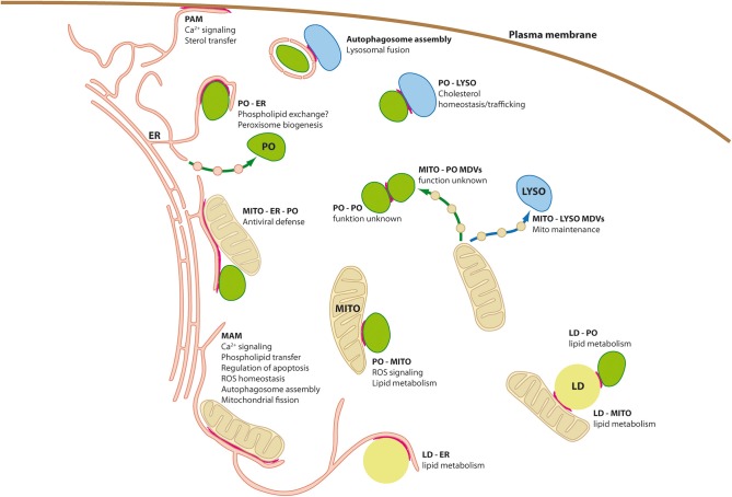
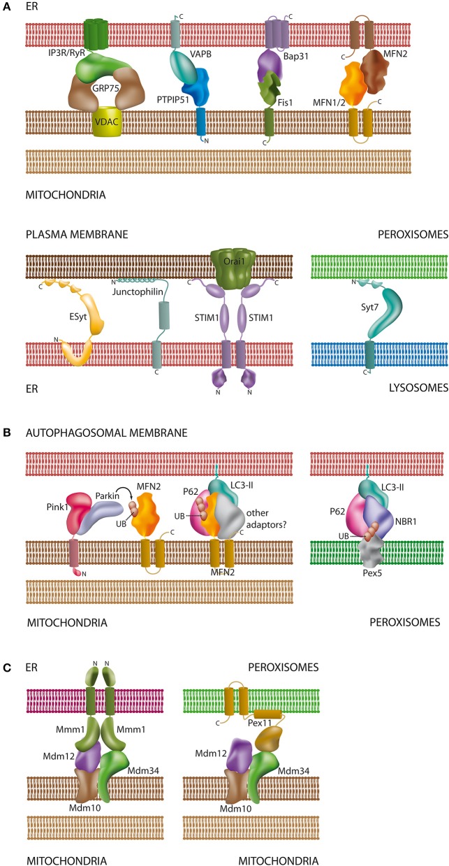
Membrane-bound organelles such as mitochondria, peroxisomes, or the endoplasmic reticulum (ER) create distinct environments to promote specific cellular tasks such as ATP production, lipid breakdown, or protein export. During recent years, it has become evident that organelles are integrated into cellular networks regulating metabolism, intracellular signaling, cellular maintenance, cell fate decision, and pathogen defence. In order to facilitate such signaling events, specialized membrane regions between apposing organelles bear distinct sets of proteins to enable tethering and exchange of metabolites and signaling molecules. Such membrane associations between the mitochondria and a specialized site of the ER, the mitochondria associated-membrane (MAM), as well as between the ER and the plasma membrane (PAM) have been partially characterized at the molecular level. However, historical and recent observations imply that other organelles like peroxisomes, lysosomes, and lipid droplets might also be involved in the formation of such apposing membrane contact sites. Alternatively, reports on so-called mitochondria derived-vesicles (MDV) suggest alternative mechanisms of organelle interaction. Moreover, maintenance of cellular homeostasis requires the precise removal of aged organelles by autophagy-a process which involves the detection of ubiquitinated organelle proteins by the autophagosome membrane, representing another site of membrane associated-signaling. This review will summarize the available data on the existence and composition of organelle contact sites and the molecular specializations each site uses in order to provide a timely overview on the potential functions of organelle interaction.

Keywords: MAM; PAM; endoplasmic reticulum; intracellular signaling; membrane contact sites; mitochondria; organelle dynamics; peroxisomes.


Figures

Figure 1
Figure 1
Organelle interplay and interorganellar contacts. Schematic diagram of a mammalian cell depicting organelle interplay and interorganellar membrane contacts (highlighted by red lines). ER, endoplasmic reticulum; LD, lipid droplets; LYSO, lysosome; MAM, mitochondria associated-membrane; MDVs, mitochondria derived vesicles; MITO, mitochondrium; PAM, plasma membrane-associated membrane; PO, peroxisome.
Figure 2
Figure 2
Schematic overview of proteins and lipids involved in the interaction of organelles. (A) Tethering complexes in mammals: unlike in yeast species only a few protein complexes have been characterized at the molecular level and involve protein-protein and protein-lipid contacts [see Sections Connections between the ER and the Plasma Membrane, The Mitochondria-associated Membrane of the ER (MAM), Interplay between Peroxisomes and Mitochondria, and Lysosomal Interactions and Autophagy]. Part of the tethering complexes shown may only comprise core complexes, which will interact with additional proteins for regulatory purposes; (B) contacts between mitochondria/peroxisomes and the autophagosomal membrane: both organelles require ubiquitination of membrane proteins for recognition by the autophagosome. In addition to MFN2 (Mitochondria) and Pex5 (Peroxisomes) other ubiquitinated organelle proteins have been described to participate in autophagosomal contacts [see Sections The Mitochondria-associated Membrane of the ER (MAM) and Lysosomal Interactions and Autophagy]; (C) ERMES as a multifunctional tethering complex in yeast: unlike mammals, yeast species possess the ERMES oligomeric complex at the mitochondrial membrane. ERMES forms complexes with the ER and peroxisomes [see Sections The Mitochondria-associated Membrane of the ER (MAM) and Interplay between Peroxisomes and Mitochondria]. In addition, a considerable number of other tethering complexes (not shown) have been described in yeast (Prinz, 2014). For molecular details and references of the depicted complexes please refer to the corresponding sections of this review. Membrane spanning α-helices in the proteins are depicted as cylindrical segments; C- and N-termini are marked with the corresponding letters.

References

    1. Agrawal G., Joshi S., Subramani S. (2011). Cell-free sorting of peroxisomal membrane proteins from the endoplasmic reticulum. Proc. Natl. Acad. Sci. U.S.A. 108, 9113–9118. 10.1073/pnas.1018749108 - DOI - PMC - PubMed
    1. Antonenkov V. D., Grunau S., Ohlmeier S., Hiltunen J. K. (2010). Peroxisomes are oxidative organelles. Antioxid. Redox Signal. 13, 525–537. 10.1089/ars.2009.2996 - DOI - PubMed
    1. Arasaki K., Shimizu H., Mogari H., Nishida N., Hirota N., Furuno A., et al. . (2015). A role for the ancient SNARE syntaxin 17 in regulating mitochondrial division. Dev. Cell 32, 304–317. 10.1016/j.devcel.2014.12.011 - DOI - PubMed
    1. Baerends R. J., Rasmussen S. W., Hilbrands R. E., Van Der Heide M., Faber K. N., Reuvekamp P. T., et al. . (1996). The Hansenula polymorpha PER9 gene encodes a peroxisomal membrane protein essential for peroxisome assembly and integrity. J. Biol. Chem. 271, 8887–8894. 10.1074/jbc.271.15.8887 - DOI - PubMed
    1. Bas Orth C., Schultz C., Müller C. M., Frotscher M., Deller T. (2007). Loss of the cisternal organelle in the axon initial segment of cortical neurons in synaptopodin-deficient mice. J. Comp. Neurol. 504, 441–449. 10.1002/cne.21445 - DOI - PubMed

LinkOut - more resources