. 2015 Nov 20;290(47):28321-28328.
doi: 10.1074/jbc.M115.683417. Epub 2015 Oct 6.

Mitogen-activated Protein Kinase (MAPK) Activated by Prostaglandin E2 Phosphorylates Connexin 43 and Closes Osteocytic Hemichannels in Response to Continuous Flow Shear Stress

Affiliations

Mitogen-activated Protein Kinase (MAPK) Activated by Prostaglandin E2 Phosphorylates Connexin 43 and Closes Osteocytic Hemichannels in Response to Continuous Flow Shear Stress

Manuel A Riquelme et al. J Biol Chem. .

Abstract

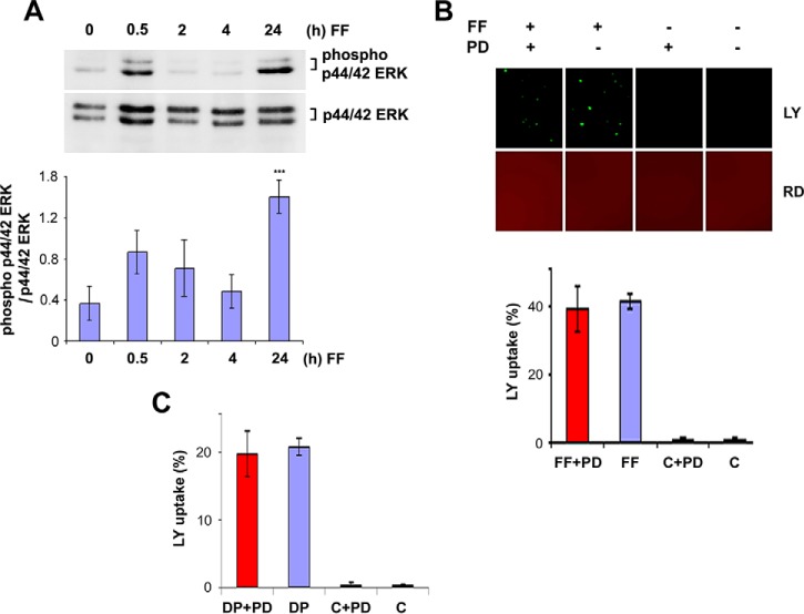
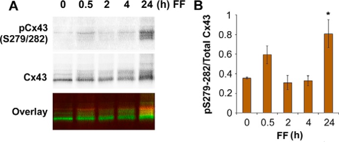
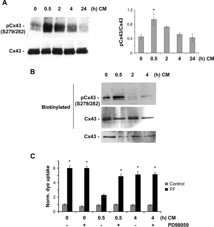
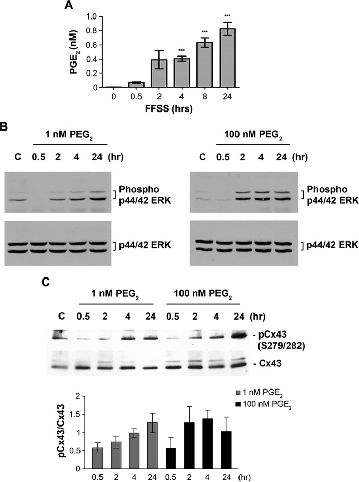
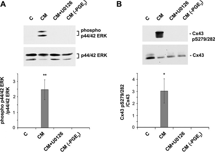
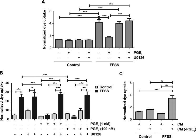
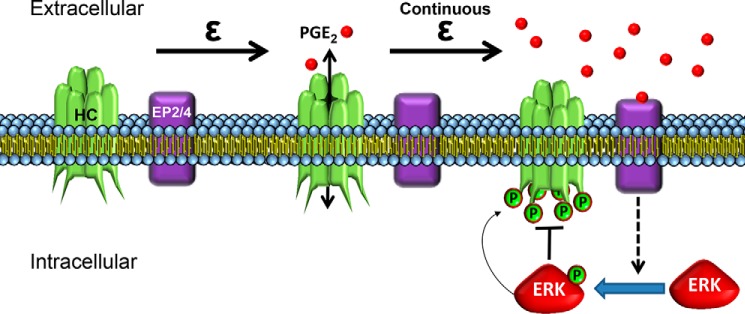
Cx43 hemichannels serve as a portal for the release of prostaglandins, a critical process in mediating biological responses of mechanical loading on bone formation and remodeling. We have previously observed that fluid flow shear stress (FFSS) opens hemichannels; however, sustained FFSS results in hemichannel closure, as continuous opening of hemichannels is detrimental to cell viability and bone remodeling. However, the mechanism that regulates the closure of the hemichannels is unknown. Here, we show that activation of p44/42 ERK upon continuous FFSS leads to Cx43 phosphorylation at Ser(279)-Ser(282), sites known to be phosphorylated sites by p44/42 MAPK. Incubation of osteocytic MLO-Y4 cells with conditioned media (CM) collected after continuous FFSS increased MAPK-dependent phosphorylation of Cx43. CM treatment inhibited hemichannel opening and this inhibition was reversed when cells were pretreated with the MAPK pathway inhibitor. We found that prostaglandin E2 (PGE2) accumulates in the CM in a time-dependent manner. Treatment with PGE2 increased phospho-p44/42 ERK levels and also Cx43 phosphorylation at Ser(279)-Ser(282) sites. Depletion of PGE2 from CM, and pre-treatment with a p44/42 ERK pathway-specific inhibitor, resulted in a complete inhibition of ERK-dependent Cx43 phosphorylation and attenuated the inhibition of hemichannels by CM and PGE2. Consistently, the opening of hemichannels by FFSS was blocked by PGE2 and CM and this blockage was reversed by U0126 and the CM depleted of PGE2. A similar observation was also obtained in isolated primary osteocytes. Together, results from this study suggest that extracellular PGE2 accumulated after continuous FFSS is responsible for activation of p44/42 ERK signaling and subsequently, direct Cx43 phosphorylation by activated ERK leads to hemichannel closure.

Keywords: Hemichannels; Mechanical stimulation; channel activation; connexin; mitogen-activated protein kinase (MAPK); osteocyte; phosphorylation.


Figures

FIGURE 1.
FIGURE 1.
Continuous FFSS for 24 h activates p44/42 MAPK, but inhibition of activated MAPK does not affect initial Cx43 opening of hemichannels by FFSS. A, MLO-Y4 cells were subjected to FFSS for 0.5, 2, 4, and 24 h, and the lysates collected were immunoblotted using anti-phospho-p44/42 ERK or p44/42 ERK (total ERK) antibody. The ratio of mean band intensities of phospho-p44/42 ERK to total p44/42 ERK was plotted as showing a significant increase in phospho-p44/42 ERK levels after 24-h FFSS. 24 h versus unstimulated cells; ***, p < 0.001, n = 3, and the data are presented as mean ± S.E. B, effect of PD98059, an inhibitor of MAPK, on Cx43 hemichannel opening was determined by treating MLO-Y4 cells with 50 μm PD98059 for 16 h and subjecting the cells to FFSS of 16 dynes/cm2 for 30 min. Untreated control (C) and untreated plus PD98059 cells (C+PD) were used as controls. An increase in dye uptake was observed in PD98059-treated cells when subjected to FFSS (FF+PD) and the percentage of dye uptake was similar to that of the untreated cells subjected to FFSS (FF). C, a similar observation was obtained when cells were mechanically stimulated by dropping medium onto the cells followed by dye uptake analyses. PD98509-treated (DP+PD) cells and untreated cells (DP) showed an increase in dye uptake percentage after stimulation when compared with that of the controls (C+PD and C). All the data are presented as mean ± S.E., n = 3.
FIGURE 2.
FIGURE 2.
Activation of p44/42 ERK and increase of Cx43 phosphorylation at Ser279-Ser282 residues after continuous FFSS for 24 h. A, MLO-Y4 cell lysates were collected after continuous FFSS for 24 h and immunoblotted using phospho-specific Cx43 Ser279-Ser282 antibody. pCx43S279/282 is indicated in red and total Cx43 in green (bottom panel). B, the ratio of mean band intensities of Cx43 phospho-Ser279-Ser282 and total Cx43 was calculated and was plotted as a graph. All data are presented as mean ± S.E. Non-treated (0 h) versus 24 h, *, p < 0.05, n = 5.
FIGURE 3.
FIGURE 3.
CM collected after 24-h FFSS promotes Cx43 phosphorylation at Ser279-Ser282 residues and inhibits FFSS-induced opening of Cx43 hemichannels. A, MLO-Y4 cells were treated with CM (collected after continuous FFSS for 24 h) for 30 min. Cell lysates were immunoblotted with antibody against Cx43 phospho-specific Ser279-Ser282 or total Cx43 protein. Band intensity was quantified and the graph was plotted using ratio of Cx43 phospho-Ser279-Ser282 and total Cx43. The data are presented as mean ± S.E. CM-treated versus untreated cells: *, p < 0.05; n = 3. B, MLO-Y4 cells were treated with CM (collected after continuous FFSS for 24 h) for 30 min and a cell surface biotinylation assay was performed. Biotinylated samples and total lysates were immunoblotted with antibody against Cx43 phospho-specific Ser279-Ser282 or total Cx43 protein. C, MLO-Y4 cells were pretreated with CM (collected after continuous FFSS for 24 h) for 0.5 and 4 h in the absence or presence of PD98059, subjected to FFSS for 30 min, and assayed for LY dye uptake. Treatment with CM for 30 min significantly blocked dye uptake induced by FFSS, whereas preincubation with CM for 4 h did not have such effect. Pretreatment of MLO-Y4 cells with 50 μm PD98059, a MAPK inhibitor, resulted in a significant reversal of inhibition of dye uptake by the pretreatment of CM for 30 min, not 4 h. With FF, 0.5 h treatment versus all other conditions, *, p < 0.05, n = 3.
FIGURE 4.
FIGURE 4.
Extracellular PGE2 is accumulated during continuous FFSS and PGE2 increases phosphorylation of p44/42 ERK and Cx43 at Ser279-Ser282 residues. A, the concentrations of PGE2 in CM collected from different periods of continuous FFSS were determined using PGE2 EIA kit. 4, 8, and 24 h versus 0 min (un-stimulated); ***, p < 0.001. B and C, MLO-Y4 cells were treated with 1 or 10 nm PGE2 for 0.5, 2, 4, and 24 h and cell lysates were immunoblotted with anti-phospho-p44/42 ERK and p44/42 ERK antibodies (B) or anti-Cx43 Ser279-Ser282 and Cx43 antibody (C). The band intensities were quantified and graphed using the ratios of phospho- versus total p44/42 ERK or Cx43 levels. All the data are presented as mean ± S.E., n = 3.
FIGURE 5.
FIGURE 5.
FF-CM depleted of PGE2 and U0126 treatment results in inhibition of p44/42 ERK and Cx43 phosphorylation. MLO-Y4 cell lysates were treated with CM, PGE2-depleted CM, or pretreated with U0126 prior to CM treatment and cell lysates were immunoblotted with anti-phospho-p44/42 ERK and total ERK (A) or anti-Cx43 Ser279-Ser282 and Cx43 antibodies (B). The band intensity was quantified and the ratio of phospho- versus total p44/42 ERK and Cx43 was calculated (A and B, lower panels). All the data are presented as mean ± S.E., n = 3. CM versus C, CM+U0126 and CM-PGE2; **, p < 0.01. *, p < 0.05.
FIGURE 6.
FIGURE 6.
Inhibition of MAPK and depletion of PGE2 from FF-CM attenuates the inhibitory effect of PGE2 or FF-CM on hemichannel opening in response to FFSS. A, MLO-Y4 cells were treated with 1 nm PGE2 for 24 h and then with or without U0126 for 30 min before subjecting cells to FFSS for 30 min or non-FFSS control. B, primary mouse osteocytes were treated with 1 or 100 nm PGE2 for 24 h and then with or without U0126 for 30 min before subjecting cells to FFSS for 30 min or non-FFSS control. C, MLO-Y4 cells were treated with FF-CM (collected after 24 h of FFSS) or FF-CM depleted of PGE2 (CM(-PGE2)) for 24 h before subjecting the cells to FFSS for 30 min or non-FF control. LY dye uptake assay was performed and quantified. All the data are presented as mean ± S.E., n = 3. ***, p < 0.001; **, p < 0.01; *, p < 0.05.
FIGURE 7.
FIGURE 7.
A schematic model showing the hemichannel closure after long FFSS is regulated by Cx43 phosphorylation by MAPK, which is activated by extracellular PGE2 released by hemichannels via feedback inhibition mechanism. During short term FFSS, Cx43 hemichannels (HC) in osteocytes are induced open, which allows the release of bone modulating factors such as PGE2. A sustained mechanical stimulation causes Cx43 hemichannel closure due to accumulation of PGE2 in the extracellular environment. The accumulated PGE2, likely through the EP2/EP4 receptor, activates p44/42 MAPK and increases Cx43 phosphorylation at Ser279-Ser282 residues thereby leading to closure of Cx43 hemichannels.

References

    1. Ajubi N. E., Klein-Nulend J., Nijweide P. J., Vrijheid-Lammers T., Alblas M. J., and Burger E. H. (1996) Pulsating fluid flow increases prostaglandin production by cultured chicken osteocytes: a cytoskeleton-dependent process. Biochem. Biophys. Res. Commun. 225, 62–68 - PubMed
    1. Cherian P. P., Siller-Jackson A. J., Gu S., Wang X., Bonewald L. F., Sprague E., and Jiang J. X. (2005) Mechanical strain opens connexin 43 hemichannels in osteocytes: a novel mechanism for the release of prostaglandin. Mol. Biol. Cell 16, 3100–3106 - PMC - PubMed
    1. Genetos D. C., Kephart C. J., Zhang Y., Yellowley C. E., and Donahue H. J. (2007) Oscillating fluid flow activation of gap junction hemichannels induces ATP release from MLO-Y4 osteocytes. J. Cell. Physiol. 212, 207–214 - PMC - PubMed
    1. Klein-Nulend J., Semeins C. M., Ajubi N. E., Nijweide P. J., and Burger E. H. (1995) Pulsting fluid flow increases nitric oxide (NO) synthesis by osteocytes but not periosteal fibroblasts-correlation with prostaglandin upregulation. Biochem. Biophys. Res. Commun. 217, 640–648 - PubMed
    1. Cherian P. P., Cheng B., Gu S., Sprague E., Bonewald L. F., and Jiang J. X. (2003) Effects of mechanical strain on the function of gap junctions in osteocytes are mediated through the prostaglandin EP2 receptor. J. Biol. Chem. 278, 43146–43156 - PubMed

Publication types

LinkOut - more resources