. 2015 Dec 4;290(49):29542-54.
doi: 10.1074/jbc.M115.691592. Epub 2015 Oct 6.

Dopamine Transporter Activity Is Modulated by α-Synuclein

Affiliations

Dopamine Transporter Activity Is Modulated by α-Synuclein

Brittany Butler et al. J Biol Chem. .

Abstract

The duration and strength of the dopaminergic signal are regulated by the dopamine transporter (DAT). Drug addiction and neurodegenerative and neuropsychiatric diseases have all been associated with altered DAT activity. The membrane localization and the activity of DAT are regulated by a number of intracellular proteins. α-Synuclein, a protein partner of DAT, is implicated in neurodegenerative disease and drug addiction. Little is known about the regulatory mechanisms of the interaction between DAT and α-synuclein, the cellular location of this interaction, and the functional consequences of this interaction on the basal, amphetamine-induced DAT-mediated dopamine efflux, and membrane microdomain distribution of the transporter. Here, we found that the majority of DAT·α-synuclein protein complexes are found at the plasma membrane of dopaminergic neurons or mammalian cells and that the amphetamine-mediated increase in DAT activity enhances the association of these proteins at the plasma membrane. Further examination of the interaction of DAT and α-synuclein revealed a transient interaction between these two proteins at the plasma membrane. Additionally, we found DAT-induced membrane depolarization enhances plasma membrane localization of α-synuclein, which in turn increases dopamine efflux and enhances DAT localization in cholesterol-rich membrane microdomains.

Keywords: addiction; amphetamine; dopamine; dopamine efflux; dopamine transporter; neurochemistry; synuclein.


Figures

FIGURE 1.
FIGURE 1.
Dopamine transporter and α-synuclein are co-localized at the plasma membrane. A, paraformaldehyde fixed but not permeabilized dopamine neurons were labeled for DAT using an antibody against the second extracellular domain of DAT (red) followed by permeabilization and labeling of α-synuclein (green). Scale bar, 50 μm. B, in CHO cells co-expressing YFP-DAT and α-synuclein (untagged), the two proteins are co-localized at the plasma membrane. In CHO cells not expressing DAT, α-synuclein is distributed uniformly within the cell. Scale bar, 20 μm. Figure shows surface membrane localization of DAT in the absence of α-synuclein. It has been shown that if α-synuclein (α-syn) is expressed in the CHO and HEK cells, its expression level is below the detection limit of Western blot and immunocytochemistry assays. C, line intensity analysis reveals an enrichment of α-synuclein at the cell surface of DAT-expressing cells. The line analyses of CHO cells expressing both DAT (green signal) and α-synuclein (red signal) show the intensity fluorescent profile of α-synuclein peaks at the plasma membrane that overlaps the intensity fluorescent profile for DAT at the plasma membrane indicated by arrowheads at the bottom of the graph. D, in the absence of DAT, the line intensity profile of α-synuclein does not peak at the plasma membrane (n = 10 cells from three independent experiments). AFU, arbitrary fluorescence unit.
FIGURE 2.
FIGURE 2.
FRET microscopy reveals an association between eCFP-α-synuclein and YFP-DAT at the plasma membrane. A, schematic of FRET microscopy according to the emission and excitation spectrum associated with each fluorophore. B, TH::RFP-positive dopamine neurons (inset) expressing YFP-DAT and eCFP-α-synuclein (α-syn). C, bar graph shows relative changes in the YFP and eCFP, fluorescence intensities in dopamine neurons. There is a significant increase in eCFP fluorescence intensity (donor) as YFP (acceptor) fluorescence intensity decreases. All values are analyzed by a paired t test, *, p < 0.05; ***, p < 0.001. AFU, arbitrary fluorescence unit. D, bar graph of FRET efficiency for control groups (positive and negative control groups) and experimental group (YFP-DAT/eCFP-α-synuclein ± AMPH). All values are analyzed by one-way ANOVA followed by Tukey post hoc analysis. *, p < 0.05; **, p < 0.01; ***, p < 0.001; ###, p < 0.001.
FIGURE 3.
FIGURE 3.
Co-immunoprecipitation and cell surface membrane biotinylation suggest DAT and α-synuclein associate at the plasma membrane. A, co-IP of YFP-DAT in CHO cells expressing vector or eCFP-α-synuclein show co-immunoprecipitation of DAT and eCFP-α-synuclein (α-syn). B, control experiments demonstrate the specificity of the DAT antibody and the signals detected in Fig. 3A. C, biotinylation of CHO cells expressing YFP-DAT and eCFP vector, eCFP-α-synuclein, or untagged α-synuclein reveals an interaction between DAT and α-synuclein at the plasma membrane. IP, immunoprecipitation.
FIGURE 4.
FIGURE 4.
Interaction between DAT and α-synuclein is transient and dynamic. The lateral mobility of eCFP-α-synuclein was examined in the presence or absence of YFP-DAT in CHO cells. A, illustration of three possible FRAP recovery profiles of two proteins in living cells. The schematic is modified from Digby et al. (36). B shows averaged normalized FRAP recovery curve for eCFP vector only, eCFP vector in the presence of YFP-DAT, eCFP-α-synuclein only, and eCFP-α-synuclein in the presence of YFP-DAT. The lateral mobility of eCFP-empty vector was not affected by the presence or absence of DAT. eCFP-α-synuclein lateral mobility and mobile fraction were significantly decreased when YFP-DAT is co-expressed in the cells. This suggests that YFP-DAT and eCFP-α-synuclein interact. C, interaction between YFP-DAT and eCFP-α-synuclein increases the t½ of the maximal recovery of eCFP signal and decreases the mobile fraction of eCFP-α-synuclein molecules during the time frame shown (data not shown). D, presence or absence of eCFP-α-synuclein molecules in the cell does not change the recovery profile of YFP-DAT molecules. The black squares show recovery profile of YFP-DAT molecules when eCFP-α-synuclein is co-expressed. The red circles depict the recovery profile of DAT molecule in the absence of eCFP-α-synuclein. E, bar graph compares the t½ of the maximal recovery of YFP signal in the presence or absence of eCFP-α-synuclein. *, p < 0.05.
FIGURE 5.
FIGURE 5.
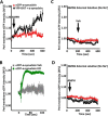
AMPH-mediated DAT activation enhances localization of eCFP-α-synuclein at or near the cell surface membrane. A, in cells expressing YFP-DAT, AMPH exposure (10 μm) increases the eCFP fluorescence intensity at or near the plasma membrane, suggesting increased localization of α-synuclein at or near the cell surface (black squares) of DAT cells. There is no change in the eCFP-fluorescence intensity at or near the surface membrane following AMPH treatment in the absence of YFP-DAT (red circle). B, there is a robust and rapid increase of α-synuclein localization at or near the plasma membrane following KCl-induced membrane depolarization (green circles). The presence of Na+ ions in the external solution is required for the AMPH-induced DAT-mediated recruitment of α-synuclein to the surface membrane. C, vehicle (Veh) treatment in the external solution containing NMDG does not affect membrane localization of eCFP-α-synuclein in the presence or absence of DAT. D, when NMDG+ is substituted for Na+ in the external solution, AMPH exposure did not increase membrane localization of eCFP-α-synuclein. AFU, arbitrary fluorescence unit.
FIGURE 6.
FIGURE 6.
Amphetamine exposure enhances the DAT and α-synuclein association at the plasma membrane. A, representative blots of co-immunoprecipitation experiments showing DAT protein is immunoprecipitated (IP) with an antibody to DAT and probed for α-synuclein following AMPH exposure. DAT immunoprecipitation pulled down significantly more α-synuclein in the presence of AMPH as compared with vehicle (Veh)-treated cells. B, bar graph depicts normalized ratio of pulled down α-synuclein to DAT (n = 3 independent experiments, p < 0.05, Student's t test). C, dopamine neurons labeled for DAT (red) and α-synuclein (green) treated with vehicle (Veh) or AMPH (right). AMPH exposure increased co-localization of two proteins at the plasma membrane (inset). Scale bar, 50 and 30 μm, respectively. D, normalized bar graph shows a significant increase in co-localization of DAT and α-synuclein at the membrane upon AMPH treatment (n = 17–20, from five independent experiments, p < 0.05, Student's t test). E, AMPH exposure increases FRET between DAT and α-synuclein at the membrane. The FRET experiments were performed as described in Fig. 2. Bar graph shows a significant increase in normalized FRET following AMPH exposure as compared with vehicle control group (n = 10–15, from three independent experiments, p < 0.05, Student's t test). *, p < 0.05; **, p < 0.01; ***, p < 0.001.
FIGURE 7.
FIGURE 7.
Elevated intracellular α-synuclein increases basal and AMPH-induced dopamine efflux. A, representative amperometric currents recorded from YFP-DAT only and YFP-DAT overexpressing eCFP-α-synuclein cells loaded with 1 μm dopamine for 20 min at 37 °C. Arrows indicate application of AMPH (10 μm). B, quantification of AMPH-induced increase in amperometric current in YFP-DAT only and YFP-DAT cells overexpressing CFP-α-synuclein (p < 0.001 by Student's t test; n = 6–8, from three independent experiments). C shows analysis of nomifensine reduction (10 μm) of basal dopamine efflux (p < 0.001 by Student's t test; n = 6–8, from three independent experiments). **, p < 0.01; ***, p < 0.001.
FIGURE 8.
FIGURE 8.
Elevation of intracellular α-synuclein increases DAT co-localization with the GM1 gangliosides in the cholesterol-rich membrane microdomains. A, dopaminergic neurons labeled with CTxB-Alexa (green) and DAT (red) without (right) and with (left) eCFP-α-synuclein overexpression. CTxB labels GM1 gangliosides that are distributed in cholesterol-rich membrane microdomains. CFP-α-synuclein overexpression increases DAT/CTx-B co-localization at the membrane (yellow) compared with control group (see merged images). B, normalized bar graph shows CTxB-647 and YFP-DAT co-localization in HEK cells ± eCFP-α-synuclein following vehicle (VEH) or amphetamine treatment.(n = 21–25 cells from three independent experiments, p < 0.05, one-way ANOVA followed by Tukey post hoc analysis). Scale bar, 10 μm. **, p < 0.01; ***, p < 0.001.

References

    1. Stefansson H., Ophoff R. A., Steinberg S., Andreassen O. A., Cichon S., Rujescu D., Werge T., Pietiläinen O. P., Mors O., Mortensen P. B., Sigurdsson E., Gustafsson O., Nyegaard M., Tuulio-Henriksson A., Ingason A., et al. (2009) Common variants conferring risk of schizophrenia. Nature 460, 744–747 - PMC - PubMed
    1. Nemani V. M., Lu W., Berge V., Nakamura K., Onoa B., Lee M. K., Chaudhry F. A., Nicoll R. A., and Edwards R. H. (2010) Increased expression of α-synuclein reduces neurotransmitter release by inhibiting synaptic vesicle reclustering after endocytosis. Neuron 65, 66–79 - PMC - PubMed
    1. Varrone A., Marek K. L., Jennings D., Innis R. B., and Seibyl J. P. (2001) [123I]β-CIT SPECT imaging demonstrates reduced density of striatal dopamine transporters in Parkinson's disease and multiple system atrophy. Mov. Disord. 16, 1023–1032 - PubMed
    1. Braak H., Del Tredici K., Rüb U., de Vos R. A., Jansen Steur E. N., and Braak E. (2003) Staging of brain pathology related to sporadic Parkinson's disease. Neurobiol. Aging 24, 197–211 - PubMed
    1. Rocha B. A., Fumagalli F., Gainetdinov R. R., Jones S. R., Ator R., Giros B., Miller G. W., and Caron M. G. (1998) Cocaine self-administration in dopamine-transporter knockout mice. Nat. Neurosci. 1, 132–137 - PubMed

Publication types

LinkOut - more resources