. 2016 May;41(6):1530-9.
doi: 10.1038/npp.2015.311. Epub 2015 Oct 7.

GABAergic Signaling within a Limbic-Hypothalamic Circuit Integrates Social and Anxiety-Like Behavior with Stress Reactivity

Affiliations

GABAergic Signaling within a Limbic-Hypothalamic Circuit Integrates Social and Anxiety-Like Behavior with Stress Reactivity

Brent Myers et al. Neuropsychopharmacology. 2016 May.

Abstract

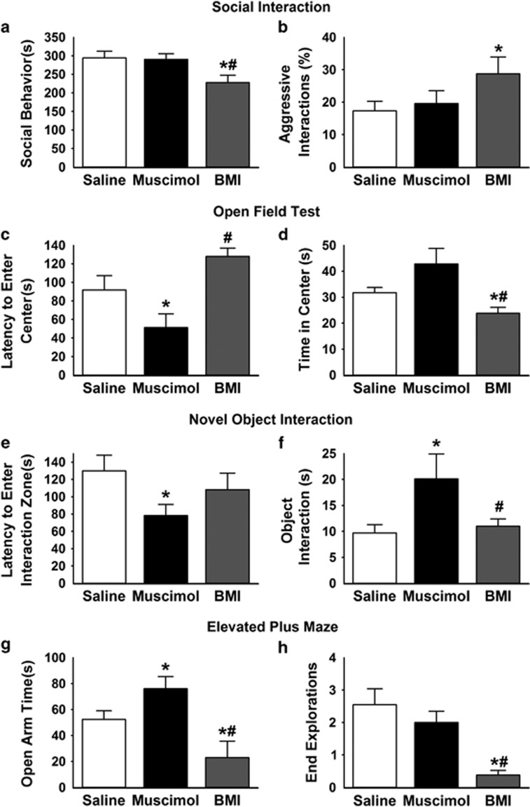
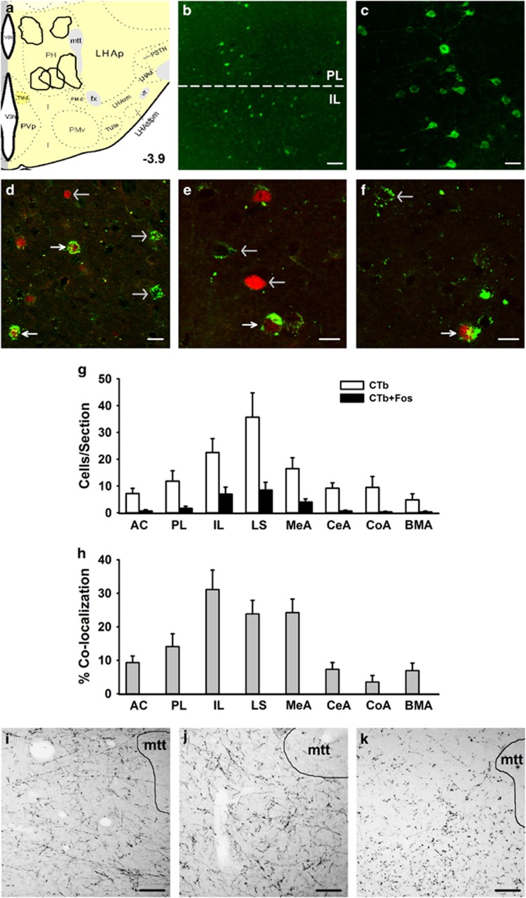
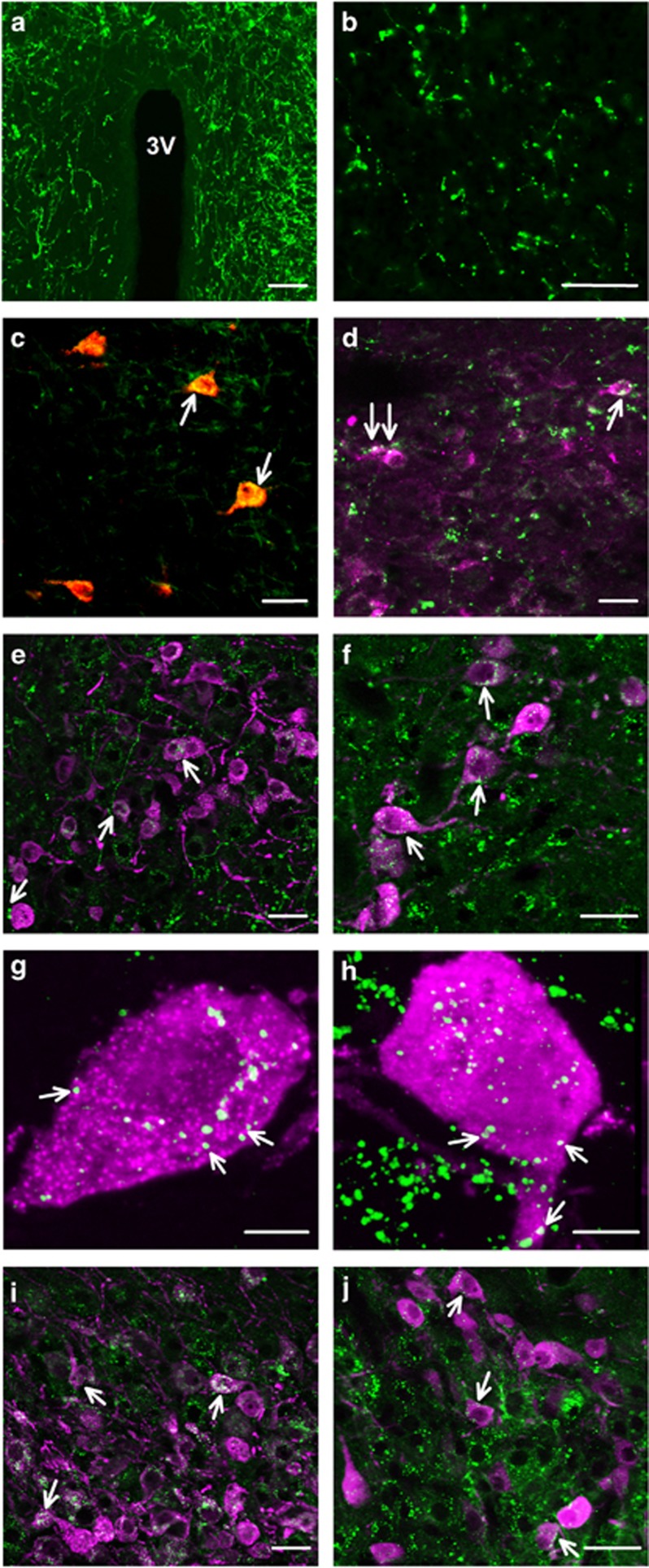
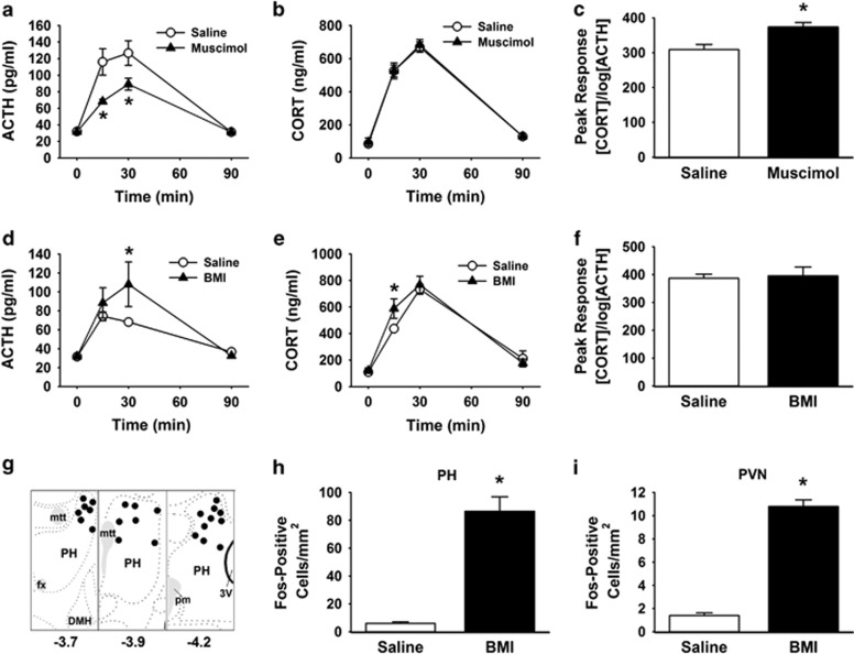
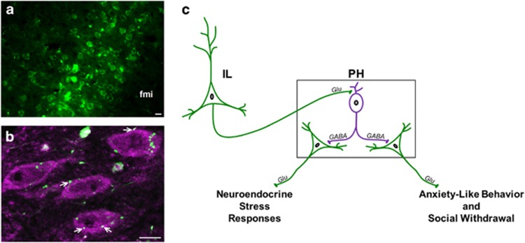
The posterior hypothalamic nucleus (PH) stimulates autonomic stress responses. However, the role of the PH in behavioral correlates of psychiatric illness, such as social and anxiety-like behavior, is largely unexplored, as is the neurochemistry of PH connectivity with limbic and neuroendocrine systems. Thus, the current study tested the hypothesis that GABAergic signaling within the PH is a critical link between forebrain behavior-regulatory nuclei and the neuroendocrine hypothalamus, integrating social and anxiety-related behaviors with physiological stress reactivity. To address this hypothesis, GABAA receptor pharmacology was used to locally inhibit or disinhibit the PH immediately before behavioral measures of social and anxiety-like behavior in rats. Limbic connectivity of the PH was then established by simultaneous co-injection of anterograde and retrograde tracers. Further, the role of PH GABAergic signaling in neuroendocrine stress responses was tested via inhibition/disinhibition of the PH. These studies determined a prominent role for the PH in the expression of anxiety-related behaviors and social withdrawal. Histological analyses revealed divergent stress-activated limbic input to the PH, emanating predominantly from the prefrontal cortex, lateral septum, and amygdala. PH projections also targeted both parvicellular and magnocellular peptidergic neurons in the paraventricular and supraoptic hypothalamus. Further, GABAA receptor pharmacology determined an excitatory effect of the PH on neuroendocrine responses to stress. These data indicate that the PH represents an important stress-integrative center, regulating behavioral processes and connecting the limbic forebrain with neuroendocrine systems. Moreover, the PH appears to be uniquely situated to have a role in stress-related pathologies associated with limbic-hypothalamic dysfunction.


Figures

Figure 1
Figure 1
Involvement of PH in social and anxiety-related behaviors. BMI injections significantly decreased social behavior in the social interaction test (a) and increased the percentage of aggressive interactions (b). In the open field test, muscimol injections in the PH significantly decreased the latency to enter the center of the field compared with saline and BMI (c), while BMI injections significantly decreased the total time spent in the center of the field compared with saline and muscimol (d). Muscimol injections also significantly decreased the latency to enter the 10 cm square interaction zone surrounding a novel object (e) and significantly increased the amount of time spent interacting (sniffing or nose-poking) with the novel object (f). Injections of muscimol into the PH significantly increased time spent in the open arms of the elevated plus maze (g). In contrast, BMI injections significantly decreased time in the open arms, as well as exploration of the ends of open arms (h). n=11–12/group, *p<0.05 compared with saline, #p<0.05 compared with muscimol. BMI, bicuculline methiodide; PH, posterior hypothalamic nucleus.
Figure 2
Figure 2
Identification of stress-activated limbic input to the PH. Injection sites for Pha-L/CTb coinjections were localized to the PH (a). Atlas image (3.9 mm posterior to bregma) was modified from Swanson 2004. CTb immunolabeling was present in the PL and IL divisions of the mPFC (b; scale bar, 100 μm). The retrograde labeling was dense in the IL, as indicated in a higher magnification view (c; scale bar 25 μm). Retrograde tracer CTb (green) co-localized with activation marker Fos (red) in the IL (d), dorsal LS (e), and anterodorsal MeA (f). Co-localization was determined in images from a single z-plane (optical section of 0.5 μm), where white arrows indicate CTb+Fos co-localization and gray arrows identify either CTb or Fos single labeling (scale bars, 15 μm). Quantification was conducted for CTb and CTb+Fos immunolabeling per section for primary limbic inputs (g) AC, PL, IL, LS, MeA, CeA, CoA, and BMA. Percent of CTb-positive cells expressing Fos was calculated to determine the relative stress-activation of PH inputs (h). Anterograde tracer Pha-L was injected in the IL (i), LS (j) and MeA (k) to verify projections to the PH (scale bars, 100 μm). mtt: mammillothalamic tract. AC, anterior cingulate; BMA, basomedial amygdala; CeA, central amygdala; CoA, cortical amygdala; IL, infralimbic cortex; LS, lateral septum; MeA, medial amygdala; mPFC, medial prefrontal cortex; PH, posterior hypothalamic nucleus; Pha-L, Phaseolus vulgaris leucoagglutinin; PL, prelimbic cortex.
Figure 3
Figure 3
Projections from the PH to peptide-producing cells. Pha-L-labeled projections from the PH densely innervated the PVN (a; scale bar, 100 μm). The PH also innervated the IL (b; scale bar, 100 μm). CTb-labeled (red) cells in the IL received input from Pha-L-labeled (green) fibers (c; scale bar, 15 μm), indicating reciprocal regional connectivity. PH projections (green) to the PVN targeted CRH- (d), AVP- (e), and oxytocin-labeled (f) neurons (magenta; scale bars, 15 μm). White arrows identify representative putative appositions. Single-cell 3-D reconstructions illustrate Pha-L (green) appositions (white arrows) onto parvicelluar AVP (magenta; g) and magnocellular oxytocin (magenta; h) neurons (scale bars, 5 μm). PH projections (green) also targeted AVP- (i; magenta) and oxytocin-labeled (j; magenta) cells in the SON (scale bars, 15 μm). AVP, arginine vasopressin; CRH, corticotropin-releasing hormone; CTb, cholera toxin beta subunit; PH, posterior hypothalamic nucleus; Pha-L, Phaseolus vulgaris leucoagglutinin; PVN, paraventricular hypothalamus; SON, supraoptic nucleus.
Figure 4
Figure 4
Role of PH in HPA axis responses to stress. The GABAA agonist muscimol injected into the PH significantly inhibited the ACTH response to restraint (a). There was no difference in the corticosterone response to stress (b). Analysis of adrenal responsivity revealed that adrenal responsiveness was enhanced during stress (c), possibly to compensate for low ACTH. Injections of the GABAA antagonist BMI into the PH significantly increased ACTH (d) and corticosterone (e) responses to stress, without altering adrenal responsivity (f). Microinjection sites were localized to the dorsal aspects of the PH (g) based on the atlas of Swanson (2004). Distances listed are posterior to bregma, mtt, fx, pm, DMH. BMI injections in the PH of unstressed rats significantly increased the number of Fos-positive cells compared with saline (h). Disinhibition of the PH also increased the number of Fos-positive cells in the PVN of unstressed animals (i). n=10–13/group, *p<0.05 compared with saline. ACTH, adrenocorticotropic hormone; BMI, bicuculline methiodide; DMH: dorsomedial hypothalamus; fx: fornix; HPA, hypothalamic-pituitary-adrenocortical; mtt: mammilothalamic tract; pm: principal mammillary tract; PH, posterior hypothalamic nucleus; PVN, paraventricular hypothalamus.
Figure 5
Figure 5
IL projections to GABAergic PH neurons. BDA was injected into the IL (a) and BDA-labeled IL terminals (green) apposed GABA-expressing neurons (magenta) in the PH (b). fmi: forceps minor of the corpus callosum. Co-localization was determined in images from a single z-plane (optical section of 0.5 μm), where white arrows indicate BDA+GABA co-localization (scale bars, 10 μm). Summary of findings and hypothesized circuitry (c). The current study details the activity of local GABA neurons (magenta) in the PH to constrain excitatory output from the PH (green) that promotes anxiety-like behavior, social withdrawal, and neuroendocrine stress responses. A potential circuit accounting for prefrontal activation of local inhibitory mechanisms within PH is based on infralimbic terminal appositions onto PH GABA neurons. BDA, biotinylated dextran amine; IL, infralimbic cortex; PH, posterior hypothalamic nucleus.

References

    1. Abrahamson EE, Moore RY (2001). The posterior hypothalamic area: chemoarchitecture and afferent connections. Brain Res 889: 1–22. - PubMed
    1. Belzung C, Griebel G (2001). Measuring normal and pathological anxiety-like behaviour in mice: a review. Behav Brain Res 125: 141–149. - PubMed
    1. Bornstein SR, Engeland WC, Ehrhart-Bornstein M, Herman JP (2008). Dissociation of ACTH and glucocorticoids. Trends Endocrinol Metab 19: 175–180. - PubMed
    1. Coolen LM, Wood RI (1998). Bidirectional connections of the medial amygdaloid nucleus in the Syrian hamster brain: simultaneous anterograde and retrograde tract tracing. J Comp Neurol 399: 189–209. - PubMed
    1. Davis M (1997). Neurobiology of fear responses: the role of the amygdala. J Neuropsychiatry Clin Neurosci 9: 382–402. - PubMed

Publication types

MeSH terms