Review
. 2015 Oct;38(10):609-620.
doi: 10.1016/j.tins.2015.08.002.

Priming the inflammatory pump of the CNS after traumatic brain injury

Affiliations
Review

Priming the inflammatory pump of the CNS after traumatic brain injury

Kristina G Witcher et al. Trends Neurosci. 2015 Oct.

Abstract

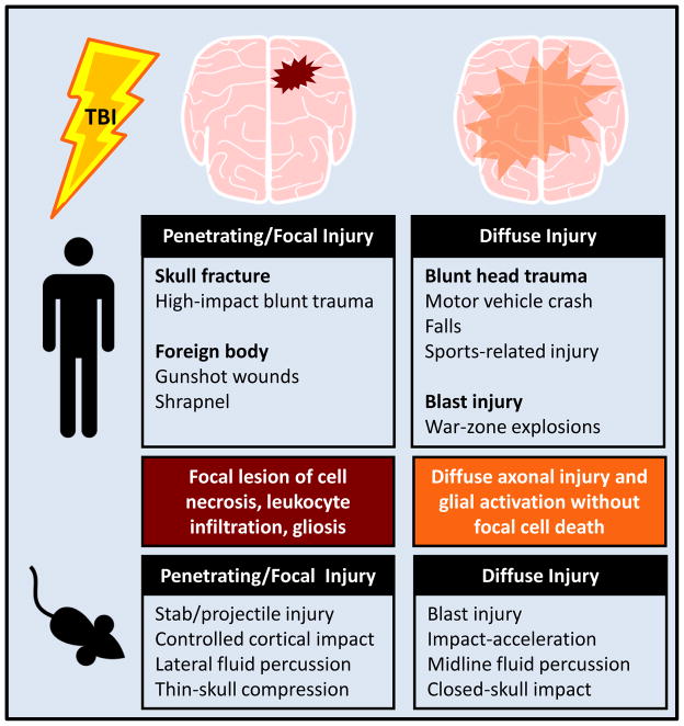
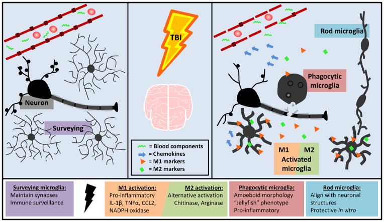
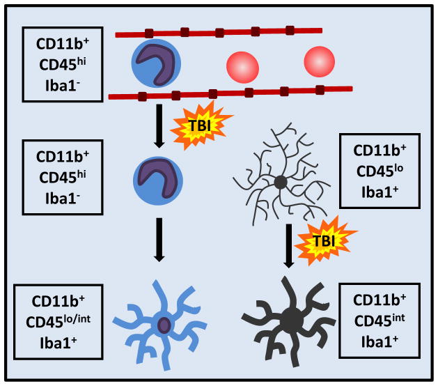
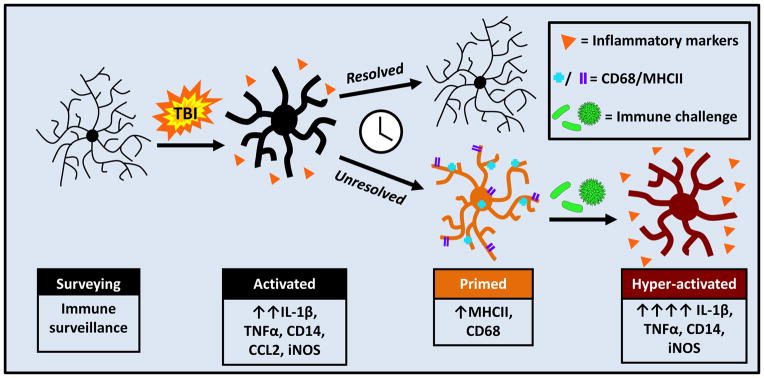
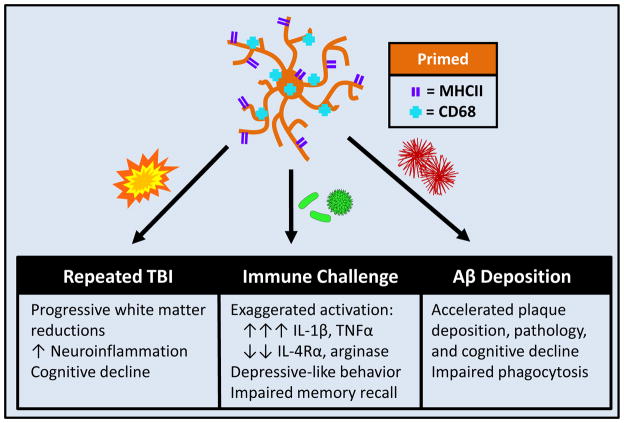
Traumatic brain injury (TBI) can lead to secondary neuropsychiatric problems that develop and persist years after injury. Mounting evidence indicates that neuroinflammatory processes progress after the initial head injury and worsen with time. Microglia contribute to this inflammation by maintaining a primed profile long after the acute effects of the injury have dissipated. This may set the stage for glial dysfunction and hyperactivity to challenges including subsequent head injury, stress, or induction of a peripheral immune response. This review discusses the evidence that microglia become primed following TBI and how this corresponds with vulnerability to a 'second hit' and subsequent neuropsychiatric and neurodegenerative complications.

Keywords: immune challenge; microglia; neuroinflammation; priming; traumatic brain injury.


Figures

FIGURE 1
FIGURE 1. Penetrating and diffuse traumatic brain injuries have varied etiologies and experimental models
Human traumatic brain injuries can either be penetrating/focal or diffuse in nature, with multiple etiologies falling into each of these categories. The animal models used to produce TBIs cause pathology that is in line with these two categories of injury.
FIGURE 2
FIGURE 2. Microglia are acutely activated following TBI and take on a variety of functional and morphological states
Microglia in their “resting” state are constantly surveying the environment and maintaining homeostasis. Following TBI, microglia are activated by cell debris, neuronal injury, and blood components. Microglia produce the chemokine CCL2, which plays a role in attracting peripheral monocytes to the CNS. Microglia upregulate both M1 and M2 cytokines and surface markers after injury. M1 activation results in production of ROS and RNS and up regulation of pro-inflammatory cytokines and surface markers including IL-1β, TNFα, and CD14. These activated cells can become phagocytic to clear debris. M2 activation is neuroprotective and indicated by upregulation of chitinase and arginase. Rod microglia align with neuronal structures after diffuse injury and are neuroprotective in vitro.
FIGURE 3
FIGURE 3. Monocytes extravasate and differentiate into brain macrophages
Following injury, circulating monocytes extravasate through activated endothelium and can traffic into the brain parenchyma. These monocytes can differentiate into macrophages. Activated microglia and ramified brain macrophages are nearly indistinguishable by both morphology and cell-surface markers.
FIGURE 4
FIGURE 4. TBI induces microglial priming and reactivity to immune challenge
Microglia are activated by TBI and respond by producing myriad cytokines and chemokines. This immediate response resolves and some microglia return to homeostasis; however a population of microglia develops a primed or pro-inflammatory profile. This profile is characterized by increased expression of MHCII and CD68. Following secondary insult, illustrated here as a peripheral immune challenge, these primed microglia become hyper-reactive and produce an amplified and prolonged pro-inflammatory response.
FIGURE 5
FIGURE 5. TBI-induced microglial priming and reactivity to secondary insults has varied consequences
Primed microglia become hyper-reactive following a secondary insult, which can include subsequent TBI, immune challenge, or pre-clinical neurodegenerative disease.

References

    1. Faul M, et al. Traumatic Brain Injury in the United States: Emergency Department Visits, Hospitalizations and Deaths 2002–2006. Centers for Disease Control and Prevention, National Center for Injury Prevention and Control; 2010.
    1. Galarneau MR, et al. Traumatic brain injury during Operation Iraqi Freedom: findings from the United States Navy-Marine Corps Combat Trauma Registry. J Neurosurg. 2008;108:950–957. - PubMed
    1. Dixon CE, et al. A controlled cortical impact model of traumatic brain injury in the rat. J Neurosci Methods. 1991;39:253–262. - PubMed
    1. Chen S, et al. Time course of cellular pathology after controlled cortical impact injury. Exp Neurol. 2003;182:87–102. - PubMed
    1. Turtzo LC, et al. Macrophagic and microglial responses after focal traumatic brain injury in the female rat. J Neuroinflammation. 2014;11:82. - PMC - PubMed

Publication types

LinkOut - more resources