Review
. 2015:49:603-25.
doi: 10.1146/annurev-genet-112414-054813. Epub 2015 Oct 5.

General Stress Signaling in the Alphaproteobacteria

Affiliations
Review

General Stress Signaling in the Alphaproteobacteria

Aretha Fiebig et al. Annu Rev Genet. 2015.

Abstract

The Alphaproteobacteria uniquely integrate features of two-component signal transduction and alternative σ factor regulation to control transcription of genes that ensure growth and survival across a range of stress conditions. Research over the past decade has led to the discovery of the key molecular players of this general stress response (GSR) system, including the sigma factor σ(EcfG), its anti-σ factor NepR, and the anti-anti-σ factor PhyR. The central molecular event of GSR activation entails aspartyl phosphorylation of PhyR, which promotes its binding to NepR and thereby releases σ(EcfG) to associate with RNAP and direct transcription. Recent studies are providing a new understanding of complex, multilayered sensory networks that activate and repress this central protein partner switch. This review synthesizes our structural and functional understanding of the core GSR regulatory proteins and highlights emerging data that are defining the systems that regulate GSR transcription in a variety of species.

Keywords: ECF sigma factor; HWE and HisKA_2 kinases; PhyR; partner switch; signal transduction; two-component signaling.


Figures

Figure 1
Figure 1
Transcriptional regulation in response to environmental change. a Simple and abundant, one-component regulators consist of a sensory input domain that regulates its adjacent DNA binding domain (DBD) (98). b Two-component signaling systems involve a sensory histidine kinase and a response regulator, the latter of which typically consists of a phosphoreceiver domain and a DNA binding output domain. The sensory domain of the histidine kinase regulates the autophosphorylation activity. The phosphoryl group is transferred from a histidine on the kinase to an aspartate on the receiver domain, which regulates the DBD output domain (17). c σ factors bind core RNA polymerase (RNAP) and confer promoter specificity to the transcriptional machinery. A host of regulatory mechanisms are employed to control alternative σ factors (46).
Figure 2
Figure 2
Alphaproteobacterial general stress response (GSR) partner switching. Under inactivating conditions, σEcfG (green) is sequestered by the anti-σ factor NepR (pink) and cannot compete with the housekeeping σ70 for binding to core RNAP (black). PhyR (purple) is unphosphorylated, in a closed confirmation, and has a low affinity for NepR. Under stress conditions, the receiver domain of PhyR (light purple) can by phosphorylated by a sensor histidine kinase(s) (white) often associated with the inner membrane (tan bar), destabilizing the interaction interface between the σ-like domain (dark purple) and the receiver domain. This open PhyR conformation reveals the NepR binding interface and thereby increases the affinity of PhyR for NepR. PhyR~P-NepR binding releases σEcfG to interact with RNAP and initiate transcription of the GSR regulon.
Figure 3
Figure 3
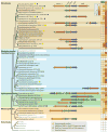
Phylogenetic distribution and organization of general stress response (GSR) loci. Organization of genomic regions surrounding phyR is overlayed on a phylogenetic tree based on analyses in Reference . PhyR-encoding loci were identified in all genomes in the SEED viewer database (using an Evalue cutoff of 1e−30) (84, 85). Pinned regions (16,000 bp) surrounding phyR were extracted to identify conserved genes in the phyR vicinity. nepR genes are not always annotated and were confirmed by manual inspection. Genes are colored as labeled at top. Closely related genomes were collapsed to a single representative. Species in a shared background color block have the same GSR locus organization. Species boxed in white lack phyR and ecfG. The color heat map on the right indicates the number of phyR, ecfG, and HisKA_2 or HWE kinase genes in each genome based on hits in SEED for phyR, annotated ECF15 σ factors in MiSTDb (99), and the sum of HisKA_2 and HWE_HK domain kinases in Pfam (29). The small size of nepR impedes reliable annotation, thus nepR paralogs were not tallied. Key indicates other annotations.
Figure 4
Figure 4
Structural features of σEcfG, NepR, and PhyR. (a) Structural model of σEcfG built using CnrH (PDB: 4CXF) as a template. The σ2 and σ4 regions are depicted in dark and light green, respectively. Subregions that interact with RNAP (2.1, 2.2, and 4.1) are indicated with black circles; regions involved in promoter recognition and binding (2.3, 2.4, and 4.2) are in white circles. The predicted NepR binding site (pink) overlaps with the conserved RNAP binding interface. (b) Promoter motifs identified upstream of genes regulated by EcfG or PhyR are highly conserved (4, 38, 40, 60, 67, 75, 78, 92). Dots represent nonconserved bases; sequences −35 and −10 base pairs from the transcriptional start site are indicated. (c) Structural representation of NepR (pink) bound across the σ2 and σ4 regions of the PhyR σ-like domain (dark purple; PDB: 3T0Y). NepR and the PhyR-receiver domain (modeled in white) compete for binding to the same surface on the σ-like domain. When NepR is bound, PhyR helix α4 (green) is repositioned and likely inhibits the receiver domain from displacing NepR. (d) NepR amino acid sequences from Caulobacter crescentus, Brucella abortus, Methylobacterium extorquens, Bradyrhizobium japonicum, Sinorhizobium meliloti, Sphingomonas melonis, and Rhodopseudomonas palustris were aligned based on the conserved core. Conservation is shown on the right (gray). Disorder disposition was predicted for each sequence using PONDR-Fit (http://www.disprot.org/pondr-fit.php) (106); the average (red) ± standard deviation (green) is plotted on the left. (e) Structural representation of PhyR in the closed conformation (PDB: 3N0R). The receiver domain (light purple) is packed against the σ-like domain (dark purple) blocking the NepR binding site (pink). Helix α4 (green) is positioned against the surface of the σ-like domain. (f) Graphical representation of the key features of PhyR that regulate partner switching.
Figure 5
Figure 5
Characteristics of the atypical HisKA_2 and HWE kinase families. (a) COG3920 encompasses the DHp (dimerization and histidine phosphorylation) and catalytic domains of HWE and HisKA_2 kinases. The Pfam domains for each kinase family domain are indicated (29). The position of the H- and N-boxes are shown as white boxes. (b) Unrooted circular tree based on alignment of COG3920 domains from kinases found in the species shown in Figure 3. Sensory input domains are not included. Tree includes 127 HisKA_2 kinases and 177 HWE kinases. Sequences were downloaded and analyzed using Geneious (http://www.geneious.com). Branches including known negative regulators or essential GSR kinases are red with the experimental species indicated. Sphingomonas PhyP branch includes the GSR locus kinases from Sphingomonas melonis FR1, Sphingomonas wittichii RW1, and Zymomonas mobilis subsp. mobilis ZM4. The Brucella branch includes the GSR locus HisKA_2 kinases from Brucella abortus 2308, Ochrobactrum anthropi ATTC 49188, Bartonella quintana str. Toulouse, and Parvibaculum lavamentivorans DS-1. The Rhodopseudomonas branch includes the GSR locus kinases from Bradyrhizobium sp. BTAi1, Nitrobacter hamburgensis X 14, Rhodopseudomonas palustris TIE-1, R. palustris BisA53, Oligotropha carboxidovorans OM5, Afipia sp. 1NLS2, and a kinase from Bradyrhizobium japonicum USDA110 encoded distal from the GSR locus. (c) Weblogos generated from the alignment of the H-box or N-box regions from the 127 HisKA_2 and 177 HWE alphaproteobacterial kinases displayed in panel b, and alignment of the 265 HisKA or 659 HATPase_c sequences in the SEED database, downloaded from Pfam (22, 29). Key conserved residues are indicated in color. Weblogos for negative regulators were generated from sequences related to known negative regulators or essential GSR kinases (red branches in panel b). For each group, only the motif that deviates from the consensus is shown; divergent residues are red.

Similar articles

Cited by

References

    1. Abromaitis S, Koehler JE. The Bartonella quintana extracytoplasmic function sigma factor RpoE has a role in bacterial adaptation to the arthropod vector environment. J Bacteriol. 2013;195:2662–74. - PMC - PubMed
    1. Akbar S, Lee SY, Boylan SA, Price CW. Two genes from Bacillus subtilis under the sole control of the general stress transcription factor σB. Microbiology. 1999;145(Pt. 5):1069–78. - PubMed
    1. Alvarez-Martinez CE, Baldini RL, Gomes SL. A Caulobacter crescentus extracytoplasmic function sigma factor mediating the response to oxidative stress in stationary phase. J Bacteriol. 2006;188:1835–46. - PMC - PubMed
    1. Alvarez-Martinez CE, Lourenco RF, Baldini RL, Laub MT, Gomes SL. The ECF sigma factor σT is involved in osmotic and oxidative stress responses in Caulobacter crescentus. Mol Microbiol. 2007;66:1240–55. - PubMed
    1. Anantharaman V, Aravind L. The CHASE domain: a predicted ligand-binding module in plant cytokinin receptors and other eukaryotic and bacterial receptors. Trends Biochem Sci. 2001;26:579–82. - PubMed

Publication types

MeSH terms

Substances

LinkOut - more resources