. 2014 May;6(1):10.1128/ecosalplus.ESP-0003-2013.
doi: 10.1128/ecosalplus.ESP-0003-2013.

DNA Methylation

Affiliations

DNA Methylation

M G Marinus et al. EcoSal Plus. 2014 May.

Abstract

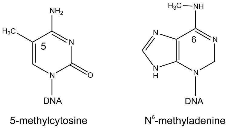
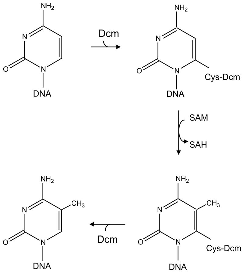
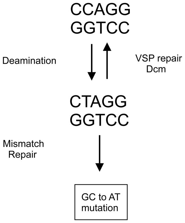
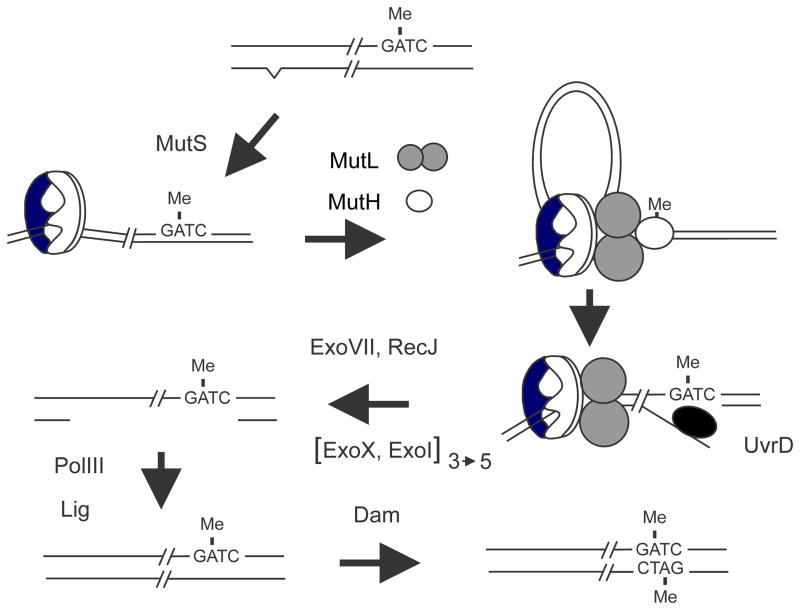
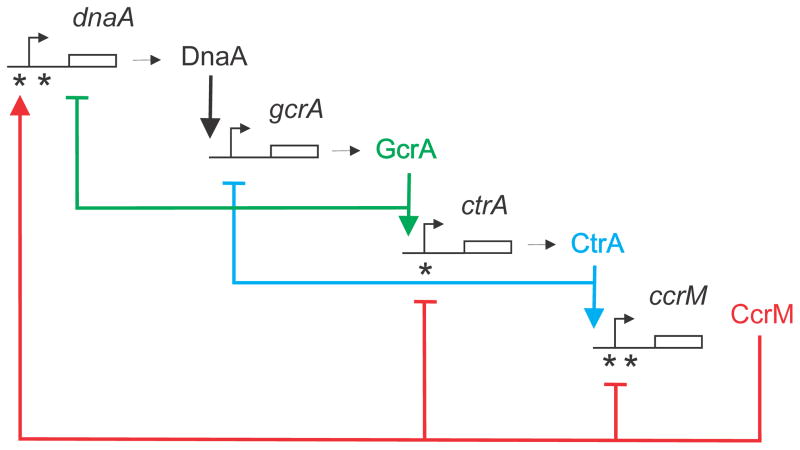
The DNA of Escherichia coli contains 19,120 6-methyladenines and 12,045 5-methylcytosines in addition to the four regular bases, and these are formed by the postreplicative action of three DNA methyltransferases. The majority of the methylated bases are formed by the Dam and Dcm methyltransferases encoded by the dam (DNA adenine methyltransferase) and dcm (DNA cytosine methyltransferase) genes. Although not essential, Dam methylation is important for strand discrimination during the repair of replication errors, controlling the frequency of initiation of chromosome replication at oriC, and the regulation of transcription initiation at promoters containing GATC sequences. In contrast, there is no known function for Dcm methylation, although Dcm recognition sites constitute sequence motifs for Very Short Patch repair of T/G base mismatches. In certain bacteria (e.g., Vibrio cholerae, Caulobacter crescentus) adenine methylation is essential, and, in C. crescentus, it is important for temporal gene expression, which, in turn, is required for coordinating chromosome initiation, replication, and division. In practical terms, Dam and Dcm methylation can inhibit restriction enzyme cleavage, decrease transformation frequency in certain bacteria, and decrease the stability of short direct repeats and are necessary for site-directed mutagenesis and to probe eukaryotic structure and function.


Figures

FIGURE 1
FIGURE 1
Structures of 5-methylcytosine and N6-methyladenine.
FIGURE 2
FIGURE 2
Organization of the dam transcriptional unit. The locations of promoters P1 through P5 are indicated, as is the transcription terminator (T) at the end of aroB. The major and growth-rate regulated promoter P2 is located 3.2 kb upstream of the dam gene.
FIGURE 3
FIGURE 3
Phylogeny of the Dam clade. TBLASTN searching was used with 4311 E. coli proteins against selected genomes with and without the dam gene. The genes encoding SeqA, MutH, HN-S, PriB and 75 other proteins were found in all selected genomes with the dam gene and in none of the genomes lacking it. Reproduced from [2] with permission from Elsevier Ltd.
FIGURE 4
FIGURE 4
Catalytic mechanism of methyl group transfer. Nucleophilic attack by cysteine-177 of Dcm at the C-6 position of cytosine leads to the formation of a covalent Dcm-DNA intermediate. This leads to activation of the C-5 position and transfer of the methyl group from SAM. The S-adenosyl-homocysteine (SAH) and the methylated cytosine are released from the covalent intermediate.
FIGURE 5
FIGURE 5
Deamination, repair and mutagenesis at a Dcm recognition site. Deamination of 5-meCyt in duplex DNA produces a T-G mismatch which is a substrate for Vsr endonuclease. After removal of the T residue, DNA polymerase I and DNA ligase reactions restore the original sequence which is re-methylated by Dcm. Failure to repair before DNA replication or if the MutHLS mismatch system acts on the mismatch before Vsr will produce a GC to AT mutation.
FIGURE 6
FIGURE 6
Dam-directed mismatch repair in E. coli. The top of the figure shows DNA immediately behind the replication fork in which the “old” top strand is methylated and the “new” strand is not and also contains a base mismatch (carat) created as a replication error. The mismatch is recognized and bound by MutS followed by recruitment of MutL and MutH to form a ternary complex. The formation of this complex is thought to involve DNA looping to bring the mismatch and a GATC sequence in close proximity but the details are unclear. In the ternary complex the latent nuclease activity of MutH is activated and it cleaves the new unmethylated strand 5′ to the GATC sequence. The nick created by MutH serves as an entry site for the UvrD helicase which unwinds the DNA exposing single-stranded DNA which is digested by one or more of the following exonucleases: RecJ, ExoVII, ExoX or ExoI. The exonuclease(s) used depends on the relative orientation of the mismatch to the GATC sequence; in the figure the direction of UvrD unwinding is 5′ to 3′ and so either ExoVII or RecJ or both are needed. If the mismatch was to the “right” of the GATC sequence, UvrD would unwind in the 3′ to 5′ direction and ExoX and/or ExoI would digest the single-stranded DNA. The gap created by nuclease digestion removes the mismatched base and is filled in by DNA polymerase III. The resulting nick is closed by DNA ligase and eventual Dam methylation precludes any further repair.
FIGURE 7
FIGURE 7
Models illustrating double-strand break formation in a dam mutant. (A) A replication fork encounters a mismatch repair (MMR) intermediate of a nick or gap on one strand leading to replication fork collapse. The MMR intermediate could arise from the processing of endogenous DNA damage or from repair of a replication error from the previous replication. Recombination between daughter chromosomal arms can restore the fork which can then be loaded with the DnaB helicase and DNA polymerase III holoenzyme. (B) MutH nicking on opposite sides of the same GATC in non-replicating DNA produces a double-strand break which can be repaired using a sister chromosome. (C) MMR processing of a replication error either by action at the same GATC as in panel B or by overlapping excision tracts from GATCs on opposite strands producing a double-strand break that can be repaired using the daughter strands as template. (D) Mismatch repair-independent double-strand break formation. Asynchronous initiation of chromosome replication in a dam mutant could lead to two initiation events close together resulting in two closely spaced forks on each chromosomal arm. If the second fork catches up to the first, replication fork collapse occurs. The exposed double-stranded end becomes a substrate for RecBCD exonuclease which, when encountering a Chi site, loads RecA on single-stranded DNA thereby generating an SOS inducing signal.
FIGURE 8
FIGURE 8
Double-strand breaks in an E. coli dam mutant detected by single-cell microgel electrophoresis showing disrupted cells with 0, 1 or 2 double-strand breaks. Reproduced from with permission from [110] copyright (2005) American Society for Microbiology.
FIGURE 9
FIGURE 9
Expression of individual genes as a function of position on the chromosome. All genes for which a significant signal was obtained were plotted relative to wild type as a function of position along the chromosome. The chromosome is linearized at a position directly opposite oriC. The replication origin has position 0 on the abscissa. (A) MG1655 seqA. (B) MG1655_pTP166 (Dam overproducer). (C) MG1655 dam-13::Tn9. Trendlines for the gene expression data are presented in A and B. All points above the trendline in panel A, i.e., genes that were derepressed in the seqA mutant are plotted as green dots, and all genes that were repressed in the seqA mutant as red dots. Expression data from individual genes in panels B and C have the same color assignment as in panel A (red and green dots). Reproduced from with permission from [49] and Copyright (2007) National Academy of Sciences, U.S.A.
FIGURE 10
FIGURE 10
Switching at the pap promoter region. Lrp binds co-operatively to either Lrp binding sites 1–3 (in the OFF-state, non-piliated) or to the Lrp binding sites 4–6 (ON-state, piliated). Lrp binding site 3 overlaps the papB promoter and Lrp binding to site 3 inhibits transcription. Lrp binding sites 2 and 5 overlaps with GATC sites and Lrp binding to either site prevents methylation of that site by DamMT. Lrp binding to sites 1–3 mutually excludes binding to sites 4–6. When in OFF-state, each DNA replication produce one hemimethylated GATC site (in Lrp site 5) and one unmethylated GATC site (in Lrp site 2) and dissociate Lrp from its binding sites. The OFF-state is preserved by rebinding of Lrp to the same binding sites around the unmethylated Lrp binding site. A shift from phase OFF to ON may occur if PapI mediates Lrp binding to the hemimethylated Lrp site 5, followed by Lrp binding to sites 4 and 6. The shift is further stabilized by full methylation of site 2 by DamMT and conversion of the hemimethylated site 5 to unmethylated by subsequent DNA replications. This figure is adapted from [275] with permission from Cell Press.
FIGURE 11
FIGURE 11
Regulation of agn43 gene transcription in E. coli. The promoter region of the agn43 gene contains three GATC sequences which must be methylated (Me) for the gene to be expressed. Replication of the gene will cause transient hemimethylation allowing one of three proteins can bind this DNA. Dam action will methylated the GATCs on the new strand thereby preserving expression of the gene. SeqA can also bind but is easily displaced by Dam resulting in methylation and continued gene expression. OxyR binding prevents Dam action and after a second round of replication the GATCs are unmethylated and transcription is prevented. This modified figure is reproduced with permission from [276] copyright (2002) John Wiley and Sons Ltd.
FIGURE 12
FIGURE 12
Activation of the tsp promoter in IS10. In wildtype bacteria both strands of IS10 are methylated (black rectangles). Upon replication, two hemimethylated forms are produced but only that with the methylated coding strand actively transcribes the tsp gene and moves to a new location while the inactive IS10 remains.
FIGURE 13
FIGURE 13
Regulatory cascade in the Caulobacter cell cycle. The genes for dnaA, gcrA, ctrA and ccrM are shown together with their respective products. The genes are shown in the sequence they are replicated on the chromosome. Asterisks indicate CcrM recognition sites (GANTC). In addition to the genes shown in this figure, DnaA, GrcA and CtrA control about 40, 50 and 95 other genes respectively. Figure modified with permission from [195] and Copyright (2007) National Academy of Sciences, U.S.A.

Similar articles

Cited by

References

    1. Marinus MG. Methylation of DNA. In: Neidhardt FC, Curtiss R III, Ingraham JL, Lin ECC, Low KB, Magasanik B, Reznikoff WS, Riley M, Schaechter M, Umbarger HE, editors. Escherichia coli and Salmonella: Cellular and Molecular Biology. 2. Vol. 1. ASM Press; Washington, DC: 1996. pp. 782–791.
    1. Løbner-Olesen A, Skovgaard O, Marinus MG. Dam methylation: coordinating cellular processes. Curr Opin Microbiol. 2005;8:154–160. - PubMed
    1. Wion D, Casadesus J. N6-methyl-adenine: an epigenetic signal for DNA-protein interactions. Nat Rev Microbiol. 2006;4:183–192. - PMC - PubMed
    1. Casadesus J, Low D. Epigenetic gene regulation in the bacterial world. Microbiol Mol Biol Rev. 2006;70:830–856. - PMC - PubMed
    1. Low DA, Casadesus J. Clocks and switches: bacterial gene regulation by DNA adenine methylation. Curr Opin Microbiol. 2008;11:106–112. - PubMed