

A splicing variant of Merlin promotes metastasis in hepatocellular carcinoma
- PMID: 26443326
- PMCID: PMC4633634
- DOI: 10.1038/ncomms9457
A splicing variant of Merlin promotes metastasis in hepatocellular carcinoma
Abstract
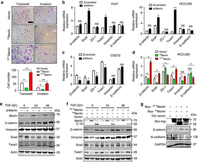
Merlin, which is encoded by the tumour suppressor gene Nf2, plays a crucial role in tumorigenesis and metastasis. However, little is known about the functional importance of Merlin splicing forms. In this study, we show that Merlin is present at low levels in human hepatocellular carcinoma (HCC), particularly in metastatic tumours, where it is associated with a poor prognosis. Surprisingly, a splicing variant of Merlin that lacks exons 2, 3 and 4 ((Δ2-4)Merlin) is amplified in HCC and portal vein tumour thrombus (PVTT) specimens and in the CSQT2 cell line derived from PVTT. Our studies show that (Δ2-4)Merlin interferes with the capacity of wild-type Merlin to bind β-catenin and ERM, and it is expressed in the cytoplasm rather than at the cell surface. Furthermore, (Δ2-4)Merlin overexpression increases the expression levels of β-catenin and stemness-related genes, induces the epithelium-mesenchymal-transition phenotype promoting cell migration in vitro and the formation of lung metastasis in vivo. Our results indicate that the (Δ2-4)Merlin variant disrupts the normal function of Merlin and promotes tumour metastasis.
Figures


Similar articles
-
Expression of mutant nuclear beta-catenin correlates with non-invasive hepatocellular carcinoma, absence of portal vein spread, and good prognosis.J Pathol. 2001 Jan;193(1):95-101. doi: 10.1002/1096-9896(2000)9999:9999<::AID-PATH720>3.0.CO;2-3. J Pathol. 2001. PMID: 11169521
-
Overexpression of forkhead box C1 promotes tumor metastasis and indicates poor prognosis in hepatocellular carcinoma.Hepatology. 2013 Feb;57(2):610-24. doi: 10.1002/hep.26029. Hepatology. 2013. PMID: 22911555
-
Molecular alterations of the NF2 gene in hepatocellular carcinoma and intrahepatic cholangiocarcinoma.Oncol Rep. 2017 Dec;38(6):3650-3658. doi: 10.3892/or.2017.6055. Epub 2017 Oct 24. Oncol Rep. 2017. PMID: 29130106
-
Merlin, the product of NF2 gene, is associated with aromatase expression and estrogen formation in human liver tissues and liver cancer cells.J Steroid Biochem Mol Biol. 2017 Sep;172:222-230. doi: 10.1016/j.jsbmb.2016.05.023. Epub 2016 Jun 8. J Steroid Biochem Mol Biol. 2017. PMID: 27289045 Review.
-
Merlin and the ERM proteins--regulators of receptor distribution and signaling at the cell cortex.Trends Cell Biol. 2009 May;19(5):198-206. doi: 10.1016/j.tcb.2009.02.006. Epub 2009 Apr 1. Trends Cell Biol. 2009. PMID: 19345106 Free PMC article. Review.
Cited by
-
Long-Read RNA Sequencing Identifies Alternative Splice Variants in Hepatocellular Carcinoma and Tumor-Specific Isoforms.Hepatology. 2019 Sep;70(3):1011-1025. doi: 10.1002/hep.30500. Epub 2019 Mar 22. Hepatology. 2019. PMID: 30637779 Free PMC article.
-
LINC01348 suppresses hepatocellular carcinoma metastasis through inhibition of SF3B3-mediated EZH2 pre-mRNA splicing.Oncogene. 2021 Jul;40(28):4675-4685. doi: 10.1038/s41388-021-01905-3. Epub 2021 Jun 17. Oncogene. 2021. PMID: 34140643
-
Alternative Splicing in Hepatocellular Carcinoma.Cell Mol Gastroenterol Hepatol. 2020;10(4):699-712. doi: 10.1016/j.jcmgh.2020.04.018. Epub 2020 May 8. Cell Mol Gastroenterol Hepatol. 2020. PMID: 32389640 Free PMC article. Review.
-
A novel BMX variant promotes tumor cell growth and migration in lung adenocarcinoma.Oncotarget. 2017 May 16;8(20):33405-33415. doi: 10.18632/oncotarget.16796. Oncotarget. 2017. PMID: 28422715 Free PMC article.
-
Repurposing fluphenazine as an autophagy modulator for treating liver cancer.Heliyon. 2023 Nov 22;9(12):e22605. doi: 10.1016/j.heliyon.2023.e22605. eCollection 2023 Dec. Heliyon. 2023. PMID: 38107270 Free PMC article.
References
-
- Ishikawa T. et al.. Percutaneous transhepatic portal vein stent placement can improve prognosis for hepatocellular carcinoma patients with portal vein tumor thrombosis. Hepatogastroenterology 61, 413–416 (2014) . - PubMed
-
- Bosch F. X., Ribes J., Diaz M. & Cleries R. Primary liver cancer: worldwide incidence and trends. Gastroenterology 127, S5–S16 (2004) . - PubMed
-
- Zhang J. et al.. RMP promotes venous metastases of hepatocellular carcinoma through promoting IL-6 transcription. Oncogene 34, 1575–1583 (2015) . - PubMed
-
- Pecina-Slaus N. Merlin, the NF2 gene product. Pathol. Oncol. Res. 19, 365–373 (2013) . - PubMed
-
- Merel P. et al.. Predominant occurrence of somatic mutations of the NF2 gene in meningiomas and schwannomas. Genes Chromosomes Cancer 13, 211–216 (1995) . - PubMed
Publication types
MeSH terms
Substances
LinkOut - more resources
Full Text Sources
Other Literature Sources
Medical
Miscellaneous
