. 2016 Jan;26(1):120-3.
doi: 10.1111/bpa.12327.

Genetics of Glioblastomas in Rare Anatomical Locations: Spinal Cord and Optic Nerve

Affiliations

Genetics of Glioblastomas in Rare Anatomical Locations: Spinal Cord and Optic Nerve

Jared Shows et al. Brain Pathol. 2016 Jan.
No abstract available


Figures

Figure 1
Figure 1
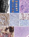
A. MRI illustrating the most uncommon anatomical location for spinal cord GBMs, that is, the lumbar region; note the expansile, nondiscrete features. Case 7 illustrated. B. At autopsy, this pediatric patient with a cervicomedullary spinal cord GBM was found to have extensive hemorrhagic features. Case 1 illustrated. C. GBMs manifested nuclear pleomorphism and hypercellularity; mitoses and vascular proliferation were present elsewhere. Case 7 illustrated. Hematoxylin and eosin, 400×. D. The lumbar GBM had no features to suggest derivation from a myxopapillary ependymoma or lower grade glioma precursor; strong GFAP fibrillary cytoplasm was present. Case 7 illustrated. 400×. E. Two of 9 cases manifested nuclear loss of ATRX; note the endothelial cell retention of the IHC (upper left), demonstrating tissue fidelity. Case 7 illustrated. 400×. F. Histone H3.F3 K27M IHC showed positive nuclear expression, paralleling mutation, in only 2/8 assessable cases. Case 7 illustrated. 400×. G. Most cases manifested no Histone H3.F3 K27M IHC expression; IHC assessment has shown good correlation with mutational status. 400×. H. Optic chiasm GBM at autopsy arising in the right optic nerve (arrowhead), with lesser involvement of the left optic nerve (arrow). Note the massive acute hemorrhage into the hypothalamus with rupture of lamina terminalis. The latter was responsible for demise 48 h after biopsy. Case 9 illustrated. I. Optic chiasm GBM demonstrating pleomorphic glial cells infiltrating nerve, albeit with preservation of fibrovascular septae. Case 8 illustrated. H&E. 100×.

References

    1. Aihara K, Mukasa A, Gotoh K, Saito K, Nagae G, Tsuji S et al (2014) H3F3A K27M mutations in thalamic gliomas from young adult patients. Neuro Oncol 16:140–146. - PMC - PubMed
    1. Buczkowicz P, Bartels U, Bouffet E, Becher O, Hawkins C (2014) Histopathological spectrum of paediatric diffuse intrinsic pontine glioma: diagnostic and therapeutic implications. Acta Neuropathol 128:573–581. - PMC - PubMed
    1. Buczkowicz P, Hoeman C, Rakopoulos P, Pajovic S, Letourneau L, Dzamba M (2014) Genomic analysis of diffuse intrinsic pontine gliomas identifies three molecular subgroups and recurrent activating ACVR1 mutations. Nat Genet 46:451–456. - PMC - PubMed
    1. Ellezam B, Theeler BJ, Walbert T, Mammoser AG, Horbinski C, Kleinschmidt‐DeMasters BK et al (2012) Low rate of R132H IDH1 mutation in infratentorial and spinal cord grade II and III diffuse gliomas. Acta Neuropathol 124:449–451. - PMC - PubMed
    1. Karsy M, Guan J, Sivakumar, Neil JA, Schmidt MH, Mahan MA (2015) The genetic basis of intradural spinal tumors and its impact on clinical treatment. Neurosurg Focus 39:E3. - PubMed

MeSH terms