. 2015 Dec;56(12):2351-67.
doi: 10.1093/pcp/pcv146. Epub 2015 Oct 6.

Arabidopsis ABA-Activated Kinase MAPKKK18 is Regulated by Protein Phosphatase 2C ABI1 and the Ubiquitin-Proteasome Pathway

Affiliations

Arabidopsis ABA-Activated Kinase MAPKKK18 is Regulated by Protein Phosphatase 2C ABI1 and the Ubiquitin-Proteasome Pathway

Filip Mitula et al. Plant Cell Physiol. 2015 Dec.

Abstract

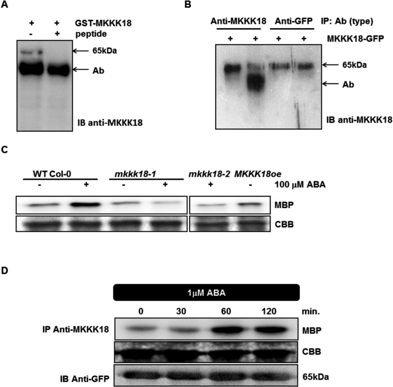
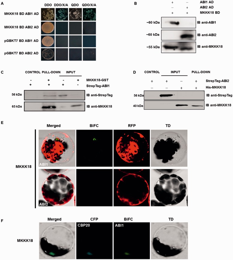
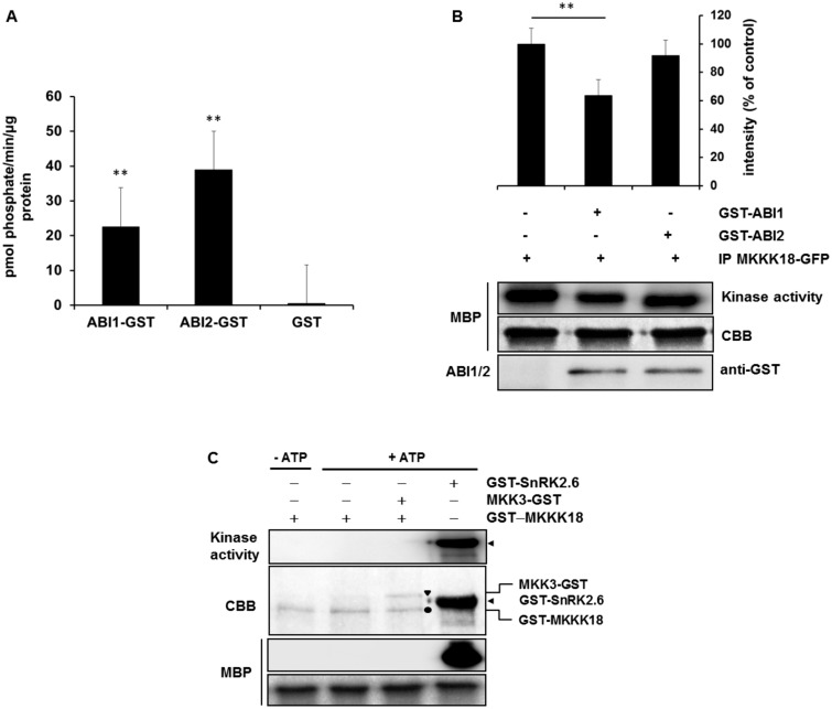
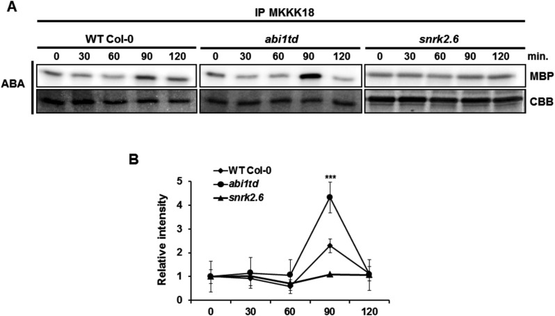
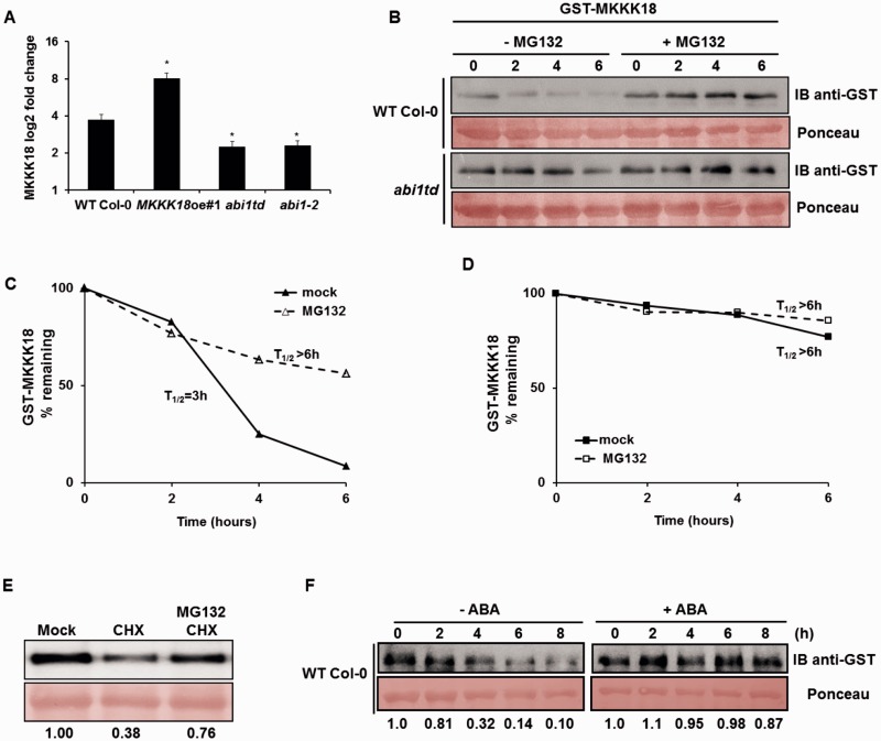
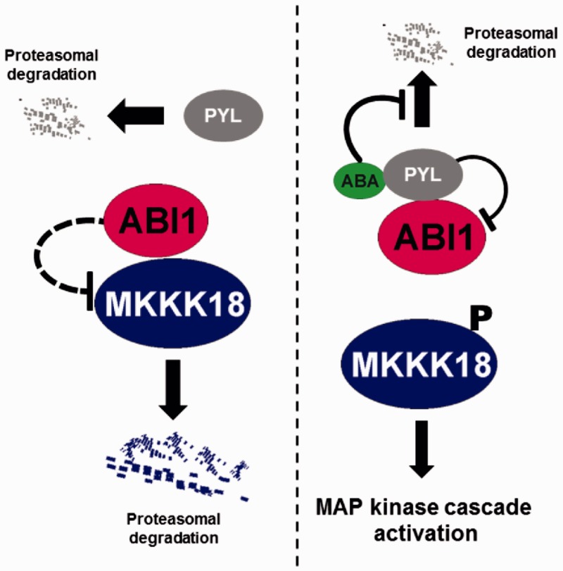
Phosphorylation and dephosphorylation events play an important role in the transmission of the ABA signal. Although SnRK2 [sucrose non-fermenting1-related kinase2] protein kinases and group A protein phosphatase type 2C (PP2C)-type phosphatases constitute the core ABA pathway, mitogen-activated protein kinase (MAPK) pathways are also involved in plant response to ABA. However, little is known about the interplay between MAPKs and PP2Cs or SnRK2 in the regulation of ABA pathways. In this study, an effort was made to elucidate the role of MAP kinase kinase kinase18 (MKKK18) in relation to ABA signaling and response. The MKKK18 knockout lines showed more vigorous root growth, decreased abaxial stomatal index and increased stomatal aperture under normal growth conditions, compared with the control wild-type Columbia line. In addition to transcriptional regulation of the MKKK18 promoter by ABA, we demonstrated using in vitro and in vivo kinase assays that the kinase activity of MKKK18 was regulated by ABA. Analysis of the cellular localization of MKKK18 showed that the active kinase was targeted specifically to the nucleus. Notably, we identified abscisic acid insensitive 1 (ABI1) PP2C as a MKKK18-interacting protein, and demonstrated that ABI1 inhibited its activity. Using a cell-free degradation assay, we also established that MKKK18 was unstable and was degraded by the proteasome pathway. The rate of MKKK18 degradation was delayed in the ABI1 knockout line. Overall, we provide evidence that ABI1 regulates the activity and promotes proteasomal degradation of MKKK18.

Keywords: ABA signaling; ABI1 PP2C; Arabidopsis thaliana; MAP kinase cascade; MKKK18; Proteasome.


Figures

Fig. 1
Fig. 1
Analysis of the activity of the MKKK18 promoter in transgenic plants expressing the ProMKKK18:GUS construct. (A) Schematic map of the MKKK18 promoter used for transformation and tissue-specific expression of MKKK18. (B) The ProMKKK18:GUS seedlings were germinated and grown on half-strength MS medium. Five-week-old plants were treated with either 0.1% methanol (mock) or 100 µM ABA. One representative of five independent lines is shown. (A) No changes in GUS activity after mock treatment. The scale bar represents 2,000 µm. (B–J) Plant samples 24 h after ABA treatment. GUS activity is visible in rosette leaves (B), flower buds and sepals (C), meristem tissues (D), pistils (E), anther filaments (F), lateral roots (G), trichomes (H), leaf guard cells (I) and hydathodes (J).
Fig. 2
Fig. 2
Subcellular localization of the MKKK18 protein in Arabidopsis protoplasts. Active MKKK18 is localized in the nucleus. (A) Microscopy images show nuclear localization of the MKKK18–GFP fusions in Arabidopsis protoplasts. Hoechst H33342 was used as nuclear localization marker. Scale bars are calibrated to 20 µm. (B) MKKK18 protein expression and activity were analyzed by immunoprecipitation using protein-specific anti-MKKK18 antibodies and immunocomplex kinase assay with MBP as a substrate. The immunocomplex assay confirmed residual kinase activity of the MKKK18K32M-GFP allele. Western blot detection using anti-GFP antibodies corroborates the presence of MKKK18–GFP fusion proteins. Coomassie Brilliant Blue (CBB) staining confirmed equal loading.
Fig. 3
Fig. 3
Germination and root growth assays. (A and B) T-DNA insertion sites derived from the sequencing of genomic DNA isolated from mkkk18-1 and mkkk18-2 mutant lines. Black boxes represent an open reading frame. Homozygous knockout mutants were verified by PCR-based genotyping using the following primers: MKKK18LP plus MKKK18RP plus LBb1 for mkkk18-1 analysis (product size for the WT, ∼960 bp; product for homozygous lines, 750 bp); MEK18F plus GKatTDNA (product for homozygous lines, 850 bp) and GwMK18F plus GwMK18R (product for the WT, 1,001 bp) for mkkk18-2 genotyping. Primer sequences are indicated in Supplementary Table S1. (C) qPCR analysis confirmed a lack of MKKK18 expression in homozygous lines. Each quantification was repeated twice with similar results. The results are given as log2 of the relative MKKK18/18S rDNA expression ratio ± SE (n = 6). (D) RD29B and RAB18 transcript levels in MKKK18 mutants. RD29B and RAB18 expression levels were determined using three biological replicates and were normalized against 18S rDNA. Each quantification was repeated twice on separate plates. The results are displayed as mean log2 fold change ± SE (n = 9) of three independent experiments with consistent results. (E and F) Germination and root growth assays. ABA-mediated inhibition of germination (E) and primary root growth (F) in WT Col-0, MKKK18oe and MKKK18 knockout lines. Both MKKK18oe #1 and #2 lines showed similar results. Values are mean ± SE for three independent experiments (n = 30). *P < 0.01; **P < 0.001; ***P < 0.0001 with respect to the control WT Col-0 line.
Fig. 4
Fig. 4
Stomatal development and movement in MKKK18 knockouts and the MKKK18-overexpressing line. (A) Stomatal development is enhanced in the MKKK18-overexpressing lines. Representative line drawings show the abaxial surface of cotyledons at day 10. Change in stomatal index and the number of guard cells (GCs) and pavement cells (PCs) per cotyledon in WT Col-0, MKKK18oe and MKKK18 knockouts. Data represent the mean ± SD (n = 300) of three independent experiments. Scale bar = 50 µm. (B) Increased stomatal aperture of mkkk18-1 and mkkk18-2 knockouts compared with the WT in standard conditions (three independent experiments, 60–80 stomatal apertures at each data point). (C) The mkkk18-1 and mkkk18-2 knockouts are insensitive to ABA- and H2O2-induced stomatal closure (two independent experiments, 80 stomatal apertures at each data point). MKKK18oe lines were hypersensitive to ABA-, CaCl2- and H2O2-induced stomatal closure (two independent experiments, 100 stomatal apertures at each data point).
Fig. 5
Fig. 5
ABA induces rapid MKKK18 activation in tobacco. (A–C) Generation of MKKK18-specific antibodies. (A) MKKK18 peptide competition assay. Recombinant GST–MKKK18 protein was incubated with anti-MKKK18 with and without 45 µg of blocking peptide. A single band of approximatley 65 kDa specific to GST–MKKK18 is absent in the immunoprecipitates containing the blocking peptide. Immunodetection was performed using anti-MKKK18 antibody. (B) MKKK18–GFP protein immunoprecipitated from tobacco total protein extracts using increasing amounts of anti-MKKK18 and anti-GFP antibodies. The MKKK18–GFP protein was precipitated by 3 µl (lane 1) or 10 µl (lane 2) of MKKK18 antiserum and 0.6 µg (lane 3) or 1.2 µg (lane 4) of anti-GFP antibody, respectively. Immunoblotting with anti-MKKK18 antibodies confirmed the presence of MKKK18–GFP fusion protein. Arrows/Ab indicate anti-MKKK18 antibodies (visible as a band in lane 2) where excess anti-MKKK18 antibody was used in the immunoprecipitation reaction. The 65 kDa band represents MKKK18–GFP protein. (C) Analysis of MKKK18 activity in the mkkk18 knockout lines. MKKK18 protein was immunoprecipitated from 600 µg of total protein extract isolated from ABA-treated WT Col-0, mkkk18-1, mkkk18-2 and MKKK18oe using specific anti-MKKK18 antibodies. Immunocomplex activity was determined using MBP (2 µg) as a substrate. Coomassie Brilliant Blue (CBB) staining of MBP confirmed equal loading. (D) Tobacco plants infiltrated with Agrobacterium strain C58C1 harboring 35S:MKKK18-GFP and 35S:p19 constructs for transient expression. Four to five days after infiltration, the tobacco plants were treated with ABA and tissue samples were collected at the indicated time points. MKKK18 activity was assessed by the immunocomplex assay using anti-MKKK18 antibody and MBP as a substrate. MKKK18–GFP was detected with anti-GFP antibody. CBB staining of MBP confirmed equal loading. The above experiments were repeated several times with similar results.
Fig. 6
Fig. 6
MKKK18 interacts with the ABI1 protein phosphatase. (A) Yeast two-hybrid analysis of the interaction between MKKK18 and ABI1/2 PP2Cs. Diploid yeast colonies were grown on double (DDO-SD medium without Leu and Trp) or quadruple selective medium (QDO-SD medium without Leu, Trp, His or Ade) with or without supplemented X-α-Gal and aureobasidin. The bait (MKKK18) did not autoactivate the reporter genes in yeast. (B) Detection of ABI1/2 and MKKK18 expression in diploid yeast strains. Expression of BD and AD fusion proteins in yeast was determined by immunoblotting using specific anti-ABI1, anti-ABI2 and anti-MKKK18 antibodies. (C and D) Pull-down assays to verify the interaction of ABI1/2 with MKKK18. Input lines represent 100% of the ABI1/2. Recombinant GST–MKKK18 or His-MKKK18 was pre-coupled to glutathione–Sepharose, and incubated with StrepTag-ABI1 or StrepTag-ABI2, respectively. Pulled-down StrepTagged ABI1 protein was detected (IB) with the epitope tag antibody. The presence of recombinant protein was confirmed using anti-MKKK18 antibody. (E and F) ABI1–MKKK18 interaction occurs within the nucleus. BiFC analysis in Arabidopsis protoplasts expressing full-length ABI1/2 and MKKK18 fused to cECFP or nVenus, respectively. RFP (E) was used as a transformation control. CFP–CBP20 (cyan fluorescent protein–Cap Binding Protein 20) (F) was used as a marker of nuclear localization.
Fig. 7
Fig. 7
ABI1 inhibits MKKK18 activity. (A) Phosphatase activity of recombinant ABI1 and ABI2 proteins. The enzyme reactions were performed in a 50 µl final volume containing 3–5 µg pf GST–ABI1 or GST–ABI2. The results presented are the means from three independent biological replicates. (B) ABI1 inactivates MKKK18. The MKKK18–GFP immunocomplex was incubated with 3 µg of GST–ABI1 and GST–ABI2 (as a negative control), after which the kinase activity was determined with MBP as a substrate. Equal loading was confirmed by Coomassie Brilliant Blue (CBB) staining of MBP. The 32P-labeled MBP bands were quantified and then normalized against the intensity of the corresponding control band using ImageJ software. Data are means ± SD of the relative band intensities from three independent experiments. An asterisk (*) indicates statistically significant changes determined using Student’s t-test. (C) Recombinant MKKK18 has no autophosphorylation activity in vitro. GST–MKKK18 was incubated with MBP or/and MKK3, without or in the presence of [32P]ATP. Bands of GST–MKKK18, MKK3–GST and GST–SnRK2.6 are indicated by a filled circle, a triangle and an arrow, respectively. Protein loading was confirmed by CBB staining. The blots shown are representative of three independent trials.
Fig. 8
Fig. 8
MKKK18 activity increases in the abi1td mutant in response to ABA treatment. (A) Seedlings of WT Col-0 and the abi1td and snrk2.6 mutants were treated with 100 µM ABA for the indicated times. A 600 µg aliquot of total protein was used for the immune complex MKKK18 activity assay using anti-MKKK18 antibody. Coomassie Brilliant Blue (CBB) staining of MBP confirmed equal loading. Due to low abundance, the endogenous MKKK18 protein was not detectable by immunoblot analysis. The results shown are representative of three independent experiments (n = 9) with consistent results. (B) 32P-labeled MBP bands were quantified using ImageJ software and normalized by taking the radioactivity of the band in the absence of ABA as 1. Data are means ± SD of the relative band intensities from three independent experiments (n = 9).
Fig. 9
Fig. 9
Proteasome-dependent degradation of MKKK18 is ABA-dependent and regulated by ABI1. (A) qPCR analysis of MKKK18 transcript accumulation in response to treatment with 50 µM ABA for 90 min in WT Col-0, MKKK18oe, abi1td and abi1-2 strains. MKKK18 expression levels were determined using three biological replicates and were normalized against 18S rDNA. The results are displayed as mean log2 fold change ± SE (n = 9) of three independent experiments with consistent results. (B) MKKK18 stability in the cell-free degradation assay. GST–MKKK18 was incubated with 100 µg of protein extract from either WT Col-0 or abi1td protoplasts incubated with or without 100 µM MG132 for 6 h in the dark. GST–MKKK18 protein levels at the indicated time points were determined by immunoblotting using anti-GST antibodies. Ponceau S staining confirmed equal loading. (C and D) Half-life plot for cell-free degradation of MKKK18 in WT Col-0 (C) and abi1td (D) extracts. Immunoblot images from each experiment were recorded simultaneously using a G:BOX Chemi XR5 fluorescence and chemiluminescence imaging system (Syngene), and the results were quantified using ImageJ software. (E) CHX treatment suppresses accumulation of MKKK18. Arabidopsis protoplasts expressing 35S:MKKK18-GFP were treated with 3 mM CHX, 3 mM CHX and 100 µM MG132, or given a mock treatment. The blot is representative of four experiments. MKKK18–GFP protein levels were determined by immunoblotting using anti-GFP antibodies. Ponceau S staining confirmed equal loading. MKKK18 protein bands were quantified using ImageJ software and normalized to the control (mock) band (set as 1). (F) MKKK18 protein levels are modified by ABA. GST–MKKK18 was incubated with 100 µg of total protein extract isolated from WT Col-0 incubated with or without 50 µM ABA for 3 h in the dark. GST–MKKK18 protein levels at the indicated time points were determined by immunoblotting using GST antibodies. Ponceau S staining confirmed equal loading. MKKK18 protein bands were quantified using ImageJ software and normalized to the control band (set as 1).
Fig. 10
Fig. 10
Regulation of MKKK18 activity and stability by ABI1 PP2C. Under standard conditions, active ABI1 protein phosphatase inhibits MKKK18 kinase activity and protein stability. ABA receptors and dephosphorylated MKKK18 are directed for degradation via the proteasome pathway. In the presence of ABA, ABI1 PP2C activity is restrained by binding of ABI1 to the ABA–PYL complex (ABA signaling is turned on). Active MKKK18 activates the downstream cascade. ABA inhibits the degradation of ABA receptors by limiting their polyubiquitination.

Similar articles

Cited by

References

    1. Babula-Skowrońska D., Ludwików A., Cieśla A., Olejnik A., Cegielska-Taras T., Bartkowiak-Broda I., et al. (2015) Involvement of genes encoding ABI1 protein phosphatases in the response of Brassica napus L. to drought stress. Plant Mol. Biol. 88: 445–457. - PMC - PubMed
    1. Bate N., Twell D. (1998) Functional architecture of a late pollen promoter: pollen-specific transcription is developmentally regulated by multiple stage-specific and co-dependent activator elements. Plant Mol. Biol. 37: 859–869. - PubMed
    1. Boudsocq M., Willmann M.R., McCormack M., Lee H., Shan L., He P., et al. (2010) Differential innate immune signaling via Ca(2+) sensor protein kinases. Nature 464: 418–422. - PMC - PubMed
    1. Brock A.K., Willmann R., Kolb D., Grefen L., Lajunen H.M., Bethke G., et al. (2010) The Arabidopsis mitogen-activated protein kinase phosphatase PP2C5 affects seed germination, stomatal aperture, and abscisic acid-inducible gene expression. Plant Physiol. 153: 1098–1111. - PMC - PubMed
    1. Cai G., Wang G., Wang L., Pan J., Liu Y., Li D. (2014) ZmMKK1, a novel group A mitogen-activated protein kinase kinase gene in maize, conferred chilling stress tolerance and was involved in pathogen defense in transgenic tobacco. Plant Sci. 214: 57–73. - PubMed

Publication types

MeSH terms