Review
. 2015 Oct 1;29(19):1981-91.
doi: 10.1101/gad.269894.115.

Mitochondrial biogenesis and dynamics in the developing and diseased heart

Affiliations
Review

Mitochondrial biogenesis and dynamics in the developing and diseased heart

Gerald W Dorn 2nd et al. Genes Dev. .

Abstract

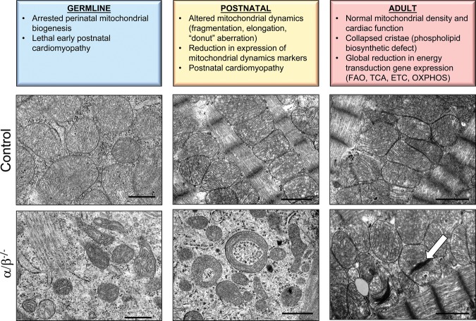
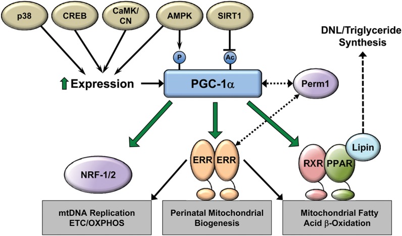
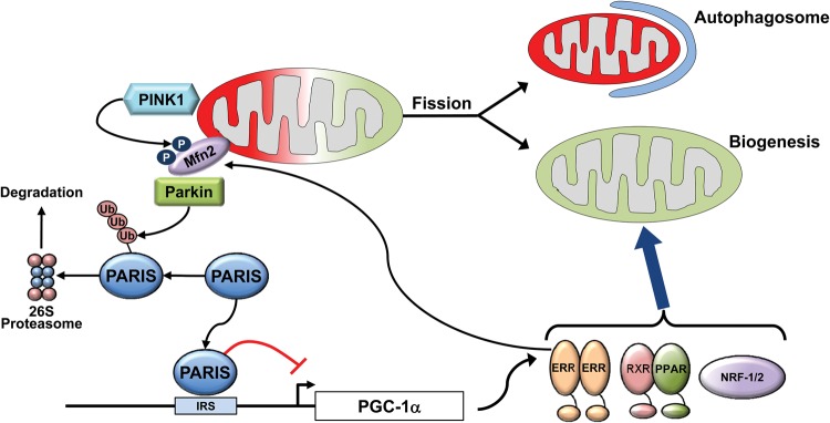
The mitochondrion is a complex organelle that serves essential roles in energy transduction, ATP production, and a myriad of cellular signaling events. A finely tuned regulatory network orchestrates the biogenesis, maintenance, and turnover of mitochondria. The high-capacity mitochondrial system in the heart is regulated in a dynamic way to generate and consume enormous amounts of ATP in order to support the constant pumping function in the context of changing energy demands. This review describes the regulatory circuitry and downstream events involved in mitochondrial biogenesis and its coordination with mitochondrial dynamics in developing and diseased hearts.

Keywords: biogenesis; mitochondria; mitochondrial dynamics; mitophagy.


Figures

Figure 1.
Figure 1.
Cardiac mitochondrial phenotypes of developmental stage-specific PGC-1 knockout mice. Electron micrographs illustrate the altered mitochondrial ultrastructure and biogenic response resultant from developmental stage- and cardiac-specific PGC-1α/β gene targeting “knockouts” (α/β−/−) (bottom row) compared with wild-type or PGC-1α−/− (adult) age-matched controls (top row). (Bottom, left) Germline disruption of PGC-1α/β genes results in a perinatal arrest of biogenesis with a profound reduction in mitochondrial content, as characterized by small numbers of immature mitochondria on day 1 following birth. (Bottom, middle) Deletion of cardiac PGC-1α/β genes in the postnatal period (using a muscle creatine kinase-driven Cre recombinase) impairs mitochondrial fusion and fission, as reflected by fragmented, elongated, and “donut”-shaped mitochondria. The micrographs were taken of cardiac ventricles at postnatal day 28. (Bottom, right) Inducible deletion of PGC-1 in adult hearts (12 wk of age) does not significantly affect mitochondrial density but results in a subset of abnormal mitochondria with collapsed cristae (white arrow), reminiscent of the phospholipid abnormalities seen with human Barth syndrome. Bars: germline micrographs, 0.5 μm; postnatal and adult micrographs, 1.0 μm. (FAO) Fatty acid oxidation.
Figure 2.
Figure 2.
The PGC-1α transcriptional regulatory cascade: upstream inputs and downstream targets. PGC-1α expression and activity are modulated by various upstream signaling pathways responsive to physiologic and metabolic stimuli to control mitochondrial biogenesis and function. As shown, PGC-1α interacts directly with and coactivates multiple DNA-binding transcription factors to control virtually all aspects of biogenesis, dynamics, and maintenance of mitochondrial protein levels. (CREB) cAMP response element-binding protein, (CaMK) calmodulin-dependent kinase, (CN) calcineurin, (AMPK) AMP-activated kinase, (SIRT1) sirtuin 1, (Perm1) PGC-1 and ERR regulator in muscle 1, (RXR) retinoid X receptor.
Figure 3.
Figure 3.
Coordinate control of mitophagy and mitochondrial biogenesis. Mitophagy and biogenesis are coordinately regulated to replace damaged mitochondria during periods of high mitochondrial turnover such as in the developing heart. Parkin is an E3 ubiquitin ligase recruited to the mitochondria through interaction with phosphorylated mitofusin 2 (Mfn2). Ubiquitination of outer mitochondrial membrane proteins by Parkin triggers partial or total engulfment by the autophagosome. Among Parkin's substrates is PARIS (Parkin-interacting substrate), a molecule that also serves as a transcriptional repressor of PGC-1α and may serve to coordinate mitophagy with biogenesis. Ubiquitination of PARIS and subsequent degradation serve to activate PGC-1α expression and the biogenic response. (IRS) Insulin response sequence.

References

    1. Ahuja P, Zhao P, Angelis E, Ruan H, Korge P, Olson A, Wang Y, Jin ES, Jeffrey FM, Portman M, et al. 2010. Myc controls transcriptional regulation of cardiac metabolism and mitochondrial biogenesis in response to pathological stress in mice. J Clin Invest 120: 1494–1505. - PMC - PubMed
    1. Akimoto T, Pohnert SC, Li P, Zhang M, Gumbs C, Rosenberg PB, Williams RS, Yan Z. 2005. Exercise stimulates Pgc-1α transcription in skeletal muscle through activation of the p38 MAPK pathway. J Biol Chem 280: 19587–19593. - PubMed
    1. Akki A, Smith K, Seymour AM. 2008. Compensated cardiac hypertrophy is characterised by a decline in palmitate oxidation. Mol Cell Biochem 311: 215–224. - PubMed
    1. Alaynick WA, Kondo RP, Xie W, He W, Dufour CR, Downes M, Jonker JW, Giles W, Naviaux RK, Giguere V, et al. 2007. ERRγ directs and maintains the transition to oxidative metabolism in the postnatal heart. Cell Metab 6: 13–24. - PubMed
    1. Andersson U, Scarpulla RC. 2001. Pgc-1-related coactivator, a novel, serum-inducible coactivator of nuclear respiratory factor 1-dependent transcription in mammalian cells. Mol Cell Biol 21: 3738–3749. - PMC - PubMed

Publication types

Substances

LinkOut - more resources