. 2015 Oct 20;112(42):13109-14.
doi: 10.1073/pnas.1512243112. Epub 2015 Oct 6.

Selective optogenetic activation of arcuate kisspeptin neurons generates pulsatile luteinizing hormone secretion

Affiliations

Selective optogenetic activation of arcuate kisspeptin neurons generates pulsatile luteinizing hormone secretion

Su Young Han et al. Proc Natl Acad Sci U S A. .

Abstract

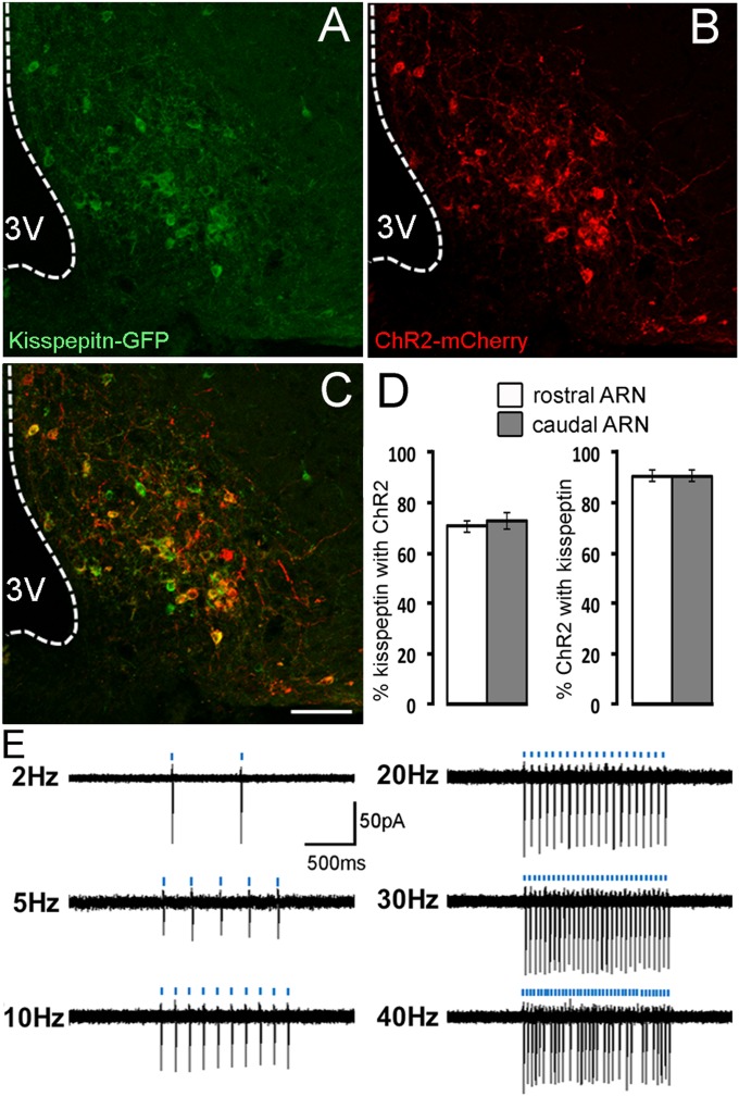
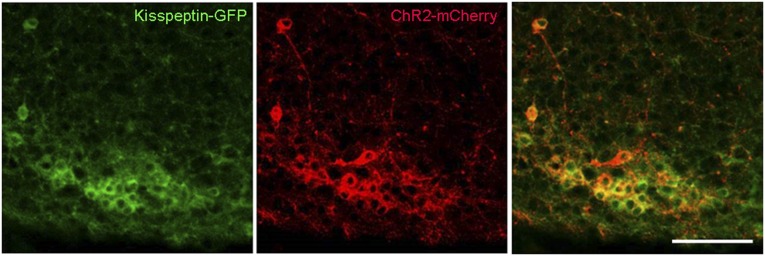
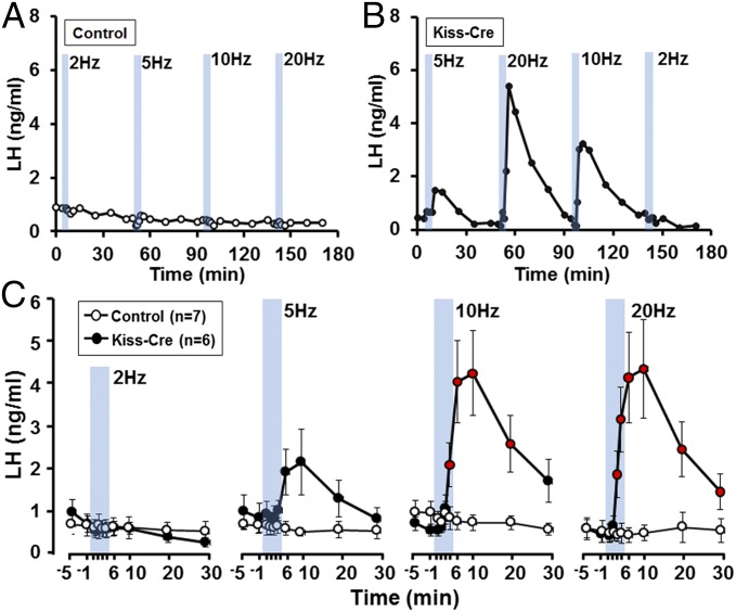
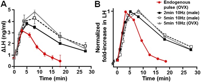
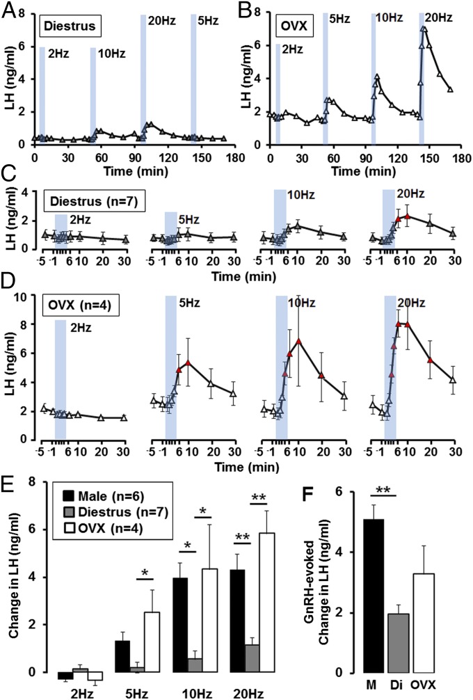
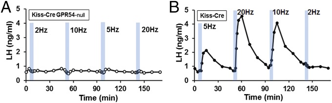
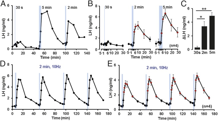
Normal reproductive functioning in mammals depends upon gonadotropin-releasing hormone (GnRH) neurons generating a pulsatile pattern of gonadotropin secretion. The neural mechanism underlying the episodic release of GnRH is not known, although recent studies have suggested that the kisspeptin neurons located in the arcuate nucleus (ARN) may be involved. In the present experiments we expressed channelrhodopsin (ChR2) in the ARN kisspeptin population to test directly whether synchronous activation of these neurons would generate pulsatile luteinizing hormone (LH) secretion in vivo. Characterization studies showed that this strategy targeted ChR2 to 70% of all ARN kisspeptin neurons and that, in vitro, these neurons were activated by 473-nm blue light with high fidelity up to 30 Hz. In vivo, the optogenetic activation of ARN kisspeptin neurons at 10 and 20 Hz evoked high amplitude, pulse-like increments in LH secretion in anesthetized male mice. Stimulation at 10 Hz for 2 min was sufficient to generate repetitive LH pulses. In diestrous female mice, only 20-Hz activation generated significant increments in LH secretion. In ovariectomized mice, 5-, 10-, and 20-Hz activation of ARN kisspeptin neurons were all found to evoke LH pulses. Part of the sex difference, but not the gonadal steroid dependence, resulted from differential pituitary sensitivity to GnRH. Experiments in kisspeptin receptor-null mice, showed that kisspeptin was the critical neuropeptide underlying the ability of ARN kisspeptin neurons to generate LH pulses. Together these data demonstrate that synchronized activation of the ARN kisspeptin neuronal population generates pulses of LH.

Keywords: GnRH; arcuate nucleus; gonadal steroids; kisspeptin; optogenetics.


Conflict of interest statement

The authors declare no conflict of interest.

Figures

Fig. 1.
Fig. 1.
Characterization of ARN kisspeptin neurons transfected with ChR2. (A–C) Photomicrographs showing (A) kisspeptin-GFP, (B) ChR2-mCherry, and (C) merged immunofluorescence in the ARN. 3V, third ventricle. (Scale bar, 100μm.) (D) Histograms showing the mean (± SEM; n = 5) percentage of rostral and caudal ARN kisspeptin neurons expressing ChR2 and percentage of ChR2 expressing neurons expressing kisspeptin. (E) Representative cell-attached, voltage-clamp recordings of ChR2-expressing kisspeptin neurons in the acute brain slice preparation activated with 5-ms blue light pulses (indicated by blue bars) given at 2, 5, 10, 20, 30, and 40 Hz over 1 s.
Fig. S1.
Fig. S1.
Photomicrographs showing GFP (kisspeptin) immunoreactivity, mCherry (ChR2), and overlay in the caudal ARN. (Scale bar, 100 μM.)
Fig. 2.
Fig. 2.
Effects of optogenetic activation of ARN kisspeptin neurons on LH secretion in male mice. (A and B) Representative examples showing LH secretion in response to blue light activation of ARN kisspeptin neurons (indicated by blue bars, 5-min each) at different frequencies in control and Kiss-Cre male mice. (C) Summary graphs showing mean ± SEM changes in LH secretion in response to 2-, 5-, 10-, and 20-Hz activation of ARN kisspeptin neurons in control male (open circle, n = 7) and Kiss-Cre male (closed circle, n = 6) mice. Circles in red indicate LH levels significantly elevated compared with prestimulation values for each frequency (P < 0.05; repeated-measures ANOVA with Dunnett’s post hoc test).
Fig. S2.
Fig. S2.
Comparison of the mean optogenetic-evoked pulse-like increases in LH secretion with endogenous LH pulses (red). (A) Dynamics of evoked increments in LH secretion following 10-Hz activation of ARN kisspeptin neurons for 2-min (black, male) or 5-min (open square, male; gray, OVX female) intervals compared with endogenous LH pulses [red; data taken from Campos and Herbison (25)]. (B) Dynamics of evoked and endogenous LH pulses after normalizing values to the peak LH responses and start time of the pulses.
Fig. 3.
Fig. 3.
Effects of optogenetic activation of ARN kisspeptin neurons on LH secretion in female mice. (A and B) Representative examples showing LH secretion in response to blue light activation of kisspeptin neurons (indicated by blue bars, 5-min each) at frequencies of 2, 5, 10, and 20 Hz, in (A) diestrous and (B) OVX female Kiss1-IRES-Cre mice. (C and D) Summary graphs showing mean ± SEM changes in LH secretion in response to 2-, 5-, 10-, and 20-Hz activation of ARN kisspeptin neurons in diestrous (n = 7) and OVX (n = 4) mice. Triangles in red indicate LH levels significantly elevated compared with prestimulation values for each frequency (P < 0.05; one-way repeated-measures ANOVA with Dunnett’s post hoc test). (E) Summary histograms showing mean + SEM increases in LH secretion evoked by 2, 5, 10, and 20 Hz of 5-min activation of ARN kisspeptin neurons in male (black), diestrous (gray), and OVX (white) mice. *P < 0.05, **P < 0.01, two-way ANOVA with Bonferroni’s post hoc test. (F) Histogram showing mean + SEM increase in LH evoked by exogenous subcutaneous GnRH in male (black), diestrous (gray), and OVX (white) mice. **P < 0.01, one-way ANOVA with Bonferroni’s post hoc test.
Fig. 4.
Fig. 4.
Effects of in vivo optogenetic activation of ARN kisspeptin neurons on LH secretion in GPR54-null mice. Representative examples of 5-min blue light activation of kisspeptin neurons (indicated by blue bars) at 2, 5, 10, and 20 Hz in (A) an AAV-injected Kiss1-IRES-Cre; Gpr54-null male mice and (B) in a control AAV-injected Kiss1-IRES-Cre;Gpr54+/− male mice.
Fig. 5.
Fig. 5.
Effects of different durations of 10-Hz activation of ARN kisspeptin neurons on LH secretion in male mice. (A) Representative example of LH secretion evoked by blue light activation of kisspeptin neurons (indicated by blue bars) at 10 Hz for 30 s, 2 min, and 5 min in Kiss-IRES-Cre male mice. (B) Summary graphs showing mean ± SEM changes in LH secretion in response to 30-s, 2-min, and 5-min activation of ARN kisspeptin neurons at 10 Hz in male mice. Circles in red indicate LH levels significantly elevated compared with prestimulation values for each time interval (P < 0.05; one-way repeated-measures ANOVA with Dunnett’s post hoc test). (C) Histogram showing mean + SEM increase in LH secretion in the first 10 min after optogenetic stimulation for different durations in male mice (n = 4). *P < 0.05, **P < 0.01, one-way ANOVA with Bonferroni’s post hoc test. (D) Shows representative example of LH secretion evoked by 2-min blue light activation of kisspeptin neurons (indicated by blue bars) at 10-Hz repeated four times over 45-min intervals in a Kiss-IRES-Cre male mice. (E) Summary graph showing mean ± SEM evoked LH secretion in male mice (n = 4). Circles in red indicate LH levels significantly elevated compared with prestimulation values before each activation (P < 0.05; one-way repeated-measures ANOVA with Dunnett’s post hoc test).

References

    1. Belchetz PE, Plant TM, Nakai Y, Keogh EJ, Knobil E. Hypophysial responses to continuous and intermittent delivery of hypopthalamic gonadotropin-releasing hormone. Science. 1978;202(4368):631–633. - PubMed
    1. Clarke IJ, Cummins JT. The temporal relationship between gonadotropin releasing hormone (GnRH) and luteinizing hormone (LH) secretion in ovariectomized ewes. Endocrinology. 1982;111(5):1737–1739. - PubMed
    1. Levine JE, Pau KY, Ramirez VD, Jackson GL. Simultaneous measurement of luteinizing hormone-releasing hormone and luteinizing hormone release in unanesthetized, ovariectomized sheep. Endocrinology. 1982;111(5):1449–1455. - PubMed
    1. Wetsel WC, et al. Intrinsic pulsatile secretory activity of immortalized luteinizing hormone-releasing hormone-secreting neurons. Proc Natl Acad Sci USA. 1992;89(9):4149–4153. - PMC - PubMed
    1. Martínez de la Escalera G, Choi AL, Weiner RI. Generation and synchronization of gonadotropin-releasing hormone (GnRH) pulses: Intrinsic properties of the GT1-1 GnRH neuronal cell line. Proc Natl Acad Sci USA. 1992;89(5):1852–1855. - PMC - PubMed

Publication types

LinkOut - more resources