. 2015 Nov 3;112(44):E5926-35.
doi: 10.1073/pnas.1515043112. Epub 2015 Oct 6.

Resting state of the human proton channel dimer in a lipid bilayer

Affiliations

Resting state of the human proton channel dimer in a lipid bilayer

Qufei Li et al. Proc Natl Acad Sci U S A. .

Abstract

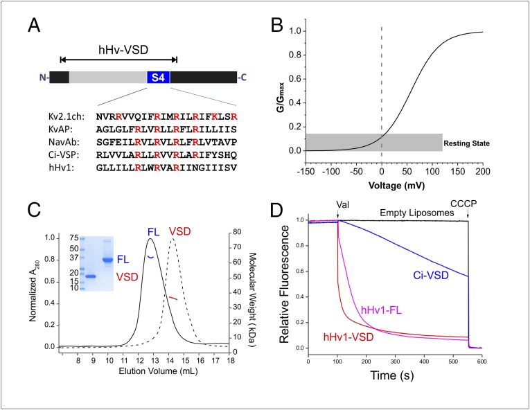
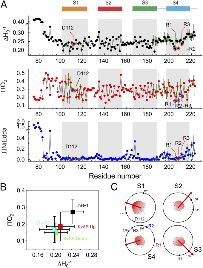
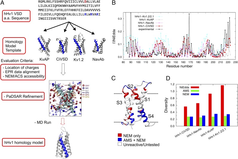
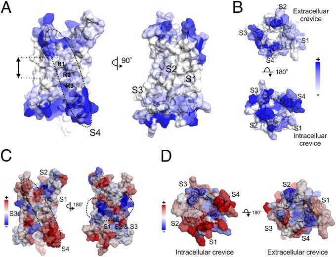
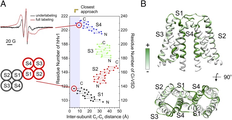
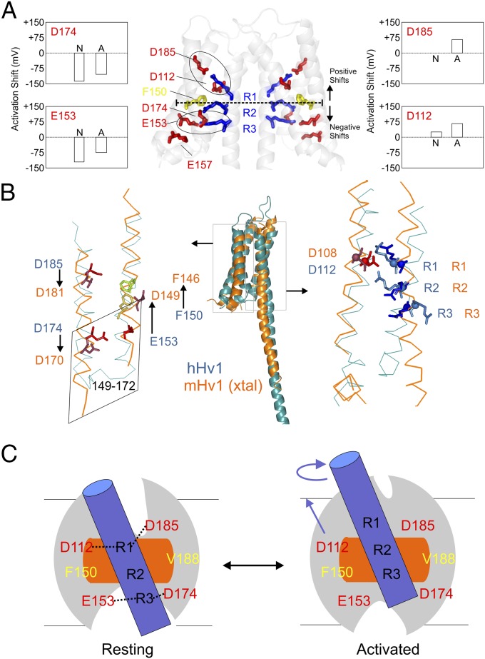
The voltage-gated proton channel Hv1 plays a critical role in the fast proton translocation that underlies a wide range of physiological functions, including the phagocytic respiratory burst, sperm motility, apoptosis, and metastatic cancer. Both voltage activation and proton conduction are carried out by a voltage-sensing domain (VSD) with strong similarity to canonical VSDs in voltage-dependent cation channels and enzymes. We set out to determine the structural properties of membrane-reconstituted human proton channel (hHv1) in its resting conformation using electron paramagnetic resonance spectroscopy together with biochemical and computational methods. We evaluated existing structural templates and generated a spectroscopically constrained model of the hHv1 dimer based on the Ci-VSD structure at resting state. Mapped accessibility data revealed deep water penetration through hHv1, suggesting a highly focused electric field, comprising two turns of helix along the fourth transmembrane segment. This region likely contains the H(+) selectivity filter and the conduction pore. Our 3D model offers plausible explanations for existing electrophysiological and biochemical data, offering an explicit mechanism for voltage activation based on a one-click sliding helix conformational rearrangement.

Keywords: EPR; MD simulation; proton channel; voltage sensing domain.


Conflict of interest statement

The authors declare no conflict of interest.

Figures

Fig. 1.
Fig. 1.
Biochemical preparation of hHv1 protein. (A) Schematic presentation of hHv1 constructs. Voltage-sensing domain (gray) and the fourth transmembrane segment (blue) were highlighted on full-length construct. The hHv1-VSD construct, which covered the entire VSD, was used as a template for cysteine scanning and EPR studies. (B) Simulated G–V curve of hHv1 with V1/2 = 58 mV and z = 0.90 (19). At 0 mV, hHv1 populates at the resting state. (C) Characterization of hHv1 full-length (FL) and hHv1-VSD (VSD) by SDS/PAGE gel, SEC (FL, solid black line; VSD, dotted black line), and MALS (FL, blue solid line; VSD, red solid line) methods. Expected molecular mass is 34 kDa for hHv1-FL and 19 kDa for hHv1-VSD. Both hHv1-FL and hHv1-VSD are dimers in the buffer of 20 mM Tris (pH 8.0), 150 mM NaCl, and 4 mM Fos-choline 12 at studied concentration (∼40 μM). (D) ACMA-based flux assay for reconstituted liposome. The truncated hHv1-VSD (red) conducts the proton as well as the full-length hHv1-FL (magenta). CiVSD (blue) has a small leakage of proton current.
Fig. 2.
Fig. 2.
Environmental parameters of hHv1 in lipids. (A) Mobility (∆Ho−1), oxygen accessibility (ΠO2), and NiEdda accessibility (ΠNi) of hHv1-VSD in POPC:POPG proteoliposome. Repeats for selected positions (n = 3–5) in S1 and S4 are shown. Error bars, SD. The putative transmembrane segments (S1, S2, S3, and S4) are shown on the top and indicated in gray in the plot. The critical residues D112, R205 (R1), R208 (R2), and R211 (R3) are indicated with circles. (B) Average mobility and oxygen accessibility of four transmembrane helices of four VSD systems hHv1-VSD (black), CiVSD (cyan), KvAP-Up (red), and KvAP-Down (green), which are studied by EPR methods, are plotted with SD. hHv1-VSD is the most dynamic VSD system in lipids by far. (C) A 2D wheel presentation of oxygen accessibility of four transmembrane helices. The vectorial sums (red arrow) of oxygen accessibility are significantly larger than values of individual residues, and preferably point to the lipid-exposing environment away from the core of the four-helix bundle. The functionally critical residues (D112, R1, R2, and R3) are on the opposite side of the lipid environment and protected by side-chain interactions. Boundaries of each transmembrane segment are shown by black dot with residue numbers for reference.
Fig. 3.
Fig. 3.
Structural modeling for hHv1-VSD at resting state. (A) Strategy of structural modeling by MD simulation. Starting from the sequence alignments, four structural models of hHv1-VSD were built on the templates of crystal structures of KvAP, CiVSD-WT, Kv1.2_chimera, and NavAb respectively. Each of the structural models was equilibrated with explicit lipid POPC and stabilized for 300 ns by MD simulation. The four MD stable models were evaluated with experimental data primarily with ΠNiEdda and NEM/AMS accessibility data. The structural model correlating best with experimental accessibilities was subjected to an additional MD simulation with constraints from present EPR spectroscopic data using an established computational method, PaDSAR (36). The constrained model was equilibrated in explicit lipid POPC for 400 ns, which serves our working model for the resting state of hHv1-VSD. (B) Predicted solvent accessibility from four structural models and the experimental NiEdda accessibility. (C) NEM/AMS accessibility data were mapped onto hHv1-VSD structural model from CiVSD-WT. (D) The correlation between predicted accessibilities and experimental accessibilities for each of the four hHv1-VSD structural models was evaluated by Kullback–Leibler divergence. The structural model from CiVSD-WT with lowest divergence was chosen to further construct the structural model for hHv1-VSD.
Fig. 4.
Fig. 4.
Mapping the EPR environmental parameters on the hHv1-VSD model. (A) ΠNiEdda data were mapped onto hHv1-VSD structural model. Apparent ΠNiEdda were observed along S4 segment (Left, dotted circle) and two turns of helix around the first two gating charges R1 and R2 (Left, black arrow), together with all other transmembrane regions in S1, S2, and S3 (Right), have minimum ΠNiEdda. (B) Both extracellular and intracellular crevices have significant ΠNiEdda. There is apparent solvent penetration through the intracellular crevice, but not obvious in the extracellular crevice. The scale bar is the same for both A and B. (C) The mapped mobility shows the S4 segment is most mobile (Left, dotted circle) among all transmembrane segments. There is a cluster of motion-restriction residues in the middle of transmembrane segments S1, S2, and S3 (Right, dotted circle); they are in the same vertical position around R1 and R2 with minimum ΠNiEdda. (D) The mapped oxygen accessibilities shows the regions with lowest oxygen accessibility are located roughly inside the four-helix bundle away from the lipid environment.
Fig. 5.
Fig. 5.
The hHv1-VSD dimer interface agrees with CiVSD-WT dimer structure. (A) (Upper Left) Strong dipolar coupling from spin-labeled hHv1-VSD-206C. (Lower Left) Mutant 121C at top of S1 can form spontaneous disulfide linkage in native hHv1 (38). (Right) Cβ distances between hHv1 monomer according to the CiVSD-WT dimer (PDB ID code 4G80). Both residues 121 and 206 are among the closest residues (<10 Å). (B) hHv1-VSD dimer model built by aligning two monomer structural models with CiVSD-WT dimer crystal structure. The cross-linking data from mHv was mapped onto the dimer model (40), and regions with high cross-linking propensities, including S4 and top of S1, are in close proximity in the dimer model.
Fig. 6.
Fig. 6.
Model comparison and mechanistic implications. (A) Functionally critical residues in the hHv1-VSD dimer model. D174 and E153 are located below F150, the putative hydrophobic gasket, and D185 and D112 are located above. Neutralization of D174 and E153 causes left-shift in the G–V curve, whereas neutralization of D185 and D112 causes right-shift. The two electrostatic clusters D185-R1-D112 and E153-R3-D174 are highlighted with black circles. (B) Structural model of hHv1 was aligned with crystal structure of mHv-chimeric by the Cα of S1 + S2 + S3a. The residues on S1 (D112) and S4 (R1, R2, and R3) agreed very well (Right) with obvious differences of the residues on S2 and S3 between two models. To match mHv1-chimeric structure, the residues on S2 (D153 and F150) need to upshift one turn of helix, whereas the residues on S3 (D174 and D185) need to downshift one turn of helix. The chimeric region from CiVSD on mHv-chimera spanned from residues 149 to 172 (corresponding to residue 153–176 in hHv1), which are highlighted in the rhomboidal box. The structures of the identical sequence in CiVSD and mHv-chimera are distinctly different. (C) Proposed gating model of hHv1 according to the one-click gating mechanism in CiVSD. At resting-state hHv1-VSD in lipid bilayer, solvent penetrates from both intracellular and extracellular crevices leaving a focused electric field around R1 and R2 region at the level of hydrophobic gasket. hHv1 scaffold is shown by the gray oval; S4 by blue cylinder; hydrophobic gasket by orange rectangle. The two salt-bridge clusters (D112-R1-D183 and E153-R3-D174) are connected by black dotted lines. Up activation: the S4 of hHv1 rotates and moves three residues up.

Comment in

  • Structural revelations of the human proton channel.
    DeCoursey TE. DeCoursey TE. Proc Natl Acad Sci U S A. 2015 Nov 3;112(44):13430-1. doi: 10.1073/pnas.1518486112. Epub 2015 Oct 14. Proc Natl Acad Sci U S A. 2015. PMID: 26466610 Free PMC article. No abstract available.

References

    1. Ramsey IS, Moran MM, Chong JA, Clapham DE. A voltage-gated proton-selective channel lacking the pore domain. Nature. 2006;440(7088):1213–1216. - PMC - PubMed
    1. Sasaki M, Takagi M, Okamura Y. A voltage sensor-domain protein is a voltage-gated proton channel. Science. 2006;312(5773):589–592. - PubMed
    1. DeCoursey TE. Voltage-gated proton channels: Molecular biology, physiology, and pathophysiology of the H(V) family. Physiol Rev. 2013;93(2):599–652. - PMC - PubMed
    1. Henderson LM, Chappell JB, Jones OT. The superoxide-generating NADPH oxidase of human neutrophils is electrogenic and associated with an H+ channel. Biochem J. 1987;246(2):325–329. - PMC - PubMed
    1. DeCoursey TE, Morgan D, Cherny VV. The voltage dependence of NADPH oxidase reveals why phagocytes need proton channels. Nature. 2003;422(6931):531–534. - PubMed

Publication types

LinkOut - more resources