. 2015 Oct 7;10(10):e0139856.
doi: 10.1371/journal.pone.0139856. eCollection 2015.

Leukotriene B4 Enhances NOD2-Dependent Innate Response against Influenza Virus Infection

Affiliations

Leukotriene B4 Enhances NOD2-Dependent Innate Response against Influenza Virus Infection

Manon Le Bel et al. PLoS One. .

Abstract

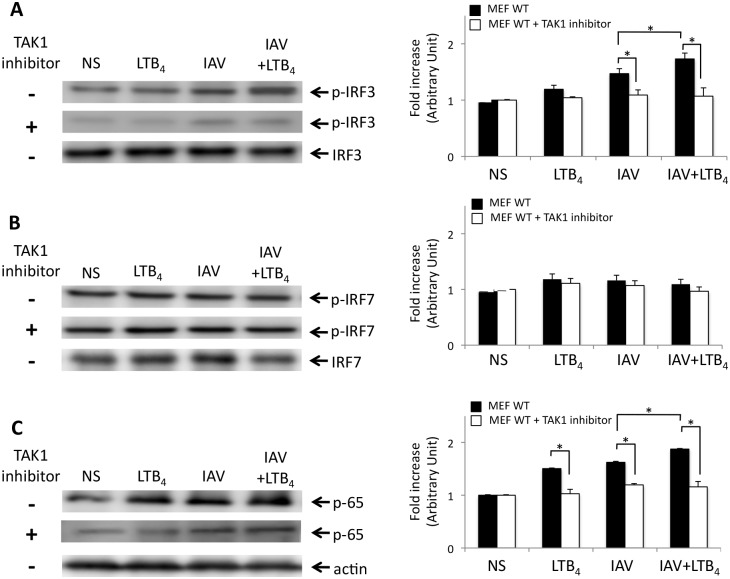
Leukotriene B4 (LTB4), a central mediator of inflammation, is well known for its chemoattractant properties on effectors cells of the immune system. LTB4 also has the ability to control microbial infection by improving host innate defenses through the release of antimicrobial peptides and modulation of intracellular Toll-like receptors (TLRs) expression in response to agonist challenge. In this report, we provide evidences that LTB4 acts on nucleotide-binging oligomerization domain 2 (NOD2) pathway to enhance immune response against influenza A infection. Infected mice receiving LTB4 show improved survival, lung architecture and reduced lung viral loads as compared to placebo-treated animals. NOD2 and its downstream adaptor protein IPS-1 have been found to be essential for LTB4-mediated effects against IAV infection, as absence of NOD2 or IPS-1 diminished its capacity to control viral infection. Treatment of IAV-infected mice with LTB4 induces an increased activation of IPS-1-IRF3 axis leading to an enhanced production of IFNβ in lungs of infected mice. LTB4 also has the ability to act on the RICK-NF-κB axis since administration of LTB4 to mice challenged with MDP markedly increases the secretion of IL-6 and TNFα in lungs of mice. TAK1 appears to be essential to the action of LTB4 on NOD2 pathway since pretreatment of MEFs with TAK1 inhibitor prior stimulation with IAV or MDP strongly abrogated the potentiating effects of LTB4 on both IFNβ and cytokine secretion. Together, our results demonstrate that LTB4, through its ability to activate TAK1, potentiates both IPS-1 and RICK axis of the NOD2 pathway to improve host innate responses.


Conflict of interest statement

Competing Interests: The authors have declared that no competing interests exist.

Figures

Fig 1
Fig 1. NOD2 contributes to the effects of LTB4 treatment on the control of IAV-infection.
WT and NOD2-/- mice (n = 6 per group) were infected with IAV and daily treated (i.v.) with placebo or LTB4 (1 μg/kg). (A) Mice were infected with a lethal dose of IAV (3000 PFU, i.n.) and survival was monitored daily for 10 days. (B) Mice were infected with IAV (50 PFU, i.n.) and lungs were harvested at days 3, 5 and 7 post-infection for viral load assessment by plaque assay. Data are representative of three independent experiments. *p<0.05 as compared to WT placebo-treated group. (C) Histological examination of mice infected with IAV (50 PFU, i.n.) and treated daily with LTB4 (i.v.). Five days post-infection, lungs were harvested and tissues were fixed and stained with hematoxylin-eosin for microscopic observation. a: alveolar and b: bronchiolar structure (original magnification ×100).
Fig 2
Fig 2. IPS–1 is required for LTB4 to control IAV infection.
WT and IPS–1-/- mice (n = 6 per group) were infected with IAV and daily treated (i.v.) with placebo or LTB4 (1 μg/kg). (A) For survival experiments, mice were infected with a lethal dose of IAV (3000 PFU, i.n.) and monitored daily for 10 days. (B) Mice were infected with IAV (50 PFU, i.n.) and lungs were harvested at days 3, 5 and 7 post-infection for viral load measurement. Data are representative of three independent experiments. *p<0.05 as compared to WT placebo-treated group.
Fig 3
Fig 3. NOD2 is involved in the enhanced effect of LTB4 on activation of IRF3 and NF-κB in IAV-infected mice.
WT, NOD2-/- and IPS–1-/- mice (n = 6 per group) were infected with IAV (50 PFU, i.n.) and treated (i.v.) with placebo or LTB4 (1 μg/kg). Six hours post-treatment, mice were sacrificed and lungs were homogenized for protein extraction. Immunoblots of (A) phosphorylated-IRF3 on serine 396 (p-IRF3), (B) phosphorylated IRF7 on serine 471/472 (p-IRF7) and (C) NF-κB-p65 proteins, as well as their respective loading control, in lung homogenates. Right panels show densitometric analysis of p-IRF3, p-IRF7 and NF-κB-p65 immunoblots. Fold increase in proteins expression is expressed relative to the respective not-stimulated (NS) group. Data are representative of three independent experiments. *p<0.05 as compared to the indicated groups.
Fig 4
Fig 4. LTB4-mediated increased production of inflammatory mediators is abrogated in IAV-infected NOD2-/- and IPS–1-/- mice.
WT, NOD2-/- and IPS–1-/- mice (n = 6 per group) were infected with IAV (50 PFU, i.n.) and treated (i.v.) daily with placebo or LTB4 (1 μg/kg). Mice were sacrificed 6 hours post-treatment and levels of (A, D) IFNβ, (B, E) TNFα and (C, F) IL–6 were determined in lung homogenates by ELISA. Data are representative of three independent experiments. *p<0.05 as compared to indicated groups.
Fig 5
Fig 5. TAK1 activation is essential to the priming effect of LTB4 on both IPS–1 and RICK axis of the NOD2 pathway.
WT, NOD2-/- and IPS–1-/- mice (n = 6 per group) were infected with IAV (50 PFU i.n.) or challenged with MDP (10 mg/kg) and treated (i.v.) with placebo or LTB4 (1 μg/kg). Six hours post-treatment, mice were sacrificed and lungs were homogenized for protein extraction. Representative immunoblots of phosphorylated TAK1 on threonine 187 and actin loading control in lung homogenates of (A) mice treated with LTB4 alone or infected with IAV and treated with LTB4 or a placebo, or (B) mice treated with MDP alone or in combination with LTB4. Right panels show densitometric analysis of p-TAK1 expression in lung homogenates. Fold increase in TAK1 is expressed relative to the respective not-stimulated (NS) group. Data are representative of two independent experiments. *p<0.05 as compared to the indicated groups.
Fig 6
Fig 6. TAK1 is required for LTB4 to enhance activation of IRF3 and NF-κB.
WT Mouse Embryonic Fibroblasts (MEFs) were treated with specific TAK1 inhibitor 5Z-7-oxozeaenol (1 μM) 30 minutes prior to IAV infection (0.5 m.o.i) and LTB4 treatment (1 μM). Cells were harvested 6 hours post-treatment and proteins were extracted for western blot analyses. Representative immunoblots of (A) phosphorylated-IRF3 on serine 396 (p-IRF3) (B) phosphorylated IRF7 on serine 471/472 (p-IRF7) and (C) NF-κB-p65 proteins and their respective IRF3, IRF7 and actin loading control in MEFs. Right panels show the folds increase in p-IRF3, p-IRF7 and NF-κB-p65 expression in MEFs. Fold increase in proteins expression is expressed relative to the respective not-stimulated (NS) group. Data are representative of two independent experiments. *p<0.05 as compared to the indicated groups.
Fig 7
Fig 7. TAK1 contributes to the potentiating effect of LTB4 on the release of IFNβ, TNFα and IL–6.
(A-C) WT Mouse Embryonic Fibroblasts (MEFs) were treated with specific TAK1 inhibitor 5Z-7-oxozeaenol (1 μM) 30 minutes prior to LTB4 (1 μM) treatment, IAV infection (0.5 m.o.i.) or IAV infection and LTB4 administration. (D-F) Mice were stimulated with MDP (10 μg/ml) alone or in combination with LTB4. Supernatants were collected 6 hours post-treatment and levels of (A, D) IFNβ, (B, E) TNFα and (C, F) IL–6 were determined by ELISA. Data are representative of three independent experiments. *p<0.05 as compared to indicated groups. n.d.: not detected.
Fig 8
Fig 8. Schematic representation of LTB4 effects on NOD2 signaling.
Following recognition of viral (IAV) or non-viral (MDP) ligands, NOD2 receptors signal through IPS–1 or RICK, respectively, to activate TAK1. Administration of LTB4 potentiates NOD2-mediated responses by acting on TAK1 (dotted arrow) at the branch of the adaptor protein IPS–1 in response to IAV infection (red arrows) or at the branch of the protein kinase RICK in response to MDP sensing (black arrows), which culminates in increased production of their respective inflammatory mediators. This boosting effect of LTB4 on TAK1 could require a bridging protein to induce an optimized NOD2-mediated innate immune response.

References

    1. Shin EH, Lee HY, Bae YS (2006) Leukotriene B4 stimulates human monocyte-derived dendritic cell chemotaxis. Biochem Biophys Res Commun 348: 606–611. - PubMed
    1. Tager AM, Bromley SK, Medoff BD, Islam SA, Bercury SD, et al. (2003) Leukotriene B4 receptor BLT1 mediates early effector T cell recruitment. Nat Immunol 4: 982–990. - PubMed
    1. Goodarzi K, Goodarzi M, Tager AM, Luster AD, von Andrian UH (2003) Leukotriene B4 and BLT1 control cytotoxic effector T cell recruitment to inflamed tissues. Nat Immunol 4: 965–973. - PubMed
    1. Gaudreault E, Gosselin J (2007) Leukotriene B4-mediated release of antimicrobial peptides against cytomegalovirus is BLT1 dependent. Viral Immunol 20: 407–420. - PubMed
    1. Gosselin J, Borgeat P, Flamand L (2005) Leukotriene B4 protects latently infected mice against murine cytomegalovirus reactivation following allogeneic transplantation. J Immunol 174: 1587–1593. - PubMed

Publication types

MeSH terms