. 2015 Oct 7;10(10):e0139506.
doi: 10.1371/journal.pone.0139506. eCollection 2015.

Annexin A2 Acts as an Adhesion Molecule on the Endometrial Epithelium during Implantation in Mice

Affiliations

Annexin A2 Acts as an Adhesion Molecule on the Endometrial Epithelium during Implantation in Mice

Bing Wang et al. PLoS One. .

Abstract

To determine the function of Annexin A2 (Axna2) in mouse embryo implantation in vivo, experimental manipulation of Axna2 activities was performed in mouse endometrial tissue in vivo and in vitro. Histological examination of endometrial tissues was performed throughout the reproduction cycle and after steroid treatment. Embryo implantation was determined after blockage of the Axna2 activities by siRNA or anti-Axna2 antibody. The expression of Axna2 immunoreactivies in the endometrial luminal epithelium changed cyclically in the estrus cycle and was upregulated by estrogen. After nidatory estrogen surge, there was a concentration of Axna2 immunoreactivities at the interface between the implanting embryo and the luminal epithelium. The phenomenon was likely to be induced by the implanting embryos as no such concentration of signal was observed in the inter-implantation sites and in pseudopregnancy. Knockdown of Axna2 by siRNA reduced attachment of mouse blastocysts onto endometrial tissues in vitro. Consistently, the number of implantation sites was significantly reduced after infusion of anti-Axna2 antibody into the uterine cavity. Steroids and embryos modulate the expression of Axna2 in the endometrial epithelium. Axna2 may function as an adhesion molecule during embryo implantation in mice.


Conflict of interest statement

Competing Interests: The authors have declared that no competing interests exist.

Figures

Fig 1
Fig 1. Axna2 expression in mouse endometrial luminal epithelium.
(A) Endometrial Axna2 expression in the estrous cycle: Axna2 expression was high in the luminal epithelium (LE) at proestrous, and absent at diestrous. (B) H-Scoring of the immunostaining. a P<0.001 with diestrus, b P = 0.002 with metestrus. c P = 0.004 with estrus. (C) Axna2 expression in mouse LE in early pregnancy. Axna2 expression was increased after estradiol surge (10:00 am—12 noon) on Day 4 of pregnancy, and was decreased on Day 6 and Day 7 of pregnancy. Magnification: 1000x.
Fig 2
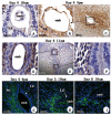
Fig 2. Expression of Axna2 at implantation sites.
On Day 4 of pregnancy, the immunoreactivities (brown) at the implantation sites were weak at 10:00 am (A). The signal increased at 4:00 pm (B). At 4:00 pm, the signal was strong at the implantation sites (B) and in the nearby stroma (Str) (C). Concentration of signal was observed at the interface between the luminal epithelium (LE) and the embryo (emb) (D, E) but not between LE (F). Immunofluorescence staining showed that Axna2 immunoreactivies (green) were on the membrane of the endometrial stroma on Day 4 (G) and Day 5 (H) of pregnancy. The immunoreactivies became cytosolic on Day 6 of pregnancy (I).
Fig 3
Fig 3. Effects of estradiol and progesterone on the expression of Axna2 in endometrial luminal epithelium of ovariectomized mice.
(A) The expression of Axna2 was increased by estradiol (E2), progesterone (P4) and their combined (E2P4) treatment. E2 had the most potent effect. (B) H-Scoring of immunohistochemical staining. a P<0.05 with control (ctrl), b P<0.05 with P4.
Fig 4
Fig 4. Effect of Axna2 on implantation.
(A) Attachment rate of mouse blastocysts in an endometrial tissue culture model for implantation. The attachment rate on Day 4 pseudopregnant endometrium was significantly suppressed after transfection of the endometrium with Axna2 siRNA. (B) The number of implantation sites (IS) was significantly decreased after uterine infusion of anti-Axna2 antibody. Right: 10 μl of normal IgG; Left: 10 μl of anti-Axna2 antibody. Bar chart shows the number of implantation sites in the anti-Axna2 antibody and IgG treated uteri. P = 0.008 between the two groups (n = 5).

References

    1. Das SK, Wang XN, Paria BC, Damm D, Abraham JA, Klagsbrun M, et al. Heparin-binding EGF-like growth factor gene is induced in the mouse uterus temporally by the blastocyst solely at the site of its apposition: a possible ligand for interaction with blastocyst EGF-receptor in implantation. Development. 1994;120(5):1071–83. - PubMed
    1. Lim H, Gupta RA, Ma WG, Paria BC, Moller DE, Morrow JD, et al. Cyclo-oxygenase-2-derived prostacyclin mediates embryo implantation in the mouse via PPARdelta. Genes Dev. 1999;13(12):1561–74. - PMC - PubMed
    1. Lim H, Paria BC, Das SK, Dinchuk JE, Langenbach R, Trzaskos JM, et al. Multiple female reproductive failures in cyclooxygenase 2-deficient mice. Cell. 1997;91(2):197–208. - PubMed
    1. Dominguez F, Garrido-Gomez T, Lopez JA, Camafeita E, Quinonero A, Pellicer A, et al. Proteomic analysis of the human receptive versus non-receptive endometrium using differential in-gel electrophoresis and MALDI-MS unveils stathmin 1 and annexin A2 as differentially regulated. Hum Reprod. 2009;24(10):2607–17. 10.1093/humrep/dep230 - DOI - PubMed
    1. Li J, Tan Z, Li M, Xia T, Liu P, Yu W. Proteomic analysis of endometrium in fertile women during the prereceptive and receptive phases after luteinizing hormone surge. Fertil Steril. 2011;95(3):1161–3. 10.1016/j.fertnstert.2010.09.033 - DOI - PubMed

LinkOut - more resources