. 2015 Oct 7;10(10):e0129008.
doi: 10.1371/journal.pone.0129008. eCollection 2015.

Proteomics Analysis Reveals Distinct Corona Composition on Magnetic Nanoparticles with Different Surface Coatings: Implications for Interactions with Primary Human Macrophages

Affiliations

Proteomics Analysis Reveals Distinct Corona Composition on Magnetic Nanoparticles with Different Surface Coatings: Implications for Interactions with Primary Human Macrophages

Carmen Vogt et al. PLoS One. .

Abstract

Superparamagnetic iron oxide nanoparticles (SPIONs) have emerged as promising contrast agents for magnetic resonance imaging. The influence of different surface coatings on the biocompatibility of SPIONs has been addressed, but the potential impact of the so-called corona of adsorbed proteins on the surface of SPIONs on their biological behavior is less well studied. Here, we determined the composition of the plasma protein corona on silica-coated versus dextran-coated SPIONs using mass spectrometry-based proteomics approaches. Notably, gene ontology (GO) enrichment analysis and Kyoto Encyclopedia of Genes and Genomes (KEGG) pathway analysis revealed distinct protein corona compositions for the two different SPIONs. Relaxivity of silica-coated SPIONs was modulated by the presence of a protein corona. Moreover, the viability of primary human monocyte-derived macrophages was influenced by the protein corona on silica-coated, but not dextran-coated SPIONs, and the protein corona promoted cellular uptake of silica-coated SPIONs, but did not affect internalization of dextran-coated SPIONs.


Conflict of interest statement

Competing Interests: The authors have declared that no competing interests exist.

Figures

Fig 1
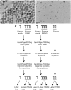
Fig 1. TEM of the silica-coated SPIONs (CSNP) (A) and dextran-coated SPIONs (nanomag-D-spio) (B). (scale bar = 50 nm). (C) Schematic overview of the protein corona analysis.
Fig 2
Fig 2. Biocompatibility assessment of SPIONs with or without a ‘hard’ corona of plasma proteins.
Human monocyte-derived macrophages were exposed for 2 h (A, D), 6 h (B, E) or 24 h (C, F) to the indicated doses (μL/mL) of CSNP or CSNP + protein corona (A-C), or to nanomag-D-spio or nanomag-D-spio + protein corona (D-F). Macrophage viability was determined using the MTT assay. Cells were cultured in the absence of FBS to exclude any confounding effects of serum proteins. Results are presented as % mitochondrial function (mean values ± S.D.) from four independent experiments using cells isolated from healthy human donors. Statistical analysis was performed using Tukey post-hoc test following one way ANOVA (**p<0.01, ***p<0.001).
Fig 3
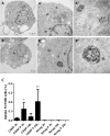
Fig 3. Macrophage internalization of SPIONs with or without a protein corona.
TEM of HMDM exposed for 2 h to 50 μg/ml of CSNP (A-A”) or to CSNP + protein corona (B-B”). The arrows in (A, B) and (A’, B’) show some of the areas where the nanoparticles are located in the cells. In the higher magnification micrographs (A”, B”) the core-shell nanoparticles can be clearly seen. C. Uptake of CSNP, CSNP + protein corona, nanomag-D-spio, and nanomag-D-spio + protein corona was quantified by using ICP-MS to determine cellular iron content. HMDM were exposed to 50 μg/ml SPIONs for 2 h and 24 h. Results are presented as % of iron concentration per 1000 cells (mean values ± S.D.) adjusted to control, from three independent experiments, using cells from healthy donors. HMDM were maintained in cell culture medium without FBS. The difference in degree of cellular uptake between the two SPIONs in the absence or presence of a protein corona was evaluated using Tukey post-hoc test following one way ANOVA (*p<0.05,**p<0.01).
Fig 4
Fig 4. Distinct plasma protein corona composition on SPIONs with different surface coating.
Cluster analysis of SPION-bound plasma proteins versus plasma proteins. Red color denotes counts higher than average when compared to other samples. Blue denotes counts lower than average (refer to legend top right corner). Clusters are numbered 1–5 (cf. S4 Table). Clusters 2 and 5 contain proteins that are enriched in the CSNP corona. Cluster 1 proteins are enriched in the nanomag-D-spio corona and clusters 3–4 contain plasma-enriched proteins. For gene ontology (GO) enrichment analysis of the corona-specific clusters, refer to Figs 5 and S5.
Fig 5
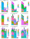
Fig 5. Plasma protein corona composition on CSNP.
Classification of identified corona proteins according to differentially enriched Gene Ontology (GO) categories and KEGG pathways. The relative abundances of the proteins, as a percentage of total estimated protein abundance in the sample, are shown and each replicate is plotted separately. A significant enrichment of plasma proteins absorbed on the CSNP surface involved (A) KEGG Complement and coagulation cascades pathway, (B) GO Regulation of coagulation, (C) GO Negative regulation of coagulation, (D) GO Positive regulation of coagulation, (E) GO Regulation of fibrinolysis, (F) GO Fibrinogen complex, (G) GO Heparin binding, (H) GO Regulation of defense response, and (I) GO Lipid biosynthetic process. For further details, refer to S3, S5 and S6 Tables.

Similar articles

Cited by

References

    1. Pouliquen D, Le Jeune JJ, Perdrisot R, Ermias A, Jallet P. Iron oxide nanoparticles for use as an MRI contrast agent: Pharmacokinetics and metabolism. MRI. 1991;9(3):275–83. 10.1016/0730-725x(91)90412-f - DOI - PubMed
    1. Mahmoudi M, Sant S, Wang B, Laurent S, Sen T. Superparamagnetic iron oxide nanoparticles (SPIONs): Development, surface modification and applications in chemotherapy. Adv Drug Deliv Rev. 2011;63(1–2):24–46. 10.1016/j.addr.2010.05.006 - DOI - PubMed
    1. Gupta AK, Gupta M. Synthesis and surface engineering of iron oxide nanoparticles for biomedical applications. Biomaterials. 2005;26(18):3995–4021. 10.1016/j.biomaterials.2004.10.012 - DOI - PubMed
    1. Kunzmann A, Andersson B, Vogt C, Feliu N, Ye F, Gabrielsson S, et al. Efficient internalization of silica-coated iron oxide nanoparticles of different sizes by primary human macrophages and dendritic cells. Toxicol and Appl Pharmacol. 2011;253(2):81–93. 10.1016/j.taap.2011.03.011 - DOI - PubMed
    1. Lynch I, Cedervall T, Lundqvist M, Cabaleiro-Lago C, Linse S, Dawson KA. The nanoparticle–protein complex as a biological entity; a complex fluids and surface science challenge for the 21st century. Adv Colloid Interface Sci. 2007;134–135:167–74. 10.1016/j.cis.2007.04.021 - DOI - PubMed

Publication types