. 2015 Oct 7;10(10):e0138787.
doi: 10.1371/journal.pone.0138787. eCollection 2015.

Cysteine-Rich Atrial Secretory Protein from the Snail Achatina achatina: Purification and Structural Characterization

Affiliations

Cysteine-Rich Atrial Secretory Protein from the Snail Achatina achatina: Purification and Structural Characterization

Sergey Shabelnikov et al. PLoS One. .

Abstract

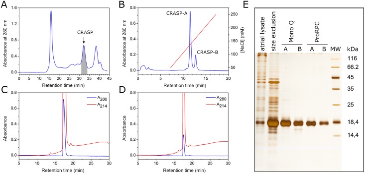
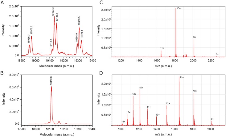
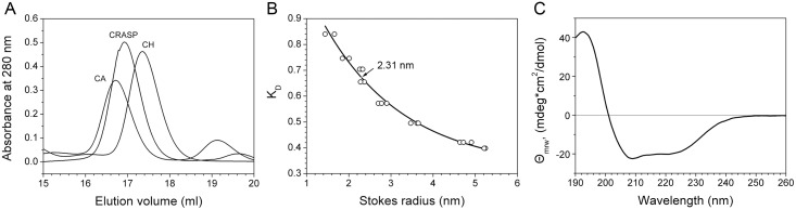
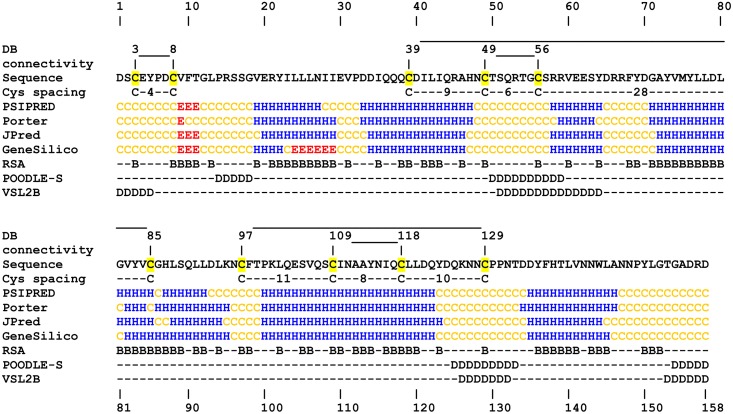
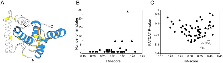
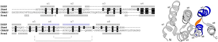
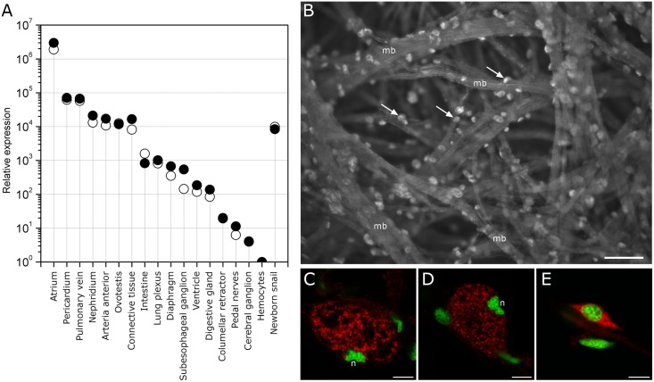
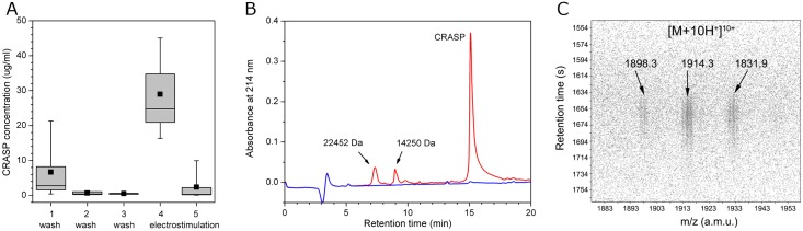
Despite extensive studies of cardiac bioactive peptides and their functions in molluscs, soluble proteins expressed in the heart and secreted into the circulation have not yet been reported. In this study, we describe an 18.1-kDa, cysteine-rich atrial secretory protein (CRASP) isolated from the terrestrial snail Achatina achatina that has no detectable sequence similarity to any known protein or nucleotide sequence. CRASP is an acidic, 158-residue, N-glycosylated protein composed of eight alpha-helical segments stabilized with five disulphide bonds. A combination of fold recognition algorithms and ab initio folding predicted that CRASP adopts an all-alpha, right-handed superhelical fold. CRASP is most strongly expressed in the atrium in secretory atrial granular cells, and substantial amounts of CRASP are released from the heart upon nerve stimulation. CRASP is detected in the haemolymph of intact animals at nanomolar concentrations. CRASP is the first secretory protein expressed in molluscan atrium to be reported. We propose that CRASP is an example of a taxonomically restricted gene that might be responsible for adaptations specific for terrestrial pulmonates.


Conflict of interest statement

Competing Interests: The authors have declared that no competing interests exist.

Figures

Fig 1
Fig 1. Purification of CRASP isoforms from the atria of A. achatina.
(A) Semi-preparative size-exclusion chromatography on a Superdex 200 column. Fraction from 31 to 34 min (shaded grey) was collected and subjected to anion exchange chromatography. (B) Separation of isoforms on a Mono-Q anion exchange column. Fractions from the two major peaks were designated CRASP-A and CRASP-B. Final purification of CRASP-A (C) and CRASP-B (D) on a ProRPC C4 reversed phase column. (E) All fractions were analysed with SDS-PAGE (15% gel) and silver staining.
Fig 2
Fig 2. Deduced amino acid sequence of CRASP.
The signal peptide is shown in red, a low compositional complexity region is shown in blue, cysteines are shaded yellow, and a potential N-glycosylation site is shaded black. The N-terminal and internal peptide sequences detected by Edman degradation are underlined.
Fig 3
Fig 3. ESI MS analysis of CRASP in water/acetonitrile with 0.25% formic acid.
(A) A typical deconvoluted ESI mass spectrum of CRASP containing N-linked glycan. Note the multiple peaks due to glycan heterogeneity. (B) The deconvoluted ESI mass spectrum of the deglycosylated protein showed a single peak of 18110.8 Da. ESI mass spectra of deglycosylated, non-reduced CRASP (C) and CRASP reduced with dithiothreitol (D). Peaks are labelled according to the charge states of the protein ion.
Fig 4
Fig 4. Biophysical characterization of CRASP.
(A) Size-exclusion chromatography showed that deglycosylated CRASP migrated unusually fast compared to carbonic anhydrase (CA) and chymotrypsinogen (CH). (B) Determination of the Stokes radius of the deglycosylated protein by size-exclusion chromatography. The elution position (arrow) and estimated Stokes radius are indicated. (C) The CD spectrum showed a strong maximum at 192 nm and two minima at 208 nm and 222 nm, the characteristic spectral features of an α-helical conformation. Only the CD spectrum of isoform A is shown because the spectra of the isoforms were almost identical.
Fig 5
Fig 5. Prediction of secondary structure, solvent accessibility and disorder.
The disulphide bonding pattern was determined experimentally. Cysteine spacing motifs are shown; the cysteine residues are shaded yellow. The secondary structure was predicted using PSIPRED, Porter, JPred and GeneSilico. Relative solvent accessibility was predicted using Spine-X. Short disordered regions were predicted using POODLE-S and VSL2B. ‘H’, ‘E’ and ‘C’ denote α-helices, β-strands and coils, respectively. ‘B’ denotes buried residues (relative solvent accessibility <25%), and ‘D’ denotes predicted disordered residues.
Fig 6
Fig 6. The use of ab initio folding simulation to detect correct templates from a pool of structures identified by threading methods.
(A) Top QUARK ab initio model refined with ModRefiner. The Cα-Cα′ distance restraints used to direct folding are satisfied (yellow). The C-terminal part of the protein shows four α-helices (blue) with right-handed superhelical topology. (B) Templates sorted according to an average TM-score calculated on the fold level, revealing SCOP a.118, an all-alpha right-handed superhelical fold (triangle). (C) Templates sorted according to the TM-score and the FATCAT p-value. A cluster of SCOP a.118.9 superfamily templates is evident (open triangles). Other templates from the SCOP a.118 fold (filled triangles) and other all-alpha templates (filled circles) are also shown.
Fig 7
Fig 7. Sequence-structure alignment of the CRASP sequence with the all-alpha, right-handed superhelical fold of the 2KM4 template.
The representative original threading alignment produced by the SP3 program (left panel) and the 3D structure of the 2KM4 template (right panel) are shown. The secondary structure is labelled ‘H’ for α-helix and ‘E’ for β-strand, where ‘DSSP’ and ‘Pred’ denote the assignment by DSSP and the prediction by PSIPRED, respectively. Alpha helices α1-α8 are shown. Residues involved in hydrophobic core packing in the experimental structure are shaded black. Note the good match in hydrophobicity and the match of the α-helical hairpin (blue), formed by the template’s sixth and seventh helices, with a long helical segment constrained by two disulphide bonds, which probably forms a similar super-secondary motif. The sequence identity is 11%.
Fig 8
Fig 8. Tissue-specific expression of CRASP mRNA.
(A) qRT-PCR analysis of CRASP mRNA expression in different tissues. Relative expression was calculated using α-tubulin (open circles) and 60S acidic ribosomal protein P0 (filled circles) as reference genes. A log scale (y-axis) was used. Data are presented as the mean of two replicate qRT-PCR reactions from pooled tissues from two snails. (B) FISH detection of CRASP mRNA in whole-mount atrium. The epifluorescent image demonstrates the distribution of transcripts within cells (arrows) located on the surface of muscle bundles (mb). The scale bar indicates 50 μm. (C, D) Confocal sections demonstrate the localization of transcripts in the intergranular space of the atrial granular cells. Note the specific eccentric location and irregular shape of the nucleus (n) in mature granular cells. (E) Rarely, transcripts were detected in small spindle-like cells. Scale bars indicate 7.5 μm. The hybrids were detected with avidin-Cy3 (red pseudo colour). DAPI was used as a general DNA dye (green pseudo colour).
Fig 9
Fig 9. CRASP is secreted by the heart and has low abundance in the haemolymph.
(A) Secretion of CRASP by the isolated heart. The protein concentration decreased significantly from the first wash to the third wash and increased in the fourth fraction collected upon electrostimulation of the heart nerve. Note the depletion of CRASP after the fourth fraction with continued stimulation. The mean (squares), median (lines), 25th to 75th percentiles (boxes) and range (whiskers) for six samples are shown. (B) Representative chromatograms of the third wash fraction (blue line) and the fraction collected upon stimulation (red line). Note that in addition to CRASP, two minor proteins are present. (C) LC-ESI MS identification of CRASP in the haemolymph. The fragment of the LC-MS 2D map shows a 10-fold charged ion species with typical heterogeneity.

Similar articles

Cited by

References

    1. Haszprunar G, Wanninger A. Molluscs. Curr Biol. 2012. July 10;22(13):R510–4. 10.1016/j.cub.2012.05.039 - DOI - PubMed
    1. J H.D.. Circulatory systems of gastropods and bivalves In: Saleuddin ASM, Wilbur KM, editors. The Mollusca. New York: Academic Press; 1983. p. 189–238.
    1. Cottrell GA, Osborne N. A neurosecretory system terminating in the Helix heart. Comp Biochem Physiol. 1969. March;28(3):1455–9.
    1. Kodirov SA. The neuronal control of cardiac functions in Molluscs. Comp Biochem Physiol A Mol Integr Physiol. 2011. October;160(2):102–16. 10.1016/j.cbpa.2011.06.014 - DOI - PMC - PubMed
    1. De With ND, van der Schors RC, Boer HH, Ebberink RHM. The sodium influx stimulating peptide of the pulmonate freshwater snail Lymnaea stagnalis. Peptides. 1993. July;14(4):783–9. - PubMed

Publication types