. 2015 Oct 7;10(10):e0139845.
doi: 10.1371/journal.pone.0139845. eCollection 2015.

Modulation of Dendritic Cell Activation and Subsequent Th1 Cell Polarization by Lidocaine

Affiliations

Modulation of Dendritic Cell Activation and Subsequent Th1 Cell Polarization by Lidocaine

Young-Tae Jeon et al. PLoS One. .

Abstract

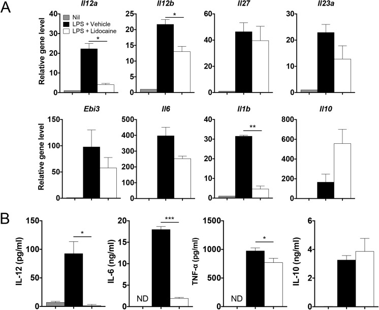
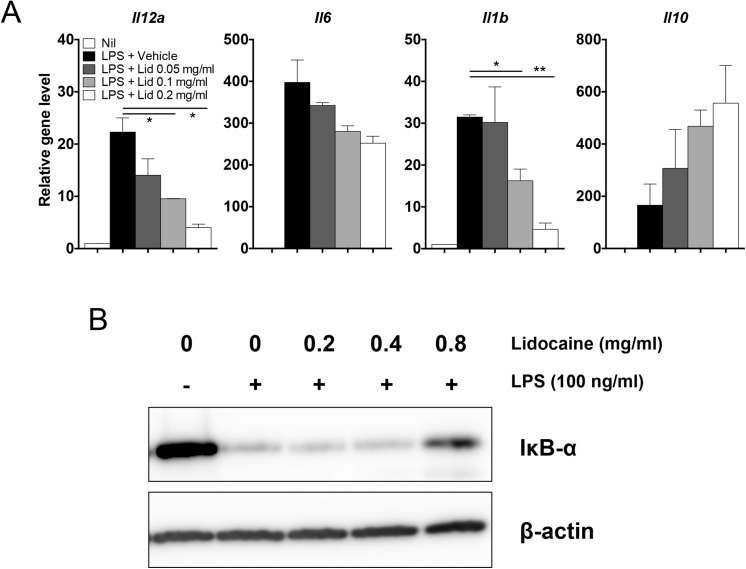
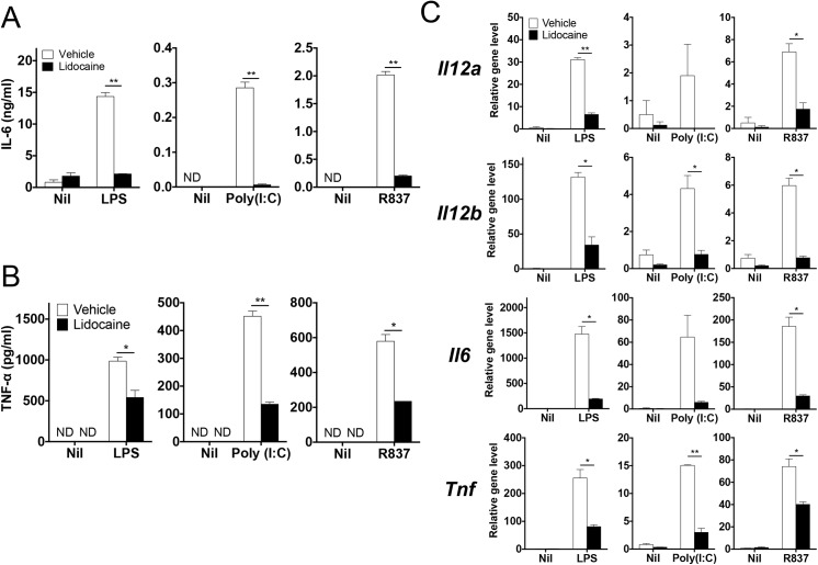
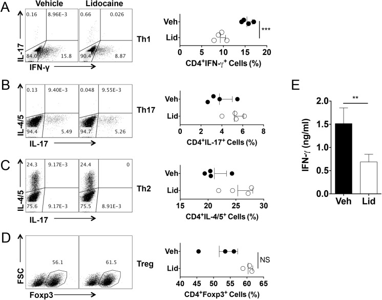
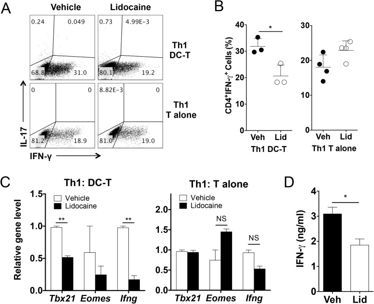
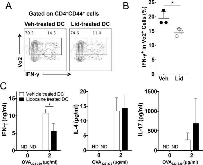
Dendritic cells play an essential role in bridging innate and adaptive immunity by recognizing cellular stress including pathogen- and damage-associated molecular patterns and by shaping the types of antigen-specific T cell immunity. Although lidocaine is widely used in clinical settings that trigger cellular stress, it remains unclear whether such treatment impacts the activation of innate immune cells and subsequent differentiation of T cells. Here we showed that lidocaine inhibited the production of IL-6, TNFα and IL-12 from dendritic cells in response to toll-like receptor ligands including lipopolysaccharide, poly(I:C) and R837 in a dose-dependent manner. Notably, the differentiation of Th1 cells was significantly suppressed by the addition of lidocaine while the same treatment had little effect on the differentiation of Th17, Th2 and regulatory T cells in vitro. Moreover, lidocaine suppressed the ovalbumin-specific Th1 cell responses in vivo induced by the adoptive transfer of ovalbumin-pulsed dendritic cells. These results demonstrate that lidocaine inhibits the activation of dendritic cells in response to toll-like receptor signals and subsequently suppresses the differentiation of Th1 cell responses.


Conflict of interest statement

Competing Interests: The authors have declared that no competing interests exist.

Figures

Fig 1
Fig 1. Effects of lidocaine on the expression of various cytokines upon LPS stimulation.
Bone marrow-derived dendritic cells were stimulated with 100 ng/ml of LPS in the presence of vehicle (EtOH) or 0.2 mg/ml lidocaine for 4 h and 24 h to examine mRNA expression and cytokine production, respectively. (A) The mRNA levels of the indicated genes were analyzed by quantitative RT-PCR. (B) The amounts of each cytokine produced were measured by ELISA. All experiments were performed at least three times. Data shown are mean ± SEM. *p<0.05; ***p<0.001; ND, not detected.
Fig 2
Fig 2. Lidocaine regulates the expression of cytokines and NF-κB signaling pathway in a dose-dependent manner.
(A) Bone marrow-derived dendritic cells were stimulated with 100 ng/ml of LPS together with increasing concentrations of lidocaine for 4 h. The mRNA levels of the indicated genes were analyzed by quantitative RT-PCR. (B) Raw 264.7 cells were treated with increasing doses of lidocaine for 2 h and stimulated with LPS for 20 min. The expression of IκB-α was examined by western blot. All experiments were performed at least three times. Data shown are mean ± SEM. *p<0.05; **p<0.01.
Fig 3
Fig 3. Regulation of cytokines expression in dendritic cells in response to various TLR ligands by lidocaine.
(A & B) Bone marrow-derived dendritic cells were stimulated with LPS (100 ng/ml), poly(I:C) (1 μg/ml) or R837 (1 μg/ml) in the presence of lidocaine (0.4 mg/ml) or vehicle. The amounts of IL–6 and TNF-α in the supernatant were measured by ELISA. (C) The mRNA levels of the indicated genes were analyzed by quantitative RT-PCR. Data represent at least two independent experiments. Data shown are mean ± SEM. *p<0.05; **p<0.01; ND, not detected.
Fig 4
Fig 4. Lidocaine inhibits dendritic cell-mediated Th1 cell differentiation while having little effects on dendritic cell-mediated Th2, Th17, regulatory T cell differentiation in vitro.
Naïve CD4+ T cells were co-cultured with bone marrow-derived dendritic cells with Th1, Th17, Th2 or regulatory T cell differentiation conditioned-media or cultured with plate-coated anti-CD3 and anti-CD28 with supernatant of dendritic cells stimulated with LPS in the presence of lidocaine (0.2 mg/ml or indicated dose) or vehicle. (A-D) The frequencies of IFN-γ, IL–17, IL–4/5 or Foxp3 positive cells among CD4+ population were measured by flow cytometer. (E) The level of IFN-γ was measured using co-cultured supernatants from Th1 differentiation condition. Data represent at least two independent experiments. Data shown are mean ± SEM. *p<0.05; **p<0.01; ***p<0.001; NS, not significant.
Fig 5
Fig 5. Lidocaine inhibits dendritic cell-mediated Th1 cell differentiation in vitro.
Naïve CD4+ T cells were either co-cultured with bone marrow-derived dendritic cells in the presence of soluble anti-CD3 and LPS, or in anti-CD3, CD28 pre-coated plates in the presence of IL–2 and IL–12 for Th1 cell differentiation. Lidocaine was added at a concentration of 0.2 mg/ml. (A & B) The frequencies of IFNγ or IL–17 producing cells among CD4+ T cells. (C) The mRNA levels of the indicated genes. (D) The levels of IFN-γ in the cultured supernatants of naïve CD4+ T cells cultured with vehicle- or lidocaine-conditioned media. Data represent at least three independent experiments. Data shown are mean ± SEM. **p<0.01; ***p<0.001; NS, not significant.
Fig 6
Fig 6. Inhibition of dendritic cell-mediated antigen-specific Th1 cell responses by lidocaine in vivo.
Bone marrow-derived dendritic cells were pulsed with OVA323-339 in the presence of lidocaine or vehicle before being transferred into OT-II TcR transgenic mice (n = 3~4). (A and B) The frequencies of IFN- γ producers among Vα2+ cells. (C) The amounts of the indicated cytokines in the supernatant of splenocyte stimulated with OVA323-339 were measured by ELISA. Data represent two independent experiments. Data shown are mean ± SEM. *p<0.05.

References

    1. Janeway CA Jr., Medzhitov R. Innate immune recognition. Annu Rev Immunol. 2002;20:197–216. Epub 2002/02/28. 10.1146/annurev.immunol.20.083001.084359 . - DOI - PubMed
    1. Rubartelli A, Lotze MT. Inside, outside, upside down: damage-associated molecular-pattern molecules (DAMPs) and redox. Trends Immunol. 2007;28(10):429–36. Epub 2007/09/12. 10.1016/j.it.2007.08.004 . - DOI - PubMed
    1. Merad M, Sathe P, Helft J, Miller J, Mortha A. The dendritic cell lineage: ontogeny and function of dendritic cells and their subsets in the steady state and the inflamed setting. Annu Rev Immunol. 2013;31:563–604. Epub 2013/03/23. 10.1146/annurev-immunol-020711-074950 - DOI - PMC - PubMed
    1. Lipscomb MF, Masten BJ. Dendritic cells: immune regulators in health and disease. Physiol Rev. 2002;82(1):97–130. Epub 2002/01/05. 10.1152/physrev.00023.2001 . - DOI - PubMed
    1. Carbone FR, Belz GT, Heath WR. Transfer of antigen between migrating and lymph node-resident DCs in peripheral T-cell tolerance and immunity. Trends Immunol. 2004;25(12):655–8. Epub 2004/11/09. 10.1016/j.it.2004.09.013 . - DOI - PubMed

Publication types

MeSH terms

LinkOut - more resources