. 2015 Oct 6;109(7):1439-45.
doi: 10.1016/j.bpj.2015.08.019.

Thermal Motion of DNA in an MspA Pore

Affiliations

Thermal Motion of DNA in an MspA Pore

Bo Lu et al. Biophys J. .

Abstract

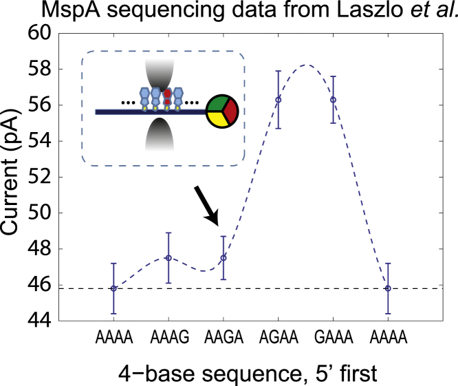
We report on an experiment and calculations that determine the thermal motion of a voltage-clamped single-stranded DNA-NeutrAvidin complex in a Mycobacterium smegmatis porin A nanopore. The electric force and diffusion constant of DNA inside a Mycobacterium smegmatis porin A pore were determined to evaluate the thermal position fluctuations of DNA. We show that an out-of-equilibrium state returns to equilibrium so quickly that experiments usually measure a weighted average over the equilibrium position distribution. Averaging over the equilibrium position distribution is consistent with results of state-of-the-art nanopore sequencing experiments. It is shown how a reduction in thermal position fluctuations can be achieved by increasing the electrophoretic force used in nanopore sequencing devices.


Figures

Figure 1
Figure 1
(A) In this schematic diagram, ssDNA is trapped inside a nanopore by a molecular stop, which opposes an electrophoretic driving force. For nanopore sequencing strategies, the impact of thermal motion on nucleotide resolution can be described by a position probability function, which depends on the driving force and diffusion constant of DNA. (B) The energy landscape for a trapped ssDNA in an MspA pore. The gray sphere is an attached NeutrAvidin molecule that prevents the ssDNA from translocating through the pore. The escape rate and time for such trapped DNA depends on the driving force and diffusion constant of DNA.
Figure 2
Figure 2
(A) Schematic figure of an ssDNA-NeutrAvidin complex being captured (i and ii) and escaping (iii and iv) from an MspA pore. (B and C) The dynamic voltage and current trace demonstrate the capture and escape process. The definition of escape time has been labeled in (C).
Figure 3
Figure 3
(A) Escape-time distribution for ssDNA at 40 mV clamping voltage. The same escape-time distribution is plotted on a log-scale axis in (B) and (C) to show the exponential decay profile at short times. (D) Plot of the average escape time as a function of clamping voltage. The red line is the first-passage calculation fit. D0 is defined as the diffusion constant for a 1.5-nm-diameter sphere in free space.
Figure 4
Figure 4
(A) The normalized DNA position probability function evolves from a delta function to an equilibrium exponential function within 10 ns at 140 mV driving voltage. F0 is 17 pN, defined as the driving force for 0.54e/base charge density ssDNA at 100 mV. D0 is defined as the diffusion constant for a 1.5-nm-diameter sphere in free space. (B) The equilibrium position probability function under different electrophoretic driving forces. (Inset) The region encompassed by 99% of the position distribution function, and its dependence on the driving force.
Figure 5
Figure 5
Data taken from Laszlo et al. (4), a previous report about MspA sequencing data. Plot shows the current signal corresponding to a 4-mer sequence with a G substitution at different locations. The error bar on each point is the standard deviation of the mean current level for the specific 4-mer sequence measured at different positions along ϕ X 174 DNA. (Inset) Orientation of AAGA inside the MspA pore.

Comment in

References

    1. Butler T.Z., Pavlenok M., Gundlach J.H. Single-molecule DNA detection with an engineered MspA protein nanopore. Proc. Natl. Acad. Sci. USA. 2008;105:20647–20652. - PMC - PubMed
    1. Derrington I.M., Butler T.Z., Gundlach J.H. Nanopore DNA sequencing with MspA. Proc. Natl. Acad. Sci. USA. 2010;107:16060–16065. - PMC - PubMed
    1. Laszlo A.H., Derrington I.M., Gundlach J.H. Detection and mapping of 5-methylcytosine and 5-hydroxymethylcytosine with nanopore MspA. Proc. Natl. Acad. Sci. USA. 2013;110:18904–18909. - PMC - PubMed
    1. Laszlo A.H., Derrington I.M., Gundlach J.H. Decoding long nanopore sequencing reads of natural DNA. Nat. Biotechnol. 2014;32:829–833. - PMC - PubMed
    1. Manrao E.A., Derrington I.M., Gundlach J.H. Reading DNA at single-nucleotide resolution with a mutant MspA nanopore and ϕ29 DNA polymerase. Nat. Biotechnol. 2012;30:349–353. - PMC - PubMed

Publication types