. 2015 Oct 7:8:513.
doi: 10.1186/s13071-015-1119-4.

SjCa8, a calcium-binding protein from Schistosoma japonicum, inhibits cell migration and suppresses nitric oxide release of RAW264.7 macrophages

Affiliations

SjCa8, a calcium-binding protein from Schistosoma japonicum, inhibits cell migration and suppresses nitric oxide release of RAW264.7 macrophages

Ji Liu et al. Parasit Vectors. .

Abstract

Background: Schistosomiasis is considered second only to malaria as the most devastating parasitic disease in tropical countries. Schistosome cercariae invade the host by penetrating the skin and migrate though the lungs and portal circulation to their final destination in the hepatic portal system and eventually the mesenteric veins. Previous studies have shown that the cytotoxic pathways that target schistosomulum in the lung-stage involve nitric oxide (NO) produced by macrophages. By contrast, skin-stage schistosomulas can evade clearance, indicating that they might be freed from macrophage NO-mediated cytotoxicity to achieve immune evasion; however, the critical molecules and mechanisms involved remain unknown.

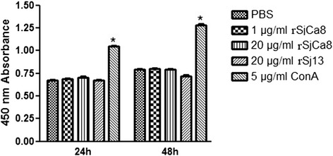
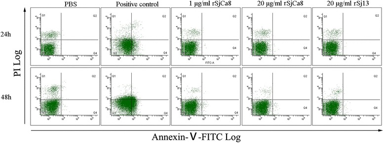
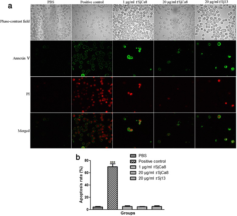
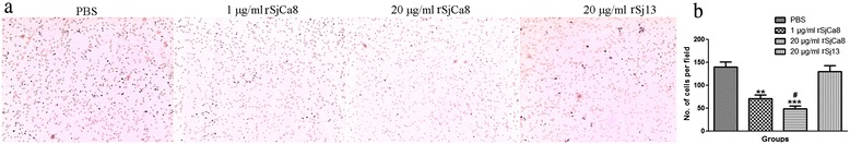
Methods: Recombinant SjCa8 (rSjCa8), an 8-kDa calcium-binding protein that is stage-specifically expressed in cercaria and early skin-stage schistosomulas of Schistosoma japonicum, was incubated with mouse RAW264.7 macrophages. Effects on macrophage proliferation were determined using Cell Counting Kit-8. Next, transwell assay was carried out to further investigate the role of rSjCa8 in macrophage migration. The effects of rSjCa8 on macrophage apoptosis were evaluated using confocal microscopy and flow cytometry. Additional impacts of rSjCa8 on NO release by lipopolysaccharide (LPS)-stimulated macrophages as well as the underlying mechanisms were explored using fluorescent probe, nitric oxide signaling pathway microarray, quantitative real-time PCR, mutagenesis, and neutralizing antibody approaches.

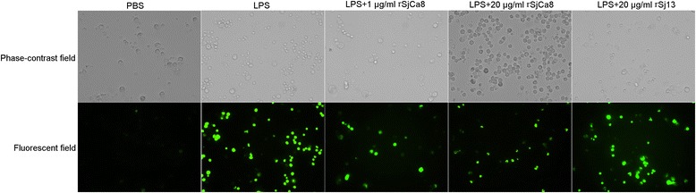
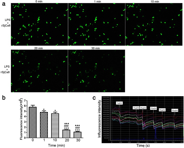
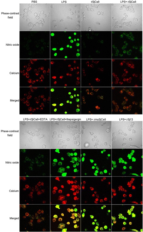
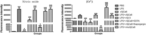
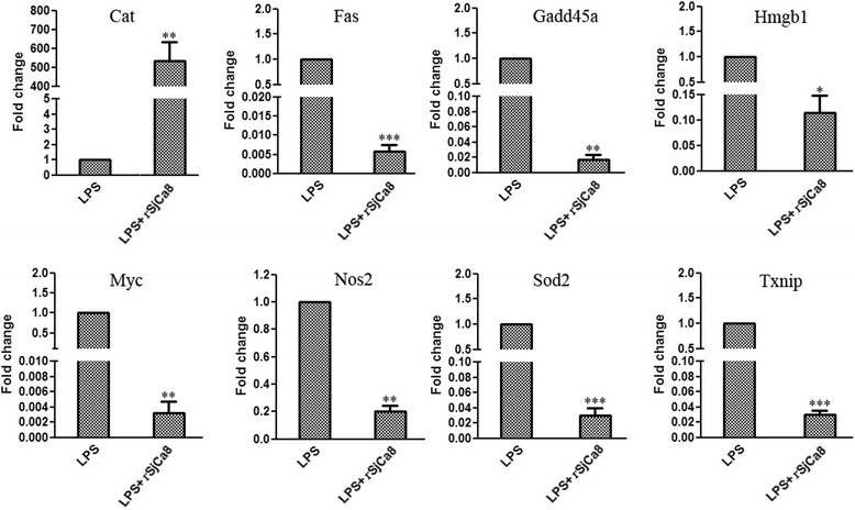
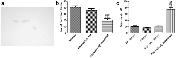
Results: rSjCa8 exhibited a striking inhibitory effect on macrophage migration, but did not markedly increase cell proliferation or apoptosis. Additionally, rSjCa8 potently inhibited NO release by LPS-stimulated macrophages in a dose- and time-dependent manner, and the inhibitory mechanism was closely associated with intracellular Ca(2+) levels, the up-regulation of catalase expression, and the down-regulation of the expression of 47 genes, including Myc, Gadd45a, Txnip, Fas, Sod2, Nos2, and Hmgb1. Vaccination with rSjCa8 increased NO concentration in the challenging skin area of infected mice and reduced the number of migrated schistosomula after skin penetration by cercariae.

Conclusions: Our findings indicate that SjCa8 might be a novel molecule that plays a critical role in immune evasion by S. japonicum cercaria during the process of skin penetration. The inhibitory impacts of rSjCa8 on macrophage migration and [Ca(2+)]i-dependent NO release suggest it might represent a novel vaccine candidate and chemotherapeutic target for the prevention and treatment of schistosomiasis.


Figures

Fig. 1
Fig. 1
Effects of rSjCa8 on the proliferation of RAW264.7 cells. RAW264.7 cells were treated or not with ConA (5 μg/ml), rSj13 (20 μg/ml), or various concentrations of rSjCa8 (1 or 20 μg/ml) for 24 or 48 h, respectively. Cell viability was assessed in triplicate using the CCK-8 method according to the manufacturer’s protocol. * P<0.05, compared with the PBS group
Fig. 2
Fig. 2
Effects of rSjCa8 on the apoptosis of RAW264.7 cells. RAW264.7 cells were treated with PBS, an apoptosis inducer (positive control), 20 μg/ml rSj13, or 1 and 20 μg/ml rSjCa8 for 24 or 48 h. Annexin-V-FITC-PI staining allows live, apoptotic, and dead cells to be identified using a flow cytometer. The x-axis indicates the Annexin V-positive cells, and the y-axis displays PI-positive cells. This experiment was performed in triplicate
Fig. 3
Fig. 3
Confirmation of the effects of rSjCa8 on RAW264.7 cell apoptosis by confocal microscopy. Cells were exposed to Annexin-V-FITC and PI for 30 min prior to analysis. The fluorescence of apoptotic cells was detected under a confocal microscope (a) and the apoptosis rate was calculated (b). The graph shows the means of three independent experiments that were performed in triplicate
Fig. 4
Fig. 4
Impaired migration of RAW264.7 cells caused by rSjCa8 in a transwell chamber assay. Filters were stained by Giemsa reagent (a) and the cells that passed through the filter were counted (b). The capacity of RAW264.7 cells to migrate through transwell filters was significantly inhibited by treatment of cells with 1 μg/ml rSjCa8 (P < 0.01) or 20 μg/ml rSjCa8 (P < 0.001), but not rSj13, compared with the PBS group. The means ± SD of three independent experiments are shown; **P < 0.01 and ***P < 0.001, compared with the PBS group; #P < 0.05, compared with the 1 μg/ml SjCa8 group
Fig. 5
Fig. 5
Inhibition of NO release by LPS-stimulated RAW264.7 cells treated with rSjCa8. RAW264.7 cells were cultured in medium containing 1 μg/ml LPS for 24 h, and were subsequently exposed for another 30 min to PBS, rSjCa8 (1 or 20 μg/ml), or rSj13 (20 μg/ml). NO generation by the cells that were loaded with the NO-specific fluorescent probe DAF-AM was detected as intracellular green fluorescence signal by laser-scanning confocal microscopy
Fig. 6
Fig. 6
Time- and dosage-dependent inhibitory effects of rSjCa8 on NO production by macrophages stimulated with LPS. Quantification of the NO signal intensity in response to treatment with 20 μg/ml rSjCa8 for 1, 10, 20, or 30 min (a). Fluorescence of the entire area of RAW264.7 cells that was captured in the microscope field of view was quantified by measuring the fluorescence intensity (b). Dynamic changes in NO release in LPS-stimulated macrophages exposed to increasing levels (0–80 μg/ml) of rSjCa8 observed by confocal microscopy were recorded (c). Data represent means ± SD of three experiments; ∗, P < 0.05; ∗∗∗, P < 0.001 compared with the group not treated with rSjCa8; ###, P < 0.001 compared with the group treated with rSjCa8 for 1 min; ▲▲▲, P < 0.001 compared with the group treated with rSjCa8 for 10 min
Fig. 7
Fig. 7
Correlations between intracellular calcium levels and reduced NO generation by rSjCa8. After incubation with or without 1 μg/ml LPS, RAW264.7 macrophages were treated with 20 μg/ml rSjCa8 plus different reagents that can alter intercellular calcium level, as described in the methods section. Cells were loaded with the NO probe DAF-AM (green) and the calcium ion probe Rhod-2 AM (red). Green and red fluorescent intensities were detected simultaneously by laser-scanning capture microscopy (600×)
Fig. 8
Fig. 8
Quantification of NO and calcium fluorescence in macrophages exposed to various in vitro stimuli. Significant differences between means are indicated; *P < 0.05, ***P < 0.001 compared with untreated cells; ###P < 0.001 compared with LPS-stimulated cells; ▲ < 0.05, ▲▲ P < 0.01, ▲▲▲P <0.001 compared with LPS-stimulated cells exposed to rSjCa8
Fig. 9
Fig. 9
Measurements of mRNA levels of the most differentially expressed genes observed in microarrays. The eight most differentially expressed genes (Cat, Fas, Gadd45a, Hmgb1, Myc, Nos2, Sod2, and Txnip) were selected to determine mRNA expression levels by quantitative real-time PCR using specific primers. Values shown represent means ± SD; ∗, P <0.05; ∗∗, P <0.01; and ∗∗∗, P <0.001 compared with the LPS-stimulated group
Fig. 10
Fig. 10
Effects of vaccination of rSjCa8 on NO production in the skin and on the number of invaded larvae. Photomicrographs (×40) of recovered schistosomula from different experimental groups as observed using a microscope (a). Harvested larvae of S. japonicum were counted (b), and NO concentrations in the skin homogenates were determined by absorbance at a 540-nm wavelength using a total nitric oxide assay kit; **P < 0.01, ***P < 0.001 compared with uninfected mice; ## P < 0.01, ###P < 0.001 compared with the adjuvant/infected group; ▲▲ P < 0.01 compared with the adjuvant + rSjCa8/infected group. Experiments were replicated three times

References

    1. Hotez PJ, Kamath A. Neglected tropical diseases in sub-saharan Africa: review of their prevalence, distribution, and disease burden. PLoS Negl Trop Dis. 2009;3:e412. doi: 10.1371/journal.pntd.0000412. - DOI - PMC - PubMed
    1. Elmorshedy H, Bergquist R, El-Ela NE, Eassa SM, Elsakka EE, Barakat R. Can human schistosomiasis mansoni control be sustained in high-risk transmission foci in Egypt? Parasit Vectors. 2015;8:372. doi: 10.1186/s13071-015-0983-2. - DOI - PMC - PubMed
    1. Xu X, Sun J, Zhang J, Wellems D, Qing X, McCutchan T, et al. Having a pair: the key to immune evasion for the diploid pathogen Schistosoma japonicum. Sci Rep. 2012;2:346. - PMC - PubMed
    1. Fishelson Z. Novel mechanisms of immune evasion by Schistosoma mansoni. Mem Inst Oswaldo Cruz. 1995;90:289–92. doi: 10.1590/S0074-02761995000200029. - DOI - PubMed
    1. Doenhoff MJ, Cioli D, Utzinger J. Praziquantel: mechanisms of action, resistance and new derivatives for schistosomiasis. Curr Opin Infect Dis. 2008;21:659–67. doi: 10.1097/QCO.0b013e328318978f. - DOI - PubMed

Publication types