Review
. 2016 Jan;131(1):49-73.
doi: 10.1007/s00401-015-1485-1. Epub 2015 Oct 7.

Propagation of alpha-synuclein pathology: hypotheses, discoveries, and yet unresolved questions from experimental and human brain studies

Affiliations
Review

Propagation of alpha-synuclein pathology: hypotheses, discoveries, and yet unresolved questions from experimental and human brain studies

Toshiki Uchihara et al. Acta Neuropathol. 2016 Jan.

Abstract

Progressive aggregation of alpha-synuclein (αS) through formation of amorphous pale bodies to mature Lewy bodies or in neuronal processes as Lewy neurites may be the consequence of conformational protein changes and accumulations, which structurally represents "molecular template". Focal initiation and subsequent spread along anatomically connected structures embody "structural template". To investigate the hypothesis that both processes might be closely associated and involved in the progression of αS pathology, which can be observed in human brains, αS amyloidogenic precursors termed "seeds" were experimentally injected into the brain or peripheral nervous system of animals. Although these studies showed that αS amyloidogenic seeds can induce αS pathology, which can spread in the nervous system, the findings are still not unequivocal in demonstrating predominant transsynaptic or intraneuronal spreads either in anterograde or retrograde directions. Interpretation of some of these studies is further complicated by other concurrent aberrant processes including neuroimmune activation, injury responses and/or general perturbation of proteostasis. In human brain, αS deposition and neuronal degeneration are accentuated in distal axon/synapse. Hyperbranching of axons is an anatomical commonality of Lewy-prone systems, providing a structural basis for abundance in distal axons and synaptic terminals. This neuroanatomical feature also can contribute to such distal accentuation of vulnerability in neuronal demise and the formation of αS inclusion pathology. Although retrograde progression of αS aggregation in hyperbranching axons may be a consistent feature of Lewy pathology, the regional distribution and gradient of Lewy pathology are not necessarily compatible with a predictable pattern such as upward progression from lower brainstem to cerebral cortex. Furthermore, "focal Lewy body disease" with the specific isolated involvement of autonomic, olfactory or cardiac systems suggests that spread of αS pathology is not always consistent. In many instances, the regional variability of Lewy pathology in human brain cannot be explained by a unified hypothesis such as transsynaptic spread. Thus, the distribution of Lewy pathology in human brain may be better explained by variable combinations of independent focal Lewy pathology to generate "multifocal Lewy body disease" that could be coupled with selective but variable neuroanatomical spread of αS pathology. More flexible models are warranted to take into account the relative propensity to develop Lewy pathology in different Lewy-prone systems, even without interconnections, compatible with the expanding clinicopathological spectra of Lewy-related disorders. These revised models are useful to better understand the mechanisms underlying the variable progression of Lewy body diseases so that diagnostic and therapeutic strategies are improved.

Keywords: Alpha-synuclein; Focal Lewy body disease; Gradient; Hyperbranching axons; Molecular template; Structural template.


Figures

Fig. 1
Fig. 1
Schematic representation of the molecular changes resulting in αS pathological inclusions. a αS is naturally predominantly an unstructured, soluble protein (green shapes) that can randomly convert to acquire a β-pleated sheet conformation (red shapes). Once in this conformation αS can polymerize into longer amyloid precursor units and eventually fibrils (shown as negative stained αS fibrils assembled in vitro and observed by electron microscopy; bar 100 nm) that coalesce to form pathological inclusions (shown as Lewy bodies: LBs, staining with an anti-αS antibody; bar 25 μm). b αS can potentially polymerize into amyloid precursor units and amyloidogenic fibrils that at the molecular level have subtle conformational differences (red shapes versus blue shapes) and these are not compatible for co-polymerizing resulting in a “strain”-like specific polymers
Fig. 2
Fig. 2
Diagrammatic representation of the mechanisms that can modulate the spread of αS inclusion pathology in WT and αS transgenic mice. a The cytoplasmic entry of PFSPs (red shapes) followed by the interaction with normally unstructured soluble αS molecules (green shapes) can induce their conversion into β-pleated sheet structures (1). In this form, αS can elongate into larger amyloidogenic polymers that coalesce into protein inclusions (2). Some of the amyloidogenic αS species may be released by neurons into the extracellular space (3), but if uptake occurs by glial cells (e.g., astrocytes) (4) that do not express αS, this extracellular αS can be terminally degraded (5). b Similarly, in αS transgenic mice with expression driven by the mouse prion protein promoter, amyloidogenic αS species can also be released by neurons into the extracellular space and taken up by glial cells. However, due to the ectopic expression of αS in transgenic glial cells, this uptake can result in αS inclusion formation that enhances the spread of αS pathology
Fig. 3
Fig. 3
Summary of biological mechanisms that may synergistically promote neurodegeneration with concomitant formation of αS inclusion formation
Fig. 4
Fig. 4
Intraneuronal gradient/progression of Lewy pathology from axon terminals to neuronal soma. Nigrostriatal dopaminergic projections are characterized by long and thin axons with hyperbranching. This structural characteristic enhances distal vulnerability by increasing the length of axons and the number of synaptic terminals, which exponentially enhance the energy burden especially at their distal ends. Normal axon terminals/distal axons (green) are gradually filled with αS (red) along disease progression as indicated by the horizontal arrow to the right. Although it remains to be clarified how such energy expenditure is related to αS deposition, such hyperbranching state enhances the probability of distal axons and terminals to be involved and induces a vicious cycle through enhancing further the progressive compromise in the metabolic support by the decreased number of axons. αS deposition in swollen axons is one of the earliest axonal changes (pale neurite a). Because deposition of αS is frequent at branching points (Lewy neurites: LNs: b, c), hyperbranching axons as in the nigrostriatal system may facilitate αS deposition. They spread toward neuronal perikarya, where mature LBs (d) are formed. The outermost layer of LBs is composed of neurofilaments (green d), while the innermost layer is composed of ubiquitin (blue d) with αS in between (red d). This three-layered structure, confirmed also by immunoelectron microscopy (e) is shared between LNs (c) and LBs (d). Furthermore, each layer is continuous when a LN is in continuity with a LB (f), suggesting that LNs evolve into LBs or Lewy pathology spreads from axon to neuronal soma. (orange arrow); f, g modified from Kanazawa et al. [93]; e courtesy, Dr. Masakuni Arima (Director, Komoro Kogen Hospital)
Fig. 5
Fig. 5
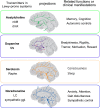
Lewy-prone systems and neurotransmitters. Although the neurotransmitters are different between associated systems, these Lewy-prone systems are characterized by widespread innervation to cerebral cortex, basal ganglia, hippocampus through hyperbranching axons. Such structural template facilitates αS deposition. Such excessive branching of axons is also related to their normal functions or dysfunctions uniformly characterized by non-focal or generalized influences without somatotopy. nbM nucleus basalis of Meynert, dmX dorsal motor nucleus of vagus, SN substantia nigra, Raphe raphe nucleus, LC locus ceruleus, ggl. ganglia; modified from Nolte and Angevine [139] with permission
Fig. 6
Fig. 6
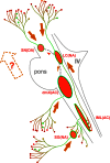
Intraaxonal progression of Lewy pathology in Lewy-prone systems. αS pathology is initiated at distal axons and spreads toward neuronal cell bodies in each Lewy-prone system as indicated by orange arrow, respectively. This axonal spread is in parallel with the direction of the so-called the “upward spread” (broken and empty arrow containing “?”) up to LC. However, the direction of this axonal spread is “downward” above LC. Additional projections form SN to LC (gray interrupted line) and that from LC to dmX (gray interrupted line) may be one of the candidate structures that may mediate transneuronal spread from dmX to LC (small arrow in orange) and that from LC to SN (arrowhead in orange), while these have not yet been documented in human brains with LB disease. DA dopamine, NA noradrenalin, IV forth ventricle, AC acetylcholine, SG sympathetic ganglia, IML intermediolateral nucleus
Fig. 7
Fig. 7
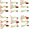
Non-connected or connected explanations for variable gradient. Relative quantity of Lewy pathology could be quite variable with predominance at the dorsal motor nucleus (dmX, a, d), the locus coeruleus (LC, b, e) or the substantia nigra (SN, c, f). Putative directions of intraneuronal spread for αS inclusion pathology are indicated with orange or red arrows. Expected directions of possible transneuronal spread of αS inclusion pathology along interrupted lines are indicated with gray arrows and arrowheads. Either dmX (d), LC (e) or SN (f) is assumed to be initially involved and subsequently spread of αS inclusion pathology through interneuronal connections. Both intra- and inter-neuronal directions are reversed according to the primary site of involvement (df). See text for details

Similar articles

Cited by

References

    1. Ahn KJ, Paik SR, Chung KC, Kim J. Amino acid sequence motifs and mechanistic features of the membrane translocation of alpha-synuclein. J Neurochem. 2006;97(1):265–279. doi: 10.1111/j.1471-4159.2006.03731.x. - DOI - PubMed
    1. Alafuzoff I, Parkkinen L, Al-Sarraj S, Arzberger T, Bell J, Bodi I, Bogdanovic N, Budka H, Ferrer I, Gelpi E, Gentleman S, Giaccone G, Kamphorst W, King A, Korkolopoulou P, Kovacs GG, Larionov S, Meyronet D, Monoranu C, Morris J, Parchi P, Patsouris E, Roggendorf W, Seilhean D, Streichenberger N, Thal DR, Kretzschmar H, BrainNet Europe C. Assessment of alpha-synuclein pathology: a study of the BrainNet Europe Consortium. J Neuropathol Exp Neurol. 2008;67(2):125–143. doi: 10.1097/nen.0b013e3181633526. - DOI - PubMed
    1. Anderson JP, Walker DE, Goldstein JM, de Laat R, Banducci K, Caccavello RJ, Barbour R, Huang J, Kling K, Lee M, Diep L, Keim PS, Shen X, Chataway T, Schlossmacher MG, Seubert P, Schenk D, Sinha S, Gai WP, Chilcote TJ. Phosphorylation of ser-129 is the dominant pathological modification of alpha-synuclein in familial and sporadic Lewy body disease. J Biol Chem. 2006;281(40):29739–29752. doi: 10.1074/jbc.M600933200. - DOI - PubMed
    1. Annerino DM, Arshad S, Taylor GM, Adler CH, Beach TG, Greene JG. Parkinson’s disease is not associated with gastrointestinal myenteric ganglion neuron loss. Acta Neuropathol. 2012;124(5):665–680. doi: 10.1007/s00401-012-1040-2. - DOI - PMC - PubMed
    1. Appel-Cresswell S, Vilarino-Guell C, Encarnacion M, Sherman H, Yu I, Shah B, Weir D, Thompson C, Szu-Tu C, Trinh J, Aasly JO, Rajput A, Rajput AH, Jon SA, Farrer MJ. Alpha-synuclein p. H50Q, a novel pathogenic mutation for Parkinson’s disease. Mov Disord. 2013;28(6):811–813. doi: 10.1002/mds.25421. - DOI - PubMed

Publication types

Substances