

Microstructure of the Midbrain and Cervical Spinal Cord in Idiopathic Restless Legs Syndrome: A Diffusion Tensor Imaging Study
- PMID: 26446110
- PMCID: PMC4712403
- DOI: 10.5665/sleep.5456
Microstructure of the Midbrain and Cervical Spinal Cord in Idiopathic Restless Legs Syndrome: A Diffusion Tensor Imaging Study
Abstract
Study objectives: Diffusion tensor imaging (DTI) allows the study of white matter microstructure in the central nervous system. The aim of this study was to examine the DTI metrics of the cervical spinal cord and the brainstem up to the midbrain in patients with idiopathic restless legs (RLS) compared to matched healthy controls.
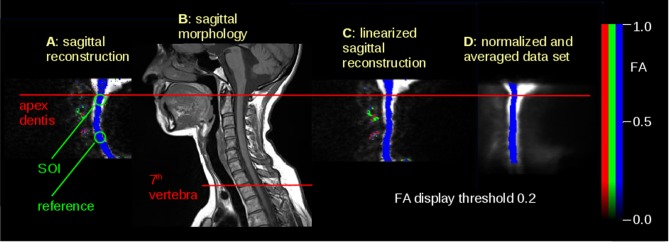
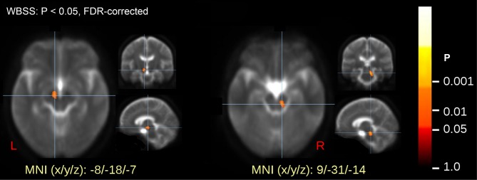
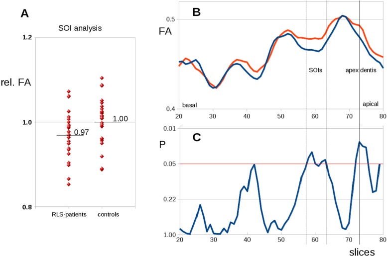
Methods: DTI analysis of the cervical spinal cord and the brainstem up into the midbrain was performed in 25 patients with idiopathic RLS and 25 matched healthy controls. Data analysis in the brain was performed by voxelwise comparison of fractional anisotropy (FA) maps at group level. Cervical spinal cord data analysis was performed by slicewise analysis of averaged FA values in axial slices along the spinal cord.
Results: Voxelwise comparison of FA maps in the brainstem showed significant microstructural alterations in two clusters in the midbrain bilaterally. Slicewise comparison of the FA maps in the cervical spinal cord showed a trend for lower FA values at the level of the second and third vertebra area in the patient sample.
Conclusions: The imaging data suggest that significant alterations in the midbrain in RLS can be visualized by DTI and might correlate to a macroscopically subtle process with changes of the tissue microstructure in the corresponding tracts. An additional area of interest is regionally clustered in the upper cervical spinal cord with a tendency toward altered diffusion metrics. These results might be addressed by further studies, e.g., at higher magnetic field strengths.
Keywords: diffusion tensor imaging; magnetic resonance imaging; midbrain; restless legs syndrome; spinal cord; substantia nigra.
© 2016 Associated Professional Sleep Societies, LLC.
Figures
References
-
- Berger K, Kurth T. RLS epidemiology - frequencies, risk factors and methods in population studies. Mov Disord. 2007;18:S420–3. - PubMed
-
- Allen RP, Picchietti D, Hening WA, Trenkwalder C, Walters AS, Montplaisir J. Restless legs syndrome: diagnostic criteria, special considerations and epidemiology. A report from the restless legs syndrome diagnosis and epidemiology workshop at the National Institutes of Health. Sleep Med. 2003;4:101–19. - PubMed
-
- Trenkwalder C, Paulus W, Walters AS. The restless legs syndrome. Lancet Neurol. 2005;4:465–75. - PubMed
-
- Ondo WG, He Y, Rajasekaran S, Le WD. Clinical correlates of 6-hydroxydopamine injections into A11 dopaminergic neurons in rats: a possible model for restless legs syndrome. Mov Disord. 2000;15:154–8. - PubMed
-
- Allen RP, Barker PB, Wehrl F, Song HK, Earley CJ. MRI measurement of brain iron in patients with restless legs syndrome. Neurology. 2001;56:263–5. - PubMed
MeSH terms
LinkOut - more resources
Full Text Sources
Other Literature Sources
Medical
