. 2015 Dec;89(24):12501-12.
doi: 10.1128/JVI.02097-15. Epub 2015 Oct 7.

Bispecific Antibodies Targeting Different Epitopes on the HIV-1 Envelope Exhibit Broad and Potent Neutralization

Affiliations

Bispecific Antibodies Targeting Different Epitopes on the HIV-1 Envelope Exhibit Broad and Potent Neutralization

M Asokan et al. J Virol. 2015 Dec.

Abstract

The potency and breadth of the recently isolated neutralizing human monoclonal antibodies to HIV-1 have stimulated interest in their use to prevent or to treat HIV-1 infection. Due to the antigenically diverse nature of the HIV-1 envelope (Env), no single antibody is highly active against all viral strains. While the physical combination of two broadly neutralizing antibodies (bNAbs) can improve coverage against the majority of viruses, the clinical-grade manufacturing and testing of two independent antibody products are time and resource intensive. In this study, we constructed bispecific immunoglobulins (IgGs) composed of independent antigen-binding fragments with a common Fc region. We developed four different bispecific IgG variants that included antibodies targeting four major sites of HIV-1 neutralization. We show that these bispecific IgGs display features of both antibody specificities and, in some cases, display improved coverage over the individual parental antibodies. All four bispecific IgGs neutralized 94% to 97% of antigenically diverse viruses in a panel of 206 HIV-1 strains. Among the bispecific IgGs tested, VRC07 × PG9-16 displayed the most favorable neutralization profile. It was superior in breadth to either of the individual antibodies, neutralizing 97% of viruses with a median 50% inhibitory concentration (IC50) of 0.055 μg/ml. This bispecific IgG also demonstrated in vivo pharmacokinetic parameters comparable to those of the parental bNAbs when administered to rhesus macaques. These results suggest that IgG-based bispecific antibodies are promising candidates for the prevention and treatment of HIV-1 infection in humans.

Importance: To prevent or treat HIV-1 infection, antibodies must potently neutralize nearly all strains of HIV-1. Thus, the physical combination of two or more antibodies may be needed to broaden neutralization coverage and diminish the possibility of viral resistance. A bispecific antibody that has two different antibody binding arms could potentially display neutralization characteristics better than those of any single parental antibody. Here we show that bispecific antibodies contain the binding specificities of the two parental antibodies and that a single bispecific antibody can neutralize 97% of viral strains with a high overall potency. These findings support the use of bispecific antibodies for the prevention or treatment of HIV-1 infection.


Figures

FIG 1
FIG 1
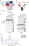
Bispecific antibodies. (A) Structure of HIV-1 BG505 SOSIP.664 Env trimer (PDB code 4TVP) showing the contact sites of the antibodies used in this study. Due to the lack of a crystal structure of PGT121, the footprint of the closely related antibody PGT122 is shown. A portion of the PGT122 binding site on the second protomer can be seen. The third binding site of PGT122 and the two other binding sites of VRC07 are on the reverse face of the trimer. (B) Schematic of the CrossMab antibody configuration. (C) Nonreduced and reduced SDS-PAGE analysis of bispecific IgGs made in the CrossMab format. HC, heavy chain; LC, light chain. (D) Analytical size exclusion profiles of the bispecific IgGs.
FIG 2
FIG 2
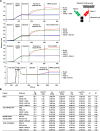
Binding characteristics of anti-Env bispecific IgGs. (A) Simultaneous ligand binding to both arms of the bispecific IgG is demonstrated by a sandwich assay using biolayer interferometry. Octet biosensors were loaded with the ligand against one arm of the bispecific IgG, either by biotinylation or by amine coupling, and then probed sequentially with the bispecific IgG and the ligand against second arm. As controls, parental IgGs were used in place of the bispecific IgG. (B) Kinetic characterization of bispecific and parental IgGs was assessed using biolayer interferometry (Octet RED384). Kinetic constants with standard error and fit parameters are summarized.
FIG 3
FIG 3
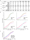
Neutralization breadth-potency of the bispecific IgGs. (A) Neutralization breadth of the parental and bispecific IgGs was tested against a panel of 206 viral strains. The antibodies were tested at a starting concentration of 25 μg/ml with serial dilutions. Breadths based on IC50s and IC80s are summarized. Potency is shown as medians and geometric mean values calculated against sensitive viruses. (B) Potency-breadth curves comparing the four bispecific IgGs to their parental IgGs. (C) Potency-breadth curves of the four bispecific IgGs.
FIG 4
FIG 4
Potencies of the bispecific IgGs compared to those of the parental IgGs. Each dot on the graph represents a virus. On the x axis, viruses are ordered 1 to 206 based on the sensitivity to the parental IgG (black) and overlaid with the potency of the bispecific IgG (red, blue, green, or purple symbols) and the second parental IgG (gray symbols) against the same virus. Dots below the black dots indicate increased potency. Number of viruses and median IC50 change of the bispecific antibody compared to those for each parent IgG are shown for viruses with decreased (above) or increased (below) susceptibility to the bispecific IgG.
FIG 5
FIG 5
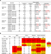
Potency comparison of bispecific and combination IgGs. (A) Viruses were classified as sensitive (S) or resistant (R) based on the parental antibodies. Within each category, the median neutralization IC50 (μg/ml) was calculated for each parental or bispecific IgG and the physical combination of parental IgGs. Parental and bispecific IgGs were started at 25 μg/ml, whereas the physical combination consisted of 12.5 μg/ml of each parental IgG (total 25 μg/ml). Fold difference in potency for the bispecific IgGs and combination was calculated as median IC50 of parental IgG divided by the median IC50 of the bispecific (or combination) IgG. Red values (values greater than 1.0) show improved potency over the parental IgG. (B) Median IC50 (nanomolar) of parental Fab and bispecific IgG is compared to that of parental IgG among monosensitive viruses. Numbers in parentheses indicate fold change of Fab or bispecific IgG compared to parental IgG.
FIG 6
FIG 6
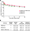
Bispecific IgG has pharmacokinetic properties similar to those of the parent IgG in rhesus macaques. (A) Endotoxin-free IgGs were administered at 10 mg/kg intravenously, and serum levels were monitored by ELISA against gp120 (ZM109F). Data from 2 or 4 animals are plotted as means ± standard deviations. (B) Pharmacokinetics parameters were calculated using a two-compartment model using WinNonlin software. Numbers are means ± standard errors.

References

    1. Mascola JR, Haynes BF. 2013. HIV-1 neutralizing antibodies: understanding nature's pathways. Immunol Rev 254:225–244. doi:10.1111/imr.12075. - DOI - PMC - PubMed
    1. Klein F, Mouquet H, Dosenovic P, Scheid JF, Scharf L, Nussenzweig MC. 2013. Antibodies in HIV-1 vaccine development and therapy. Science 341:1199–1204. doi:10.1126/science.1241144. - DOI - PMC - PubMed
    1. Burton DR, Mascola JR. 2015. Antibody responses to envelope glycoproteins in HIV-1 infection. Nat Immunol 16:571–576. doi:10.1038/ni.3158. - DOI - PMC - PubMed
    1. Pancera M, Zhou T, Druz A, Georgiev IS, Soto C, Gorman J, Huang J, Acharya P, Chuang GY, Ofek G, Stewart-Jones GB, Stuckey J, Bailer RT, Joyce MG, Louder MK, Tumba N, Yang Y, Zhang B, Cohen MS, Haynes BF, Mascola JR, Morris L, Munro JB, Blanchard SC, Mothes W, Connors M, Kwong PD. 2014. Structure and immune recognition of trimeric pre-fusion HIV-1 Env. Nature 514:455–461. doi:10.1038/nature13808. - DOI - PMC - PubMed
    1. Lyumkis D, Julien JP, de Val N, Cupo A, Potter CS, Klasse PJ, Burton DR, Sanders RW, Moore JP, Carragher B, Wilson IA, Ward AB. 2013. Cryo-EM structure of a fully glycosylated soluble cleaved HIV-1 envelope trimer. Science 342:1484–1490. doi:10.1126/science.1245627. - DOI - PMC - PubMed

Publication types