Review
. 2015 Nov;56(6):1478-91.
doi: 10.3349/ymj.2015.56.6.1478.

Treatment of Retinoblastoma: The Role of External Beam Radiotherapy

Affiliations
Review

Treatment of Retinoblastoma: The Role of External Beam Radiotherapy

Joo-Young Kim et al. Yonsei Med J. 2015 Nov.

Abstract

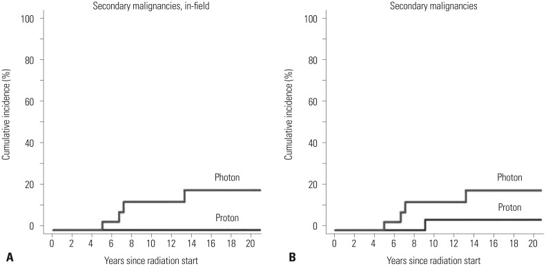
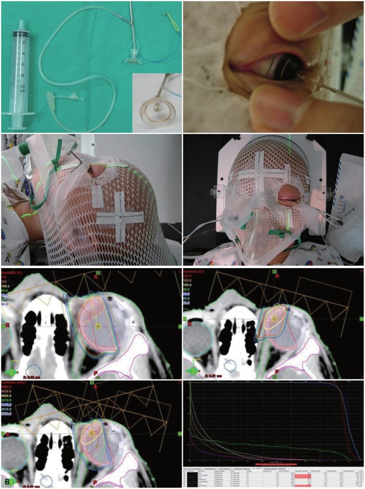
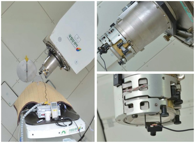
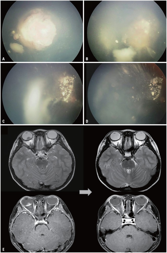
The risk of radiotherapy-related secondary cancers in children with constitutional retinoblastoma 1 (RB1) mutations has led to reduced use of external beam radiotherapy (EBRT) for RB. Presently, tumor reduction with chemotherapy with or without focal surgery (chemosurgery) is most commonly undertaken; EBRT is avoided as much as possible and is considered only as the last treatment option prior to enucleation. Nevertheless, approximately 80% of patients are diagnosed at a locally advanced stage, and only 20-25% of early stage RB patients can be cured with a chemosurgery strategy. As a whole, chemotherapy fails in more than two-thirds of eyes with advanced stage disease, requiring EBRT or enucleation. Radiotherapy is still considered necessary for patients with large tumor(s) who are not candidates for chemosurgery but who have visual potential. When radiation therapy is indicated, the lowest possible radiation dose combined with systemic or local chemotherapy and focal surgery may yield the best clinical outcomes in terms of local control and treatment-related toxicity. Proton beam therapy is one EBRT method that can be used for treatment of RB and reduces the radiation dose delivered to the adjacent orbital bone while maintaining an adequate dose to the tumor. To maximize the therapeutic success of treatment of advanced RB, the possibility of integrating radiotherapy at early stages of treatment may need to be discussed by a multidisciplinary team, rather than considering EBRT as only a last treatment option.

Keywords: Retinoblastoma; external beam radiotherapy; treatment.


Conflict of interest statement

The authors have no financial conflicts of interest.

Figures

Fig. 1
Fig. 1. Cumulative rates of (A) "in-field" or "radiation-induced" secondary malignancies and (B) all secondary malignancies in patients treated with proton beam therapy and photon radiotherapy.
Fig. 2
Fig. 2. Practices at National Cancer Center, Korea. (A) Under anesthesia, a small suction cup is placed on the cornea and the eyeball is rotated so that the proton beam can maximally avoid the orbital bone while covering the retinal target. (B) Dose distribution in proton beam therapy-initial field (left upper), boost field (right upper), summation of both fields (left lower) and corresponding dose volume histogram for the entire plan (right lower).
Fig. 3
Fig. 3. Cosmetic outcomes in a patient treated with the radiotherapy plan described in Fig. 2. The patient's right eye was enucleated and the left eye was treated with PBT. Left to right: photos taken prior to PBT, 3 months after completion of PBT, and 6 years after PBT. PBT, proton beam therapy.
Fig. 4
Fig. 4. Treatment monitoring system used at National Cancer Center, Korea. The position of the eye is monitored in the control room through a CC camera attached to the aperture during treatment. CC, closed-circuit.
Fig. 5
Fig. 5. Treatment outcome of unilateral RB after PBT. Fundoscopic examination of a 5-year-old boy diagnosed with RB in the right eye. (A) Pre-PBT: endophytic mass obscuring the posterior pole with two satellite masses. (B) During PBT: following the delivery of 2340 cGy, the main mass at the posterior pole has partially regressed, whereas the smaller seeding masses have slightly increased in size. (C) Two weeks after PBT: the main mass has regressed while the seeding masses have remained stable. (D) Three months after PBT: masses have overall remained stable. (E) MRI findings before (left) and after (right) PBT: a lobulated contoured intraocular mass of the right orbit, present before PBT, absent after PBT. RB, retinoblastoma; PBT, proton beam therapy.
Fig. 6
Fig. 6. Treatment outcome of PBT in a patient with bilateral RB. The patient was diagnosed with bilateral RB and received chemotherapy and transpupillary thermotherapy (TTT) for 1 year. After treatment, the masses regressed and have remained stable for 1.5 years. However, new masses developed in the right eye. PBT was delivered to the mass refractory to TTT. (A) Pre-PBT, showing the re-growth of a solid mass (arrow) at the temporal margin of a previous mass that had regressed. (B, C, and D) Views 1 (B), 2.5 (C), and 8 (D) months after PBT, showing that the main mass had regressed and remained stable for 8 months. Five months after PBT, a hemorrhage developed at the site of the previously regressed mass treated with TTT, and persisted until 8 months after PBT. There has been no evidence of disease to date, but glaucoma developed as a result of hemorrhage. RB, retinoblastoma; PBT, proton beam therapy.

References

    1. Halperin EC, Constine LS, Tarbell NJ, Kun LE. Pediatric Radiation Oncology. Philadelphia: Wolters Kluwer Health; 2012.
    1. Abramson DH. Retinoblastoma: saving life with vision. Annu Rev Med. 2014;65:171–184. - PubMed
    1. Munier FL, Verwey J, Pica A, Balmer A, Zografos L, Abouzeid H, et al. New developments in external beam radiotherapy for retinoblastoma: from lens to normal tissue-sparing techniques. Clin Experiment Ophthalmol. 2008;36:78–89. - PubMed
    1. Dimaras H, Kimani K, Dimba EA, Gronsdahl P, White A, Chan HS, et al. Retinoblastoma. Lancet. 2012;379:1436–1446. - PubMed
    1. Dimaras H, Dimba EA, Gallie BL. Challenging the global retinoblastoma survival disparity through a collaborative research effort. Br J Ophthalmol. 2010;94:1415–1416. - PubMed

Publication types