. 2015 Nov 27;290(48):29022-34.
doi: 10.1074/jbc.M115.670224. Epub 2015 Oct 7.

Distinct Acute Lymphoblastic Leukemia (ALL)-associated Janus Kinase 3 (JAK3) Mutants Exhibit Different Cytokine-Receptor Requirements and JAK Inhibitor Specificities

Affiliations

Distinct Acute Lymphoblastic Leukemia (ALL)-associated Janus Kinase 3 (JAK3) Mutants Exhibit Different Cytokine-Receptor Requirements and JAK Inhibitor Specificities

Elisabeth Losdyck et al. J Biol Chem. .

Abstract

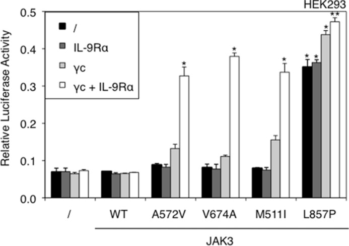
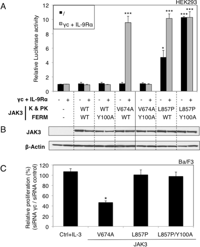
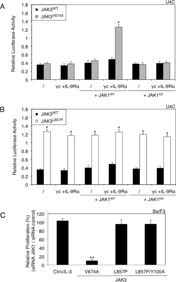
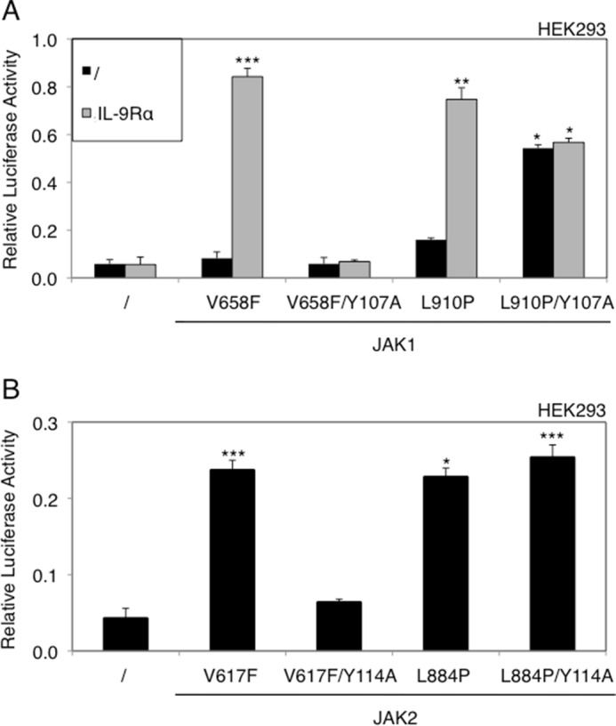
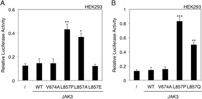
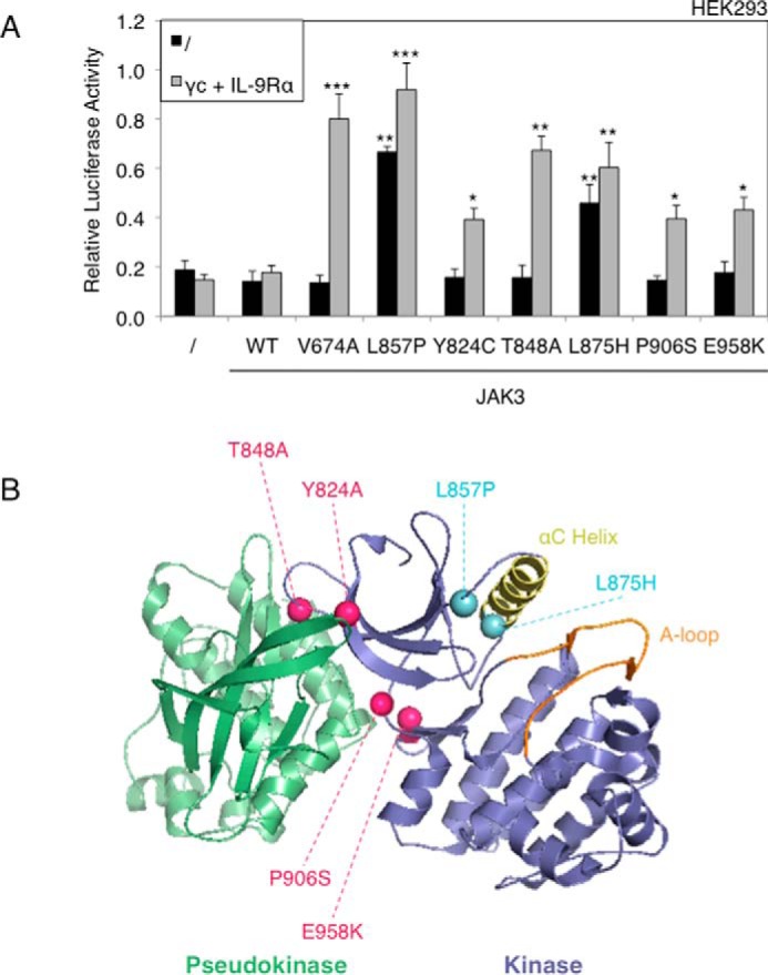
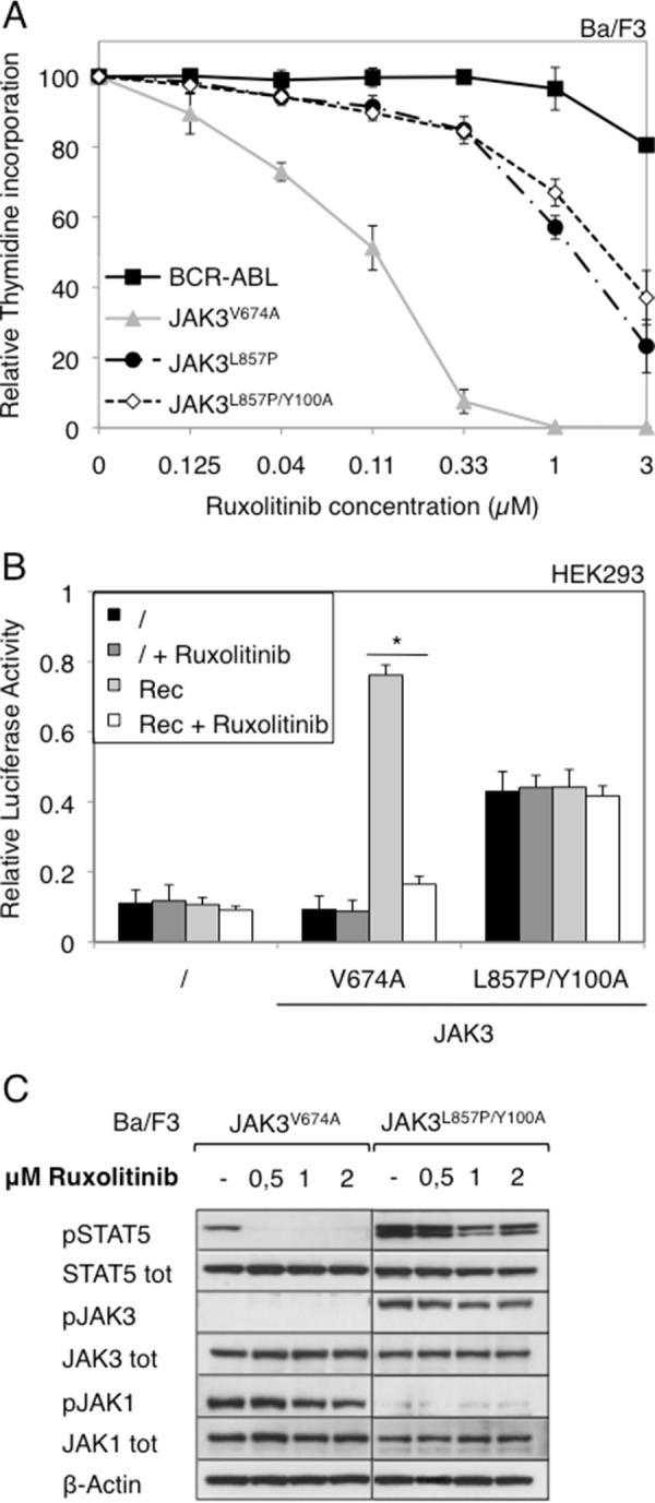
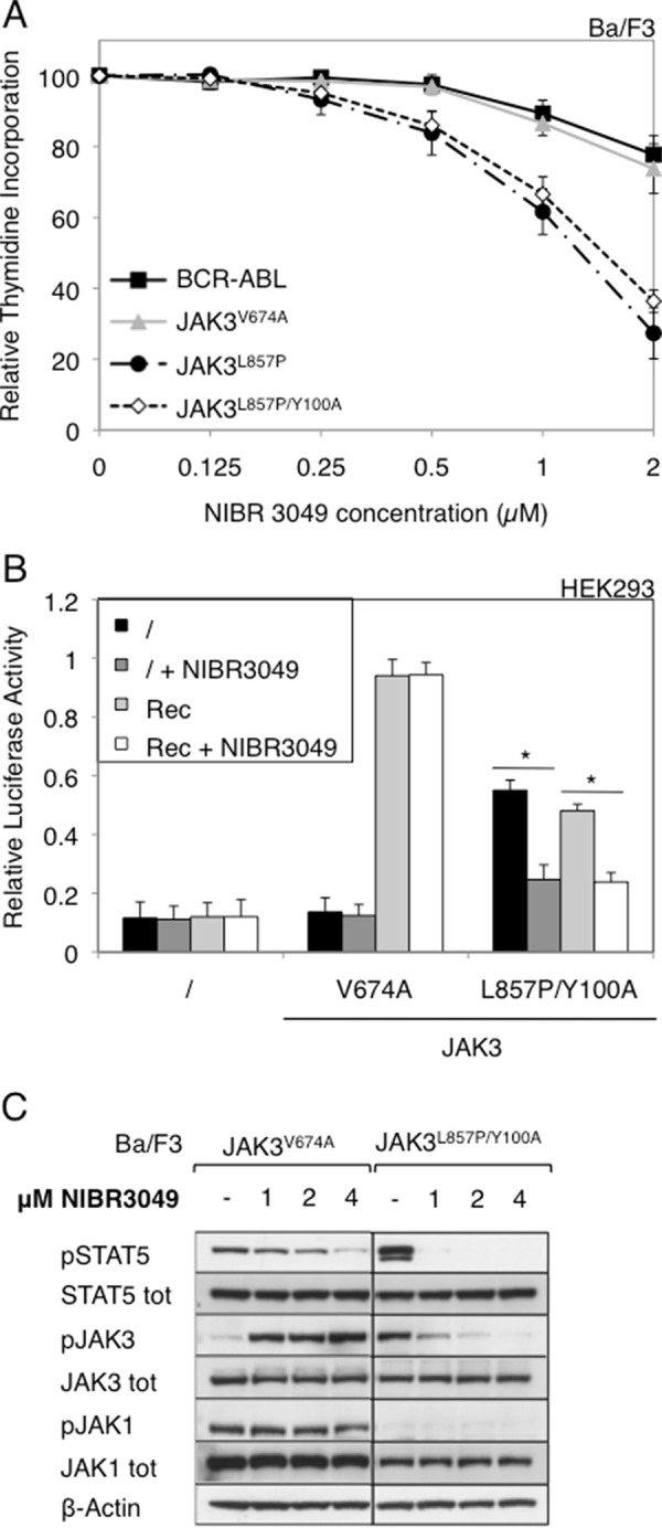
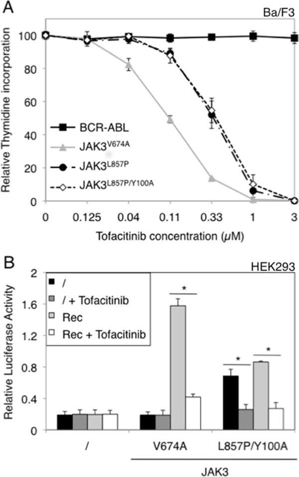
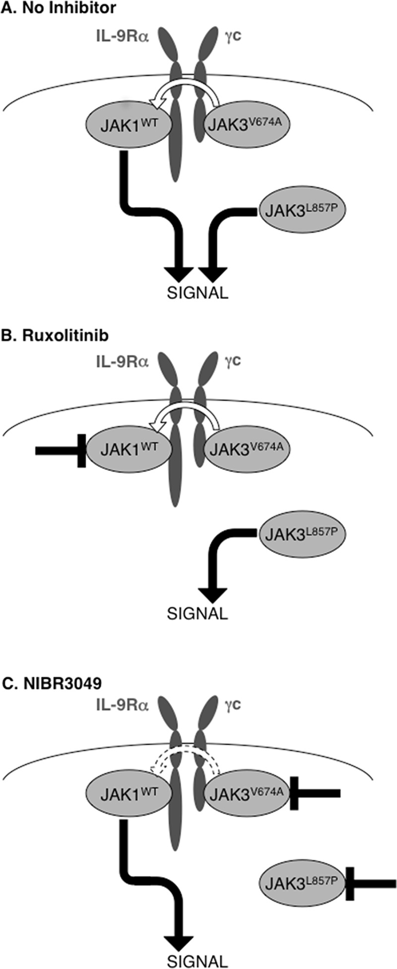
JAK1 and JAK3 are recurrently mutated in acute lymphoblastic leukemia. These tyrosine kinases associate with heterodimeric cytokine receptors such as IL-7 receptor or IL-9 receptor, in which JAK1 is appended to the specific chain, and JAK3 is appended to the common gamma chain. Here, we studied the role of these receptor complexes in mediating the oncogenic activity of JAK3 mutants. Although JAK3(V674A) and the majority of other JAK3 mutants needed to bind to a functional cytokine receptor complex to constitutively activate STAT5, JAK3(L857P) was unexpectedly found to not depend on such receptor complexes for its activity, which was induced without receptor or JAK1 co-expression. Introducing a mutation in the FERM domain that abolished JAK-receptor interaction did not affect JAK3(L857P) activity, whereas it inhibited the other receptor-dependent mutants. The same cytokine receptor independence as for JAK3(L857P) was observed for homologous Leu(857) mutations of JAK1 and JAK2 and for JAK3(L875H). This different cytokine receptor requirement correlated with different functional properties in vivo and with distinct sensitivity to JAK inhibitors. Transduction of murine hematopoietic cells with JAK3(V674A) led homogenously to lymphoblastic leukemias in BALB/c mice. In contrast, transduction with JAK3(L857P) induced various types of lymphoid and myeloid leukemias. Moreover, ruxolitinib, which preferentially blocks JAK1 and JAK2, abolished the proliferation of cells transformed by the receptor-dependent JAK3(V674A), yet proved much less potent on cells expressing JAK3(L857P). These particular cells were, in contrast, more sensitive to JAK3-specific inhibitors. Altogether, our results showed that different JAK3 mutations induce constitutive activation through distinct mechanisms, pointing to specific therapeutic perspectives.

Keywords: JAK inhibitor; Janus kinase (JAK); leukemia; oncogene; signal transduction; tyrosine-protein kinase (tyrosine kinase).


Figures

FIGURE 1.
FIGURE 1.
STAT5 activation by JAK3 activating mutants is receptor-dependent, except for the L857P mutant. JAK3- and γc-deficient HEK293 cells were transiently co-transfected with JAK3WT or ALL-associated A572V, V674A, M511I, and L857P JAK3 mutants and γc and/or IL-9Rα, in addition to the STAT5-responsive luciferase reporter pLHRE-luc (firefly luciferase) and the pRLTK plasmid (Renilla luciferase) as transfection control. 24 h post-transfection, the cells were subjected to a luciferase assay. The relative luciferase activity corresponds to the firefly luciferase light emission values divided by the Renilla luciferase light emission values. The results are means ± standard deviation of triplicates. Similar results were obtained in three independent experiments. A Kruskal-Wallis test with Dunn correction was performed to determine p values between the control condition without JAK3 and the WT or mutant forms of JAK3 for each condition (with or without IL-9R complex). *, p < 0.05; **, p < 0.01.
FIGURE 2.
FIGURE 2.
JAK3 FERM domain integrity is required for the activity of the V674A mutant, but not for the L857P mutant. A, HEK293 cells were transiently co-transfected either with JAK3WT, or different JAK3 mutants (Y100A, V674A, and L857P) or double mutants (V674A/Y100A and L857P/Y100A) together with γc and IL-9Rα or not, in addition to the STAT5-responsive luciferase reporter pLHRE-luc (firefly luciferase) and the pRLTK plasmid (Renilla luciferase) as transfection control. 24 h post-transfection, the cells were subjected to a luciferase assay. The relative luciferase activity corresponds to the firefly luciferase light emission values divided by the Renilla luciferase light emission values. The results are means ± standard deviation of three different experiments, each performed in triplicate. A Kruskal-Wallis test with Dunn correction was performed to determine p values between the control condition without JAK3 and the WT or mutant forms of JAK3 for each condition (with or without IL-9R complex). *, p < 0.05; ***, p < 0.001. K, kinase domain; PK, pseudokinase domain. B, in parallel of the luciferase assay, transfected HEK293 cells were lysed 24 h post-transfection and subjected to Western blot analysis using an anti-JAK3 antibody and an anti-β-actin antibody as loading control. C, relative proliferation of IL-3-dependent Ba/F3 cells or autonomous Ba/F3 cells obtained after transduction with ALL-associated JAK3 mutants (V674A and L857P) and double mutant (L857P/Y100A) after knockdown of endogenous γc compared with the proliferation observed with an irrelevant control siRNA. After 72 h, tritiated thymidine incorporation was measured. The results are means ± standard deviation of three different experiments, each performed in triplicate. A Kruskal-Wallis test with Dunn correction was performed to determine p values between the IL-3-dependent control Ba/F3 cells and the transformed Ba/F3 cells. *, p < 0.05.
FIGURE 3.
FIGURE 3.
JAK1 expression is required for the activity of the V674A mutant, but not for the L857P mutant. A, JAK1-deficient U4C cells were transiently co-transfected with JAK3WT or JAK3V674A, γc, and IL-9Rα with or without JAK1WT or JAK1KD, in addition to the STAT5-responsive luciferase reporter pLHRE-luc (firefly luciferase) and the pRLTK plasmid (Renilla luciferase) as transfection control. 24 h post-transfection, the cells were subjected to a luciferase assay. The relative luciferase activity corresponds to the firefly luciferase light emission values divided by the Renilla luciferase light emission values. The results are means ± standard deviation of three different experiments, each performed in triplicate. A Kruskal-Wallis test with Dunn correction was performed to determine p values. *, p < 0.05. B, JAK1-deficient U4C cells were transiently co-transfected with JAK3WT or JAK3L857P, γc, and IL-9Rα with or without JAK1WT or JAK1KD, in addition to the STAT5-responsive luciferase reporter pLHRE-luc and the pRLTK plasmid as transfection control. 24 h post-transfection, the cells were subjected to a luciferase assay. The results are means ± standard deviation of three different experiments, each performed in triplicate. A Kruskal-Wallis test with Dunn correction was performed to determine p values. *, p < 0.05. C, relative proliferation of IL-3-dependent Ba/F3 cells or autonomous Ba/F3 cells obtained after transduction with ALL-associated JAK3 mutants (V674A and L857P) and double mutant (L857P/Y100A) after knockdown of endogenous JAK1 compared with the proliferation observed with an irrelevant control siRNA. After 72 h, tritiated thymidine incorporation was measured. The results are means ± standard deviation of three different experiments, each performed in triplicate. A Kruskal-Wallis test with Dunn correction was performed to determine p values between the IL-3-dependent control Ba/F3 cells and the transformed Ba/F3 cells. **, p < 0.01.
FIGURE 4.
FIGURE 4.
Integrity of the FERM domain is not required for activation of JAK1 and JAK2 through homologous mutation of JAK3L857P. A, HEK293 cells were transiently co-transfected with ALL-associated JAK1V658F, JAK1L910P (homologous to JAK3 L857P), or JAK1L910P/Y107A, with or without IL-9Rα, in addition to the STAT5-responsive luciferase reporter pLHRE-luc (firefly luciferase) and the pRLTK plasmid (Renilla luciferase) as transfection control. 24 h post-transfection, the cells were subjected to a luciferase assay. The relative luciferase activity corresponds to the firefly luciferase light emission values divided by the Renilla luciferase light emission values. The results are means ± standard deviation of means of three different experiments, each performed in triplicate. A Kruskal-Wallis test with Dunn correction was performed to determine p values between the control condition without JAK1 and the mutant forms of JAK1 for each condition (with or without IL-9Rα). *, p < 0.05; **, p < 0.01; ***, p < 0.001. B, HEK293 cells were transiently co-transfected with myeloproliferative neoplasm-associated JAK2V617F, JAK2L884P (homologous to JAK3 L857P), or JAK2L884P/Y114A, in addition to the STAT5-responsive luciferase reporter pLHRE-luc and the pRLTK plasmid as transfection control. 24 h post-transfection, the cells were subjected to a luciferase assay. The results are means ± standard deviation of means of triplicates of three different experiments. A Kruskal-Wallis test with Dunn correction was performed to determine p values between the control condition without JAK2 and the mutant forms of JAK2. *, p < 0.05; ***, p < 0.001.
FIGURE 5.
FIGURE 5.
Substitution of Leu857 of JAK3 with different residues can confer receptor-independent constitutive activity. HEK293 cells were transiently transfected either with WT JAK3, or different JAK3 mutants: JAK3WT, JAK3V674A, JAK3L857P, JAK3L857A, and JAK3L857E in pMX-IRES-CD4 (A) and JAK3WT, JAK3V674A, JAK3L857P, and JAK3L857Q (B) in pMSCV-GFP. The STAT5-responsive luciferase reporter pLHRE-luc (firefly luciferase) and the pRLTK plasmid (Renilla luciferase) were co-transfected. 24 h post-transfection, the cells were subjected to a luciferase assay. The relative luciferase activity corresponds to the firefly luciferase light emission values divided by the Renilla luciferase light emission values. The results are means ± standard deviation of means of three different experiments, each performed in triplicate. A Kruskal-Wallis test with Dunn correction was performed to determine p values between the control condition without JAK3 and the WT or mutant forms of JAK3 for each condition. *, p < 0.05; **, p < 0.01; ***, p < 0.001.
FIGURE 6.
FIGURE 6.
The majority of kinase domain mutants are receptor-dependent, except JAK3L857P and JAK3L875H. A, HEK293 cells were transiently co-transfected either with JAK3WT or different JAK3 mutants (V674A, L857P, Y824A, T848A, L875H, P906S, and E958K), in addition to the STAT5-responsive luciferase reporter pLHRE-luc (firefly luciferase) and the pRLTK plasmid (Renilla luciferase) as transfection control. 24 h post-transfection, the cells were subjected to a luciferase assay. The relative luciferase activity corresponds to the firefly luciferase light emission values divided by the Renilla luciferase light emission values. The results are means ± standard deviation of means of three different experiments, each performed in triplicate. A Kruskal-Wallis test with Dunn correction was performed to determine p values between the control condition without JAK3 and the WT or mutant forms of JAK3 for each condition. *, p < 0.05; **, p < 0.01; ***, p < 0.001. B, localization of ALL-associated JAK3 mutants of the kinase domain. The figure represents the kinase and pseudo-kinase domains of JAK3 based on the recent TYK2 JH1/JH2 crystal structure (33). The kinase domain is shown in indigo, with the αC helix in yellow and the activation loop in orange. The adjacent pseudokinase domain is shown in green. Mutated residues close to the pseudokinase domain are indicated with pink balls, and mutated residues close to the αC helix are indicated with light blue balls.
FIGURE 7.
FIGURE 7.
Cells expressing JAK3V674A mutant are more sensitive to ruxolitinib than cells expressing JAK3L857P or JAK3L857P/Y100A. A, autonomous Ba/F3 cells stably transduced with ALL-associated JAK3 mutants (V674A and L857P) and double mutant (L857P/Y100A) or BCR-ABL as a control were treated with increasing concentrations of JAK1/JAK2 inhibitor ruxolitinib (0–3 μm). After 72 h, tritiated thymidine incorporation was measured. The results are means ± standard deviation of three different experiments, each performed in triplicate, represented as percentages of the proliferation of the respective untreated cells. B, HEK293 cells were transiently co-transfected either with JAK3V674A or JAK3L857P/Y100A, with a receptor complex (Rec = γc, IL-9Rα, and JAK1WT), in addition to the STAT5-responsive luciferase reporter pLHRE-luc (firefly luciferase) and the pRLTK plasmid (Renilla luciferase) as transfection control. Cells were treated with ruxolitinib (1 μm). 24 h post-transfection, the cells were subjected to a luciferase assay. The relative luciferase activity corresponds to the firefly luciferase light emission values divided by the Renilla luciferase light emission values. The results are means ± standard deviation of three different experiments, each performed in triplicate. A Kruskal-Wallis test with Dunn correction was performed to determine p values. *, p < 0.05. C, 106 autonomous Ba/F3 cells stably transduced with ALL-associated JAK3 mutant V674A or double mutant L857P/Y100A were treated with increasing concentration of ruxolitinib (0–2 μm). Two hours after treatment, the cells were lysed and subjected to Western blot analysis. Phosphorylation of STAT5, JAK3, and JAK1 was detected using specific anti-pY694 STAT5, anti-pY980/81 JAK3, and anti-pY1034/35 JAK1 antibodies. Membranes were reprobed with anti-STAT5, anti-JAK3, anti-JAK1, and anti-β-actin antibodies as loading controls.
FIGURE 8.
FIGURE 8.
Cells expressing JAK3 L857P or L857P/Y100A mutants are more sensitive to NIBR3049 than cells expressing JAK3 V674A. A, autonomous Ba/F3 cells stably transduced with ALL-associated JAK3 mutants (V674A and L857P) and double mutant (L857P/Y100A) or BCR-ABL as a control were with increasing concentrations of JAK3 inhibitor NIBR3049 (0–2 μm). After 72 h, tritiated thymidine incorporation was measured. The results are means ± standard deviation of three different experiments, each performed in triplicate, represented as percentages of the proliferation of the respective untreated cells. B, HEK293 cells were transiently co-transfected either with JAK3V674A or JAK3L857P/Y100A, with a receptor complex (Rec = γc, IL-9Rα, and JAK1WT), in addition to the STAT5-responsive luciferase reporter pLHRE-luc (firefly luciferase) and the pRLTK plasmid (Renilla luciferase) as transfection control. Cells were treated with NIBR3049 (0.5 μm). 24 h post-transfection, the cells were subjected to a luciferase assay. The relative luciferase activity corresponds to the firefly luciferase light emission values divided by the Renilla luciferase light emission values. The results are means ± standard deviation of three different experiments, each performed in triplicate. A Kruskal-Wallis test with Dunn correction was performed to determine p values. *, p < 0.05. C, 106 autonomous Ba/F3 cells stably transduced with ALL-associated JAK3 mutant V674A or double mutant L857P/Y100A were treated with increasing concentration of NIBR3049 (0–4 μm). Two hours after treatment, the cells were lysed and subjected to Western blot analysis. Phosphorylation of STAT5, JAK3, and JAK1 was detected using specific anti-pY694 STAT5, anti-pY980/81 JAK3, and anti-pY1034/35 JAK1 antibodies. Membranes were reprobed with anti-STAT5, anti-JAK3, anti-JAK1, and anti-β-actin antibodies as loading controls.
FIGURE 9.
FIGURE 9.
Cells expressing JAK3 V674A, L857P, and L857P/Y100A mutants are sensitive to tofacitinib. A, autonomous Ba/F3 cells stably transduced with ALL-associated JAK3 mutants (V674A and L857P) and double mutant (L857P/Y100A) or BCR-ABL as a control were treated with increasing concentrations of JAK1/JAK3 inhibitor tofacitinib (0–3 μm). After 72 h, tritiated thymidine incorporation was measured. The results are means ± standard deviation of three different experiments, each performed in triplicate, represented as percentages of the proliferation of the respective untreated cells. B, HEK293 cells were transiently co-transfected either with JAK3V674A or JAK3L857P/Y100A, with a receptor complex (Rec = γc, IL-9Rα, and JAK1WT), in addition to the STAT5-responsive luciferase reporter pLHRE-luc (firefly luciferase) and the pRLTK plasmid (Renilla luciferase) as transfection control. Cells were treated with tofacitinib (0.5 μm). 24 h post-transfection, the cells were subjected to a luciferase assay. The relative luciferase activity corresponds to the firefly luciferase light emission values divided by the Renilla luciferase light emission values. The results are means ± standard deviation of three different experiments, each performed in triplicate. A Kruskal-Wallis test with Dunn correction was performed to determine p values. *, p < 0.05.
FIGURE 10.
FIGURE 10.
Expression of JAK3 mutants in bone marrow cells of Balb/c mice leads to distinct leukemia phenotypes. Spleen and bone marrow cells of diseased animals were analyzed by flow cytometry with anti-CD4, anti-CD8, anti-Gr1, and anti-B220 antibodies. Cells were gated for GFP expression. FACS analysis representative of a CD8+ T-lymphoproliferation in a JAK3V674A mouse (A), a CD4/8+ T-lymphoproliferation in a JAK3L857P mouse (B), a Gr1+ myeloproliferation in a JAK3L857P/Y100A mouse (C), a CD8+ T-lymphoproliferation in a JAK3L857P/Y100A mouse (D), and a B220+ B-lymphoproliferation in a JAK3L857P mouse (E) are shown, either as dot plots (spleen) or as stacked column charts representing the percentage of cells positive for the different markers (bone marrow).
FIGURE 11.
FIGURE 11.
Schematic representation of the activity of JAK3 mutants under JAK inhibitor treatment.

References

    1. Haan C., Rolvering C., Raulf F., Kapp M., Drückes P., Thoma G., Behrmann I., and Zerwes H. G. (2011) Jak1 has a dominant role over Jak3 in signal transduction through γc-containing cytokine receptors. Chem. Biol. 18, 314–323 - PubMed
    1. Saharinen P., and Silvennoinen O. (2002) The pseudokinase domain is required for suppression of basal activity of Jak2 and Jak3 tyrosine kinases and for cytokine-inducible activation of signal transduction. J. Biol. Chem. 277, 47954–47963 - PubMed
    1. Haan C., Is'harc H., Hermanns H. M., Schmitz-Van De Leur H., Kerr I. M., Heinrich P. C., Grötzinger J., and Behrmann I. (2001) Mapping of a region within the N terminus of Jak1 involved in cytokine receptor interaction. J. Biol. Chem. 276, 37451–37458 - PubMed
    1. Tang W., Huo H., Zhu J., Ji H., Zou W., Xu L., Sun L., Zheng Z., Theze J., and Liu X. (2001) Critical sites for the interaction between IL-2Rγ and JAK3 and the following signaling. Biochem. Biophys. Res. Commun. 283, 598–605 - PubMed
    1. Cacalano N. A., Migone T. S., Bazan F., Hanson E. P., Chen M., Candotti F., O'Shea J. J., and Johnston J. A. (1999) Autosomal SCID caused by a point mutation in the N-terminus of Jak3: mapping of the Jak3-receptor interaction domain. EMBO J. 18, 1549–1558 - PMC - PubMed

Publication types

MeSH terms