. 2015 Dec 15;26(25):4646-59.
doi: 10.1091/mbc.E15-09-0626. Epub 2015 Oct 7.

Cell type-dependent mechanisms for formin-mediated assembly of filopodia

Affiliations

Cell type-dependent mechanisms for formin-mediated assembly of filopodia

Lorna E Young et al. Mol Biol Cell. .

Abstract

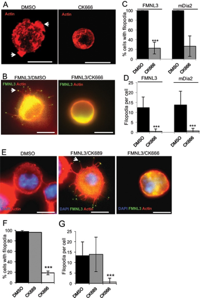
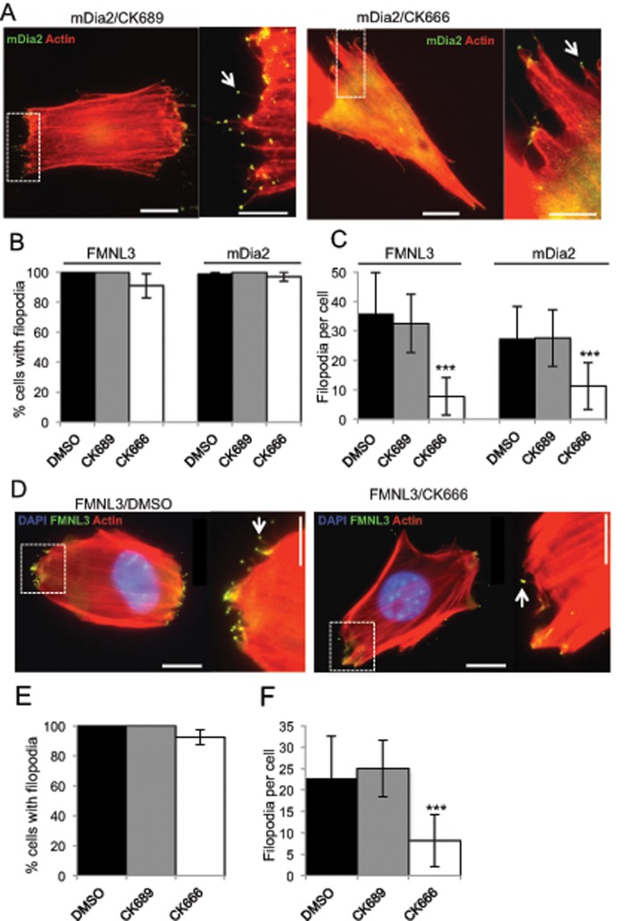
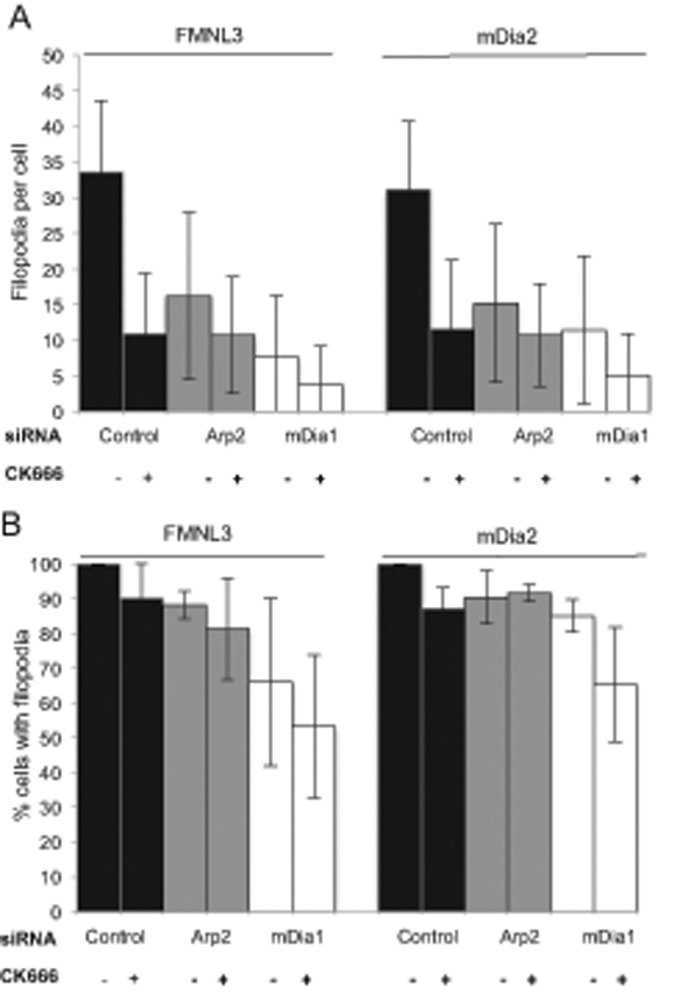
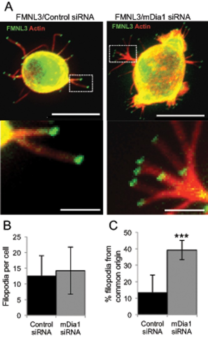
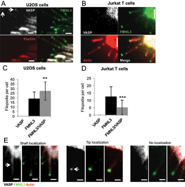
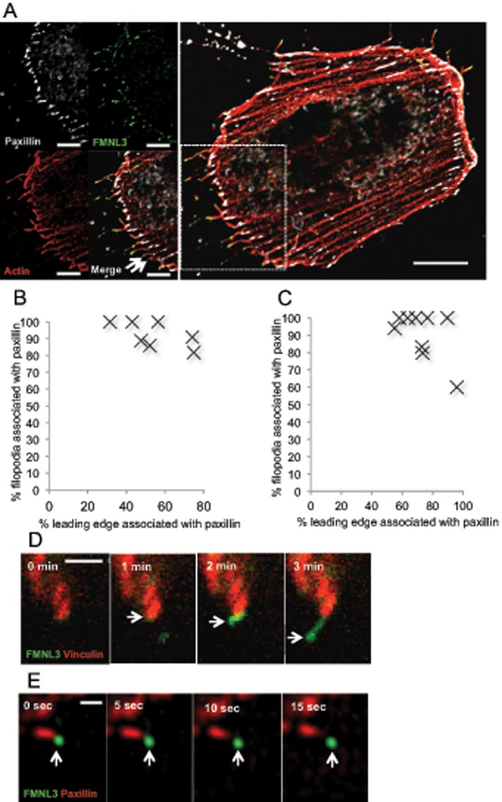
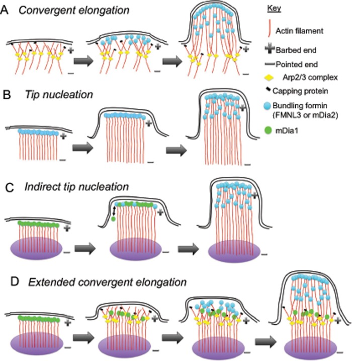
Filopodia are finger-like protrusions from the plasma membrane and are of fundamental importance to cellular physiology, but the mechanisms governing their assembly are still in question. One model, called convergent elongation, proposes that filopodia arise from Arp2/3 complex-nucleated dendritic actin networks, with factors such as formins elongating these filaments into filopodia. We test this model using constitutively active constructs of two formins, FMNL3 and mDia2. Surprisingly, filopodial assembly requirements differ between suspension and adherent cells. In suspension cells, Arp2/3 complex is required for filopodial assembly through either formin. In contrast, a subset of filopodia remains after Arp2/3 complex inhibition in adherent cells. In adherent cells only, mDia1 and VASP also contribute to filopodial assembly, and filopodia are disproportionately associated with focal adhesions. We propose an extension of the existing models for filopodial assembly in which any cluster of actin filament barbed ends in proximity to the plasma membrane, either Arp2/3 complex dependent or independent, can initiate filopodial assembly by specific formins.


Figures

FIGURE 1:
FIGURE 1:
Arp2/3 complex inhibition effectively ablates formin-mediated filopodia in suspension cells. (A) Representative micrographs of untransfected Jurkat T-cells treated in suspension for 2 h with DMSO (left) or 200 μM Arp2/3 complex inhibitor CK666 (right) and stained with TRITC-phalloidin (red). Cells were fixed in suspension before adhesion to PLL-coated coverslips. Arrowheads represent actin-rich membrane ruffles in DMSO control. Scale bar, 10 μm. (B) Representative micrographs of GFP-FMNL3-FF–transfected Jurkat T-cells treated in suspension for 2 h with DMSO (left) or 200 μM CK666 (right). Similar micrographs for mDia2-FFC are shown in Supplemental Figure S1B. Cells were adhered to PLL-coated coverslips for 10 min before fixation. Actin is stained with TRITC-phalloidin (red), and GFP-FMNL3-FF is green. Arrowhead indicates FMNL3 enriched at filopodial tip. Filopodia number in examples: DMSO, 13; and CK666, 0. Scale bar, 10 μm. (C) Quantification of percentage of transfected cells displaying filopodia after indicated treatment. (D) Quantification of filopodia per transfected cell after indicated treatment. Results are pooled from three to five independent experiments, 55–80 cells (FMNL3) and 34–68 cells (mDia2). (E) Representative micrographs of 300.19 cells untransfected and treated with DMSO showing no filopodia (left) and transfected with GFP-FMNL3-FF and treated with 200 μM inactive CK689 control (middle) or 200 μM Arp2/3 complex inhibitor CK666 (right). All cells were treated in suspension for 2 h, followed by 10 min of adhesion onto PLL-coated coverslips before paraformaldehyde fixation. Actin is stained with TRITC-phalloidin (red) and DNA with DAPI (blue), and GFP-FMNL3-FF is green. Arrowhead indicates FMNL3 enriched at the filopodial tip. Filopodia number in examples: CK689, 15; and CK666, 0. Scale bar, 5 μm. (F) Quantification of percentage of transfected cells displaying filopodia after the indicated treatment. (G) Quantification of filopodia number per transfected cell treated as indicated. Results are pooled from three to five independent experiments (except for CK689, one experiment); 51–133 cells. Error bars, SD. ***p < 0.001.
FIGURE 2:
FIGURE 2:
Arp2/3 complex inhibition reduces formin-mediated filopodia in adherent cells. (A) Representative micrographs of U2OS cells transfected with GFP-mDia2-FFC and treated for 2 h with 200 μM CK689 (left) or 200 μM CK666 (right). Actin is stained with TRITC-phalloidin (red), and GFP-mDia2-FFC is green. Arrowheads indicate mDia2 enriched at filopodial tips. Filopodia number in insets: GFP/DMSO, 0; mDia2/CK689, 10; and mDia2/CK666, 5. Examples of untreated U2OS cells transfected with GFP alone in Supplemental Figure S3. (B) Quantification of percentage of transfected cells displaying filopodia after the indicated treatment. (C) Quantification of filopodia number on transfected cells treated as indicated. Results are pooled from three or four independent experiments, 24–37 cells (FMNL3) and 92–141 cells (mDia2). (D) Representative micrographs of NIH 3T3 cells transfected with GFP-FMNL3-FF and then treated for 2 h with DMSO (left) or 200 μM CK666 (right). Actin is stained with TRITC-phalloidin (red) and DNA with DAPI (blue), and GFP-FMNL3-FF is green. Arrowheads indicate FMNL3 enriched at filopodial tips. Filopodia numbers in insets: DMSO, 9; and CK666, 2. Scale bar, 10 μm (main image) and 5 μm (insets). (E) Quantification of percentage of transfected cells displaying filopodia after indicated treatment. (F) Quantification of filopodia number on each transfected cell after the indicated treatment. Results are pooled from three to five independent experiments; 48–118 cells. Scale bars: 10 μm (main image) and 5 μm (insets). Error bars, SD. ***p < 0.001.
FIGURE 3:
FIGURE 3:
mDia1 depletion reduces formin-mediated filopodia in adherent cells. U2OS cells were transfected for 72 h with the indicated siRNA (control, mDia1 targeting, or Arp2 targeting). At 48 h after siRNA transfection, cells were transfected with either GFP-FMNL3-FF or GFP-mDia2-FFC and treated for 2 h with DMSO or 200 μM CK666. (A) Quantification of filopodia number per transfected cell after the indicated treatment. Results with CK666 differ from DMSO control with p < 0.001 for all but the Arp2 siRNA, mDia2-transfected results (p = 0.003). (B) Percentage of GFP-FMNL3-FF or GFP-mDia2-FFC transfected cells displaying filopodia after the indicated treatment. Results with CK666 differ from DMSO control with p > 0.1 for all conditions. Results are pooled from three of four independent experiments; 88–124 cells (FMNL3) and 79–127 cells (mDia2). Error bar, SD.
FIGURE 4:
FIGURE 4:
mDia1 depletion does not reduce FMNL3-mediated filopodia in Jurkat T-cells. (A) Representative images of Jurkat T-cells transfected for 72 h with control (left) or mDia1-targeting siRNA (right), followed by transfection with GFP-FMNL3-FF for the final 24 h. Cells were adhered to PLL-coated coverslips for 10 min before paraformaldehyde fixation. Actin is stained with TRITC-phalloidin (red), and FMNL3 is green. Insets provide examples of filopodia originating from a common base. Images are maximum intensity projections from 25 × 0.2–μm Z-slices. Scale bar, 10 μm (main image) and 2 μm (insets). (B) Quantification of filopodia per transfected cell after the indicated treatment. Results are pooled from four independent experiments; 104–118 cells. Error bar, SD. p = 0.09. (C) Quantification of percentage of filopodia that originate from a common base after the indicated treatment. Results are pooled from four independent experiments; 1317–1688 filopodia. Error bar, SD. ***p < 0.001.
FIGURE 5:
FIGURE 5:
Effect of VASP on formin-mediated filopodial assembly differs between suspension and adherent cells. (A, B) Endogenous VASP (white) staining by immunofluorescence microscopy of U2OS cells (A) or Jurkat cells (B) transfected with GFP-FMNL3-FF (green). Red for U2OS cells is endogenous paxillin and for Jurkat cells indicates actin filaments (TRITC-phalloidin). Arrows indicate two filopodia with some degree of VASP at tips. Scale bar, 2 μm. (C) Quantification of filopodia number per transfected U2OS cell after transfection with mApple-VASP, GFP-FMNL3-FF, or mApple-VASP and GFP-FMNL3-FF; 20–25 cells. Error bar, SD. **p < 0.01. (D) Quantification of filopodia number per transfected Jurkat cell after transfection with mApple-VASP, GFP-FMNL3-FF, or mApple-VASP and GFP-FMNL3-FF; 30–87 cells. Error bar, SD. ***p < 0.001. (E) Fluorescence micrographs of mApple-VASP (white), GFP-FMNL3-FF (green), and BFP-Lifeact (red) in U2OS cells, showing examples of VASP enrichment in filopodial shaft (left), VASP enrichment in filopodial tip (middle), or no VASP enrichment in filopodia (right). Scale bar, 1 μm.
FIGURE 6:
FIGURE 6:
A subset of FMNL3-FF–mediated filopodia associates with focal adhesions. (A) Representative micrograph of the leading-edge region of a U2OS cell cotransfected with RFP-paxillin (white), BFP-Lifeact (red), and GFP-FMNL3-FF (green). Images are maximum intensity projections from 10 × 0.2–μm Z-slices. Arrowheads indicate two filopodia that are not associated with focal adhesions in this example. Scale bar, 0.5 μm. (B) Quantification of percentage of FMNL3-FF–enriched filopodia that are associated with focal adhesions (judged from fixed cells stained with anti-paxillin) as a function of percentage of focal adhesion coverage at the leading edge; 7 cells. See Materials and Methods for description of quantification. (C) Quantification of focal adhesion–associated filopodia from confocal movies similar to D. (D) Time-lapse montage of FMNL3 punctum emanating from a focal adhesion (individual time points from Supplemental Movie S3). U2OS cells cotransfected with mApple-vinculin (red) and GFP-FMNL3-FF (green). mApple and GFP micrographs were acquired sequentially (<1 s between colors) every 30 s. Scale bar, 2 μm. (E) Structured illumination microscopy time-lapse montage of FMNL3 punctum emanating from a focal adhesion (individual time points from Supplemental Movie S4). U2OS cells cotransfected with RFP-paxillin (red) and GFP-FMNL3 (green). RFP and GFP micrographs were acquired sequentially (<1 s between colors) every 5 s. Scale bar, 0.5 μm.
FIGURE 7:
FIGURE 7:
Myosin II inhibition does not reduce filopodia number but increases filopodial length. U2OS cells or NIH 3T3 cells were transfected with GFP-FMNL3-FF (both cell types) or GFP-mDia2-FFC (U2OS only) and treated for 2 h with DMSO or 50 μM blebbistatin. (A) Quantification of filopodia number per transfected cell treated as indicated. Results are pooled from three or four independent experiments; 31–103 U2OS cells, 83–117 NIH 3T3 cells. Error bar, SD. *p < 0.05, ***p < 0.001. (B) Quantification of filopodial length in GFP-FMNL3-FF– or GFP-mDia2-FFC–transfected cells. Results are pooled from three or four independent experiments; 40–95 filopodia in U2OS cells, 297–423 filopodia in 3T3 cells. Error bar, SD. ***p < 0.001. (C) Representative micrographs of U2OS cells transfected with GFP-FMNL3 and treated with DMSO (left) or 50 μM blebbistatin (right). Scale bar, 10 μm (main image) and 5 μm (insets).
FIGURE 8:
FIGURE 8:
Models for filopodial assembly. Models A (convergent elongation) and B (tip nucleation) were proposed previously. We propose models C and D from our results in this work. (C) Indirect tip nucleation model. A nucleation factor (e.g., mDia1) assembles new filaments at a specific site near the leading edge (purple oval), such as a focal adhesion. During filament elongation, the barbed end–bound nucleation factor is displaced by a bundling formin (e.g., FMNL3 or mDia2), which allows further elongation, as well as bundling, to drive productive filopodial growth. (D) Extended convergent elongation model. Actin filaments are assembled at a specific site (purple oval) by a nucleation factor such as mDia1 and serve as mother filaments for Arp2/3 complex–mediated nucleation. Bundling formins such as FMNL3 and mDia2 then mediate convergent elongation on the Arp2/3 complex–nucleated barbed ends. We do not depict VASP in these models because its role is still unclear, but it could in principle be enhancing the initial assembly of filaments in models A, C, and D.

References

    1. Applewhite DA, Barzik M, Kojima S-I, Svitkina TM, Gertler FB, Borisy GG. Ena/VASP proteins have an anti-capping independent function in filopodia formation. Mol Biol Cell. 2007;18:2579–2591. - PMC - PubMed
    1. Aratyn-Schaus Y, Oakes PW, Gardel ML. Dynamic and structural signatures of lamellar actomyosin force generation. Mol Biol Cell. 2011;22:1330–1339. - PMC - PubMed
    1. Bachmann C, Fischer L, Walter U, Reinhard M. The EVH2 domain of the vasodilator-stimulated phosphoprotein mediates tetramerization, F-actin binding, and actin bundle formation. J Biol Chem. 1999;274:23549–23557. - PubMed
    1. Barzik M, Kotova TI, Higgs HN, Hazelwood L, Hanein D, Gertler FB, Schafer DA. Ena/VASP proteins enhance actin polymerization in the presence of barbed end capping proteins. J Biol Chem. 2005;280:28653–28662. - PMC - PubMed
    1. Barzik M, McClain LM, Gupton SL, Gertler FB. Ena/VASP regulates mDia2-initiated filopodial length, dynamics, and function. Mol Biol Cell. 2014;25:2604–2619. - PMC - PubMed

Publication types

MeSH terms

LinkOut - more resources