Review
. 2015 Dec 1;309(11):R1326-43.
doi: 10.1152/ajpregu.00178.2015. Epub 2015 Oct 7.

Increased risk for the development of preeclampsia in obese pregnancies: weighing in on the mechanisms

Affiliations
Review

Increased risk for the development of preeclampsia in obese pregnancies: weighing in on the mechanisms

Frank T Spradley et al. Am J Physiol Regul Integr Comp Physiol. .

Abstract

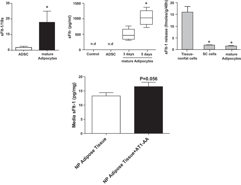
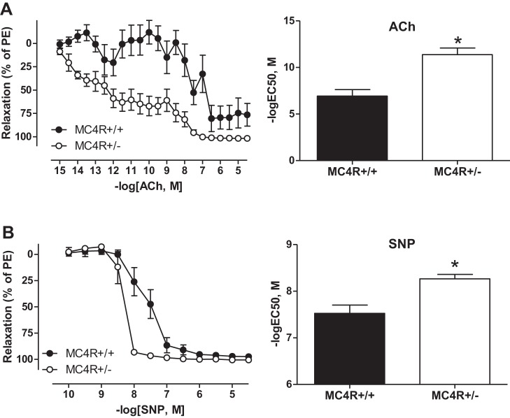
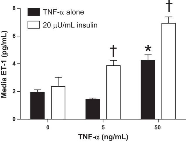
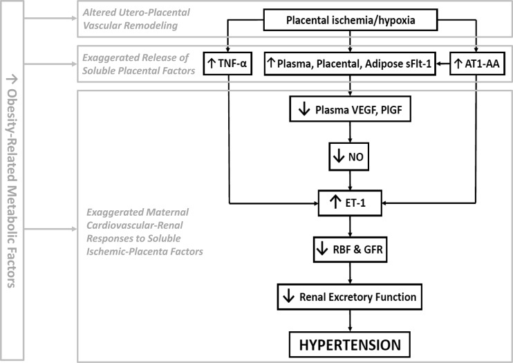
Preeclampsia (PE) is a pregnancy-specific disorder typically presenting as new-onset hypertension and proteinuria. While numerous epidemiological studies have demonstrated that obesity increases the risk of PE, the mechanisms have yet to be fully elucidated. Growing evidence from animal and human studies implicate placental ischemia in the etiology of this maternal syndrome. It is thought that placental ischemia is brought about by dysfunctional cytotrophoblast migration and invasion into the uterus and subsequent lack of spiral arteriole widening and placental perfusion. Placental ischemia/hypoxia stimulates the release of soluble placental factors into the maternal circulation where they cause endothelial dysfunction, particularly in the kidney, to elicit the clinical manifestations of PE. The most recognized of these factors are the anti-angiogenic sFlt-1 and pro-inflammatory TNF-α and AT1-AA, which promote endothelial dysfunction by reducing levels of the provasodilator nitric oxide and stimulating production of the potent vasoconstrictor endothelin-1 and reactive oxygen species. We hypothesize that obesity-related metabolic factors increase the risk for developing PE by impacting various stages in the pathogenesis of PE, namely, 1) cytotrophoblast migration and placental ischemia; 2) release of soluble placental factors into the maternal circulation; and 3) maternal endothelial and vascular dysfunction. This review will summarize the current experimental evidence supporting the concept that obesity and metabolic factors like lipids, insulin, glucose, and leptin affect placental function and increase the risk for developing hypertension in pregnancy by reducing placental perfusion; enhancing placental release of soluble factors; and by increasing the sensitivity of the maternal vasculature to placental ischemia-induced soluble factors.

Keywords: RUPP; body mass index; inflammation; placental ischemia; pregnancy; sFlt-1.


Figures

Fig. 1.
Fig. 1.
Rates of preeclampsia (PE) increase with increasing body mass index (BMI). From Mbah AK et al. (110).
Fig. 2.
Fig. 2.
High-fat diet (HFD) increases maternal body weight (A) and reduces placental volume blood flow (B), which is accompanied by increased plasma leptin (C) and insulin (D) levels in the third trimester of pregnancy in baboons. Control, control diet; R, HFD resistant; S, HFD sensitive; cQutv, placental volume blood flow. *P < 0.05 vs. control and HFD R. From Frias AE et al. (52).
Fig. 3.
Fig. 3.
Euglycemic (A) hyperinsulinemia (B) elicits hypertension (C) in Sprague-Dawley pregnant rats. Insulin was infused at 1.5 mU·kg−1·day−1 from gestational day 14–19 while rats were provided with 20% glucose in drinking water. *P < 0.05 vs. pregnant. From Palei AC et al. (128).
Fig. 4.
Fig. 4.
The soluble fms-like tyrosine kinase 1 (sFlt-1) levels in obese MC4R+/− versus MC4R+/+ pregnant rats are slightly but not significantly elevated the circulation (A), elevated in adipose tissue (B); but similar in placental tissue (C). The sFlt-1 was quantified using a mouse Flt-1 ELISA from R&D Systems. *P < 0.05 vs. MC4R+/+ pregnant rats. From Spradley FT et al. (171).
Fig. 5.
Fig. 5.
The release of sFlt-1 is greater under hypoxic conditions in placental villous explants isolated from obese MC4R+/− (n = 10) versus lean MC4R+/+ (n = 6) pregnant rats. Placental explant studies were conducted as previously described (55). The sFlt-1 was quantified using a mouse Flt-1 ELISA from R&D Systems. Data are means ± SE. *P < 0.05 vs. MC4R+/+ pregnant rats. These data are unpublished.
Fig. 6.
Fig. 6.
Top, left: mRNA expression of sFlt-1 in isolated adipose-derived stem cells (ADSCs) and isolated mature adipocytes from human adipose tissue; *P < 0.01. Middle: release of sFlt-1 from ADSCs incubated for 5 days and mature adipocytes incubated for 3 or 5 days; *P < 0.05 for 3 vs. 5 days; n.d., not detectable. Right: release of sFlt-1 from adipose tissue nonfat cells, stromal vascular cells (SV cells) and mature adipocytes; *P < 0.01 vs. adipose tissue nonfat cells. From Herse et al. (66). Bottom: stimulation with AT1-AA promotes sFlt-1 release from adipose tissue explants isolated from normal pregnant (NP) Sprague-Dawley rats. Adipose explant (∼100 mg) were incubated in F12K supplemented with media (DMEM containing 4 mM l-glutamin, 4,500 mg/l glucose, 1 mM sodium pyruvate, 1,500 mg/l sodium bicarbonate, 4 g/l BSA, and 1% PenStrep) in the presence or absence of AT1-AA (1:100 dilution) for 48 h, at which time media were collected and assayed for sFlt-1 as previously described (171). n = 3 rats/group. Data are means ± SE. Data in bottom panel are unpublished.
Fig. 7.
Fig. 7.
Endothelial-dependent responses to acetylcholine (ACh) (A; left: concentration-response curve, right: logEC50 sensitivity) and endothelial-independent responses to sodium nitroprusside (SNP) (B; left: concentration-response curve, right: logEC50 sensitivity) are enhanced in third-order mesenteric arteries from obese MC4R+/− pregnant versus lean MC4R+/+ pregnant rats. *P < 0.05 vs. MC4R+/+ pregnant rats. From Spradley et al. (171).
Fig. 8.
Fig. 8.
Insulin exaggerates tumor nerosis factor-α (TNF-α)-induced release of endothelin-1 (ET-1) from human umbilical vein endothelial cells (HUVECs). One million cells were seeded into 6-well plates and allowed to grow until 70% confluent at which time they were serum starved for 36 h then treated with TNF-α or in combination with insulin at levels observed in obese preeclamptic women (20 μU/ml) for 8 h. ET-1 levels were measured in HUVEC-conditioned media as previously described (89). n = 3–6 wells/group. Data are means ± SE. *P < 0.05 vs. 0 TNF-α; †P < 0.05 vs. corresponding TNF-α group. These data are unpublished.
Fig. 9.
Fig. 9.
Hypothetical scheme whereby obesity and obesity-related metabolic factors act in the cascade of events leading to enhancement of placental ischemia-induced hypertension. We propose that these metabolic factors may act individually or in combination to potentiate dysfunctional uteroplacental vascular remodeling, exaggerate the release of soluble placental factors into the maternal circulation, and to exaggerate the maternal cardiovascular-renal responses to these soluble placental-ischemia factors. This may occur in a feed-forward fashion subsequently exaggerating the downstream actions of these metabolic factors.

References

    1. Ajne G, Nisell H, Jansson T. Effects of blockade of the endothelin receptor A and inhibition of nitric oxide synthesis on uteroplacental and renal blood flow in awake pregnant rats. Am J Obstet Gynecol 192: 295–301, 2005. - PubMed
    1. Akyol A, Langley-Evans SC, McMullen S. Obesity induced by cafeteria feeding and pregnancy outcome in the rat. Br J Nutr 102: 1601–1610, 2009. - PubMed
    1. Alexander BT, Cockrell KL, Massey MB, Bennett WA, Granger JP. Tumor necrosis factor-alpha-induced hypertension in pregnant rats results in decreased renal neuronal nitric oxide synthase expression. Am J Hypertens 15: 170–175, 2002. - PubMed
    1. American College of Obstetricians and Gynecologists; Task Force on Hypertension in Pregnancy. Hypertension in pregnancy. Report of the American College of Obstetricians and Gynecologists' Task Force on Hypertension in Pregnancy. Obstet Gynecol 122: 1122–1131, 2013. - PubMed
    1. Athanassiades A, Hamilton GS, Lala PK. Vascular endothelial growth factor stimulates proliferation but not migration or invasiveness in human extravillous trophoblast. Biol Reprod 59: 643–654, 1998. - PubMed

Publication types

MeSH terms