. 2015 Nov 3;6(34):36731-45.
doi: 10.18632/oncotarget.5481.

The axon guidance molecule semaphorin 3F is a negative regulator of tumor progression and proliferation in ileal neuroendocrine tumors

Affiliations

The axon guidance molecule semaphorin 3F is a negative regulator of tumor progression and proliferation in ileal neuroendocrine tumors

Julien Bollard et al. Oncotarget. .

Abstract

Gastro-intestinal neuroendocrine tumors (GI-NETs) are rare neoplasms, frequently metastatic, raising difficult clinical and therapeutic challenges due to a poor knowledge of their biology. As neuroendocrine cells express both epithelial and neural cell markers, we studied the possible involvement in GI-NETs of axon guidance molecules, which have been shown to decrease tumor cell proliferation and metastatic dissemination in several tumor types. We focused on the role of Semaphorin 3F (SEMA3F) in ileal NETs, one of the most frequent subtypes of GI-NETs.SEMA3F expression was detected in normal neuroendocrine cells but was lost in most of human primary tumors and all their metastases. SEMA3F loss of expression was associated with promoter gene methylation. After increasing endogenous SEMA3F levels through stable transfection, enteroendocrine cell lines STC-1 and GluTag showed a reduced proliferation rate in vitro. In two different xenograft mouse models, SEMA3F-overexpressing cells exhibited a reduced ability to form tumors and a hampered liver dissemination potential in vivo. This resulted, at least in part, from the inhibition of mTOR and MAPK signaling pathways.This study demonstrates an anti-tumoral role of SEMA3F in ileal NETs. We thus suggest that SEMA3F and/or its cellular signaling pathway could represent a target for ileal NET therapy.

Keywords: proliferation; semaphorin; small intestine neuroendocrine tumor; tumor progression.


Conflict of interest statement

CONFLICTS OF INTEREST

The authors disclose no potential conflicts of interest.

Figures
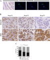
Figure 1
Figure 1. SEMA3F is differentially expressed in human endocrine cells
A. Immunohistochemical staining of human ileal sections (SEMA3F positivity in brown, black arrows, X200), (Scale bar: 25 μm), and confocal analysis of double-immunofluorescence staining (SEMA3F+: magenta; CgA+: green; DAPI) (Scale bar: 10 μm). B. SEMA3F and NRP2 immunohistochemical staining of human multifocal ileal tumors. C. SEMA3F expression according to tumor stage: data represent the percent of SEMA3F positive or negative cases; *P < 0.05, **P < 0 .005.
Figure 2
Figure 2. SEMA3F expression level correlates with proliferation rate and promoter methylation
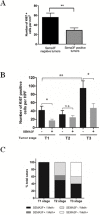
A. KI67 expression in human multifocal carcinoid tumors: mean ± SEM; **P < 0 .01. B. The number of Ki67-positive tumor cells is significantly reduced in SEMA3F-positive tumors compared to SEMA3F-negative tumors within T1 and T3 stages; *P < 0.05 and significantly increases with tumor stage; **P < 0.05. C. SEMA3F promoter methylation correlates with loss of SEMA3F expression.
Figure 3
Figure 3. Expression of SEMA3F in intestinal endocrine tumoral cell lines
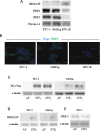
A. Immunoblot analysis of SEMA3F, NRP1, NRP2 and PLEXINA1 expression in both STC-1, GluTag and INS-1E cells. B. Confocal immunostaining of NRP-2 in STC-1, GluTag and INS-1E cells (NRP-2+: green; DAPI). Scale bar: 10 μm. C. Immunoblot analysis of MycTag expression in STC-S3F2 and GluTag-S3F2 clones. D. Immunoblot analysis of SEMA3F expression in STC-S3F2 and GluTag-S3F2 clones. E. Immunoblot analysis of NRP2 expression in STC-S3F2 clone.
Figure 4
Figure 4. SEMA3F decreases cell viability and proliferation of tumoral cell lines
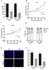
A. Cell viability (MTT) assay on STC-S3F2 and GluTag-S3F2 clones (n = 4, ***P < 0 .0005). B–C. Kinetics study of cell proliferation using MTT assay at 72, 96, and 120 hours following seeding. (n = 3, ***P < 0.0005). D. Cell cycle analysis by FACS of STC-1 and GluTag S3F2 clones (n = 3, *P < 0.05). E. Immunofluorescent staining of phospho-Histone H3 (pH-H3 +: red; DAPI) (×200), Scale bar: 10 μm. F. Quantification of cells positive for pH-H3 staining, (n = 3, **P < .005). (mean ± SEM).
Figure 5
Figure 5. SEMA3F inhibits STC-1 cells intrahepatic dissemination in vivo
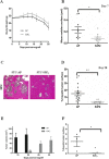
A. Body weight curve in STC-AP and STC-S3F2 xenografted mice (n = 12). B. Morphometric analyses of intrahepatic nodules 7 days after intrasplenic injection of STC-AP and STC-S3F2 cell lines (n = 21, *P < 0.05). C–D. Histologic and morphometric analysis at day 28 of intrahepatic nodules following STC-AP and STC-S3F2 cell lines grafting. (n = 31, **P < 0.005) (×100) Scale bar: 100 μm. E. Volume comparison of subcutaneous tumors developed after STC-AP or STC-S3F2 cells grafting. F. Morphometric analyses of intrahepatic nodules 8 weeks after intracaecal graft of either STC-AP or STC-S3F2 derived-subcutaneous tumors. (n = 7, *P < 0.05); (mean ± SEM).
Figure 6
Figure 6. SEMA3F reduces the proliferation rate of tumor cells in vivo and inhibits tumor vascularization
A–B. KI67 immunohistochemical staining and quantification of intrahepatic nodules derived from STC-AP or STC-S3F2 intrasplenic grafted cells (n = 31, ***P < 0.0005, ×100). Scale bar: 50 μm. C–D. CD31 immunohistochemical staining and quantification of intrahepatic nodules derived from STC-AP or STC-S3F2 intrasplenic grafted cells (n = 31, ***P < 0.0005200). Scale bar: 25 μm.
Figure 7
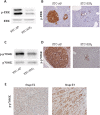
Figure 7. SEMA3F expression decreases activation of MAPK and mTOR signaling pathways
A–C. STC-AP and STC-S3F2 cell lines were subjected to immunoblot analysis of ERK and p70S6K (n = 3). B–D. Phospho-ERK and phospho-p70S6K immunohistochemical staining on intrahepatic nodules derived from STC-AP or STC-S3F2 intrasplenic grafted cells (X400), Scale bar: 50 μm. E. Phospho-p70S6K immunohistochemical staining on human multifocal ileal tumors (T1 and T3 stages) (X200).

Similar articles

Cited by

References

    1. Yao JC, Hassan M, Phan A, Dagohoy C, Leary C, Mares JE, Abdalla EK, Fleming JB, Vauthey J-N, Rashid A, Evans DB. One hundred years after “carcinoid”: epidemiology of and prognostic factors for neuroendocrine tumors in 35,825 cases in the United States. J Clin Oncol. 2008;26:3063–3072. - PubMed
    1. Cohen T, Gluzman-Poltorak Z, Brodzky A, Meytal V, Sabo E, Misselevich I, Hassoun M, Boss JH, Resnick M, Shneyvas D, Eldar S, Neufeld G. Neuroendocrine cells along the digestive tract express neuropilin-2. Biochem Biophys Res Commun. 2001;284:395–403. - PubMed
    1. Villasenor A, Marty-Santos L, Dravis C, Fletcher P, Henkemeyer M, Cleaver O. EphB3 marks delaminating endocrine progenitor cells in the developing pancreas. Dev Dyn. 2012;241:1008–1019. - PMC - PubMed
    1. Yang YHC, Szabat M, Bragagnini C, Kott K, Helgason CD, Hoffman BG, Johnson JD. Paracrine signalling loops in adult human and mouse pancreatic islets: netrins modulate beta cell apoptosis signalling via dependence receptors. Diabetologia. 2011;54:828–842. - PubMed
    1. Chédotal A, Kerjan G, Moreau-Fauvarque C. The brain within the tumor: new roles for axon guidance molecules in cancers. Cell Death Differ. 2005;12:1044–1056. - PubMed

Publication types

MeSH terms