Comparative Study
. 2015 Oct 8;10(10):e0140292.
doi: 10.1371/journal.pone.0140292. eCollection 2015.

Metabolic and Phenotypic Differences between Mice Producing a Werner Syndrome Helicase Mutant Protein and Wrn Null Mice

Affiliations
Comparative Study

Metabolic and Phenotypic Differences between Mice Producing a Werner Syndrome Helicase Mutant Protein and Wrn Null Mice

Lucie Aumailley et al. PLoS One. .

Abstract

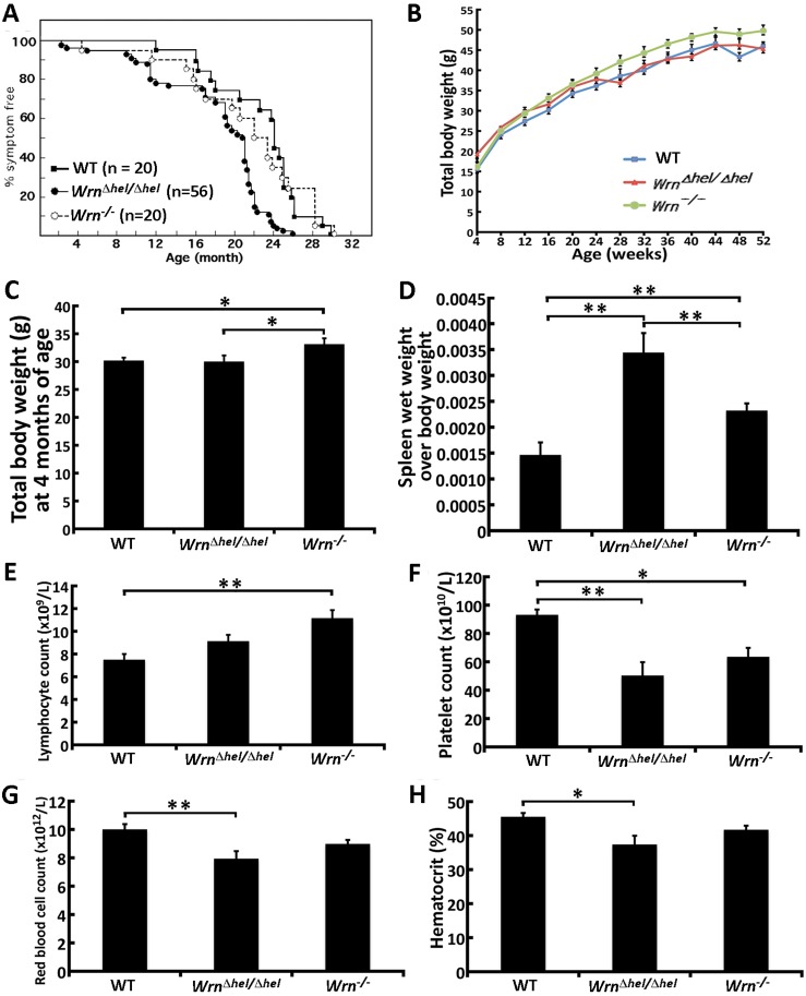
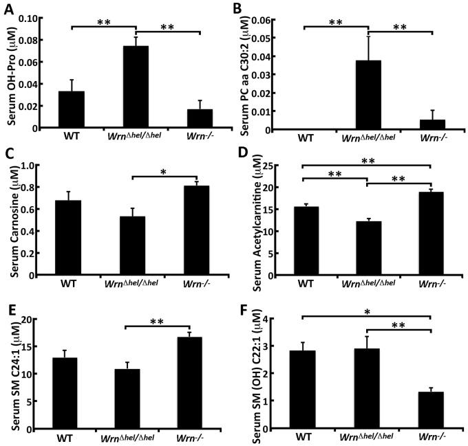
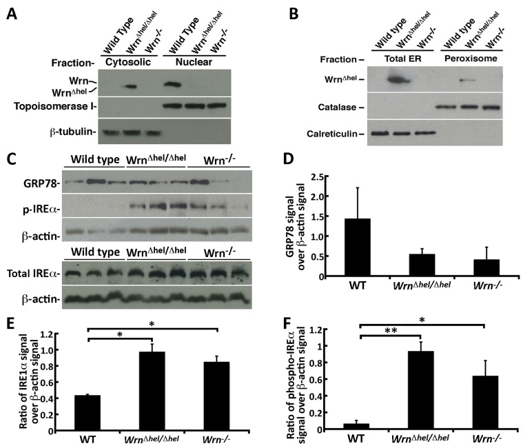
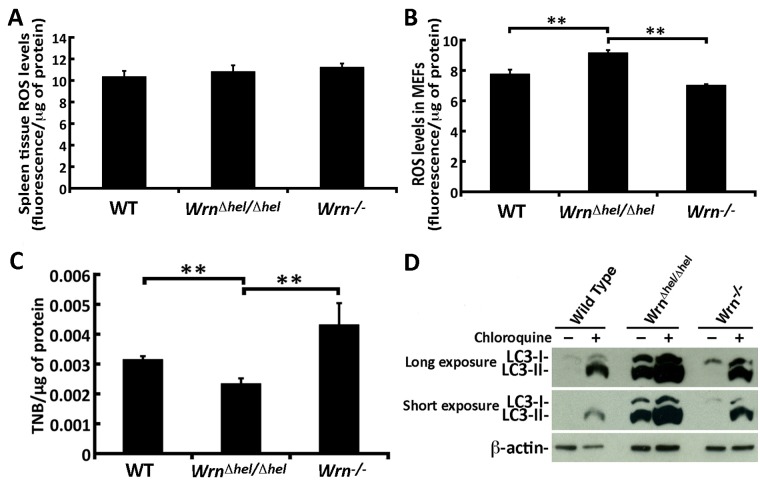
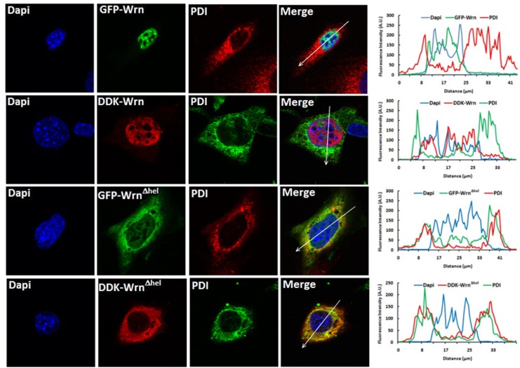
Werner syndrome (WS) is a premature aging disorder caused by mutations in a RecQ-family DNA helicase, WRN. Mice lacking part of the helicase domain of the WRN orthologue exhibit many phenotypic features of WS, including metabolic abnormalities and a shorter mean life span. In contrast, mice lacking the entire Wrn protein (i.e. Wrn null mice) do not exhibit a premature aging phenotype. In this study, we used a targeted mass spectrometry-based metabolomic approach to identify serum metabolites that are differentially altered in young Wrn helicase mutant and Wrn null mice. An antibody-based quantification of 43 serum cytokines and markers of cardiovascular disease risk complemented this study. We found that Wrn helicase mutants exhibited elevated and decreased levels, respectively, of the anti-inflammatory cytokine IL-10 and the pro-inflammatory cytokine IL-18. Wrn helicase mutants also exhibited an increase in serum hydroxyproline and plasminogen activator inhibitor-1, markers of extracellular matrix remodeling of the vascular system and inflammation in aging. We also observed an abnormal increase in the ratio of very long chain to short chain lysophosphatidylcholines in the Wrn helicase mutants underlying a peroxisome perturbation in these mice. Remarkably, the Wrn mutant helicase protein was mislocalized to the endoplasmic reticulum and the peroxisomal fractions in liver tissues. Additional analyses with mouse embryonic fibroblasts indicated a severe defect of the autophagy flux in cells derived from Wrn helicase mutants compared to wild type and Wrn null animals. These results indicate that the deleterious effects of the helicase-deficient Wrn protein are mediated by the dysfunction of several cellular organelles.


Conflict of interest statement

Competing Interests: The authors have declared that no competing interests exist.

Figures

Fig 1
Fig 1. Phenotypic traits of Wrn Δhel/Δhel and Wrn -/- mice.
(A) Percentage of disease-free animals with age. The number of animals in each group is indicated (50% males + 50% females). (Log rank tests: WT vs. Wrn Δhel/Δhel mice, P = 6.8x10⁻⁵; WT vs. Wrn -/- mice, P = 0.001; Wrn Δhel/Δhel vs. Wrn -/- mice, P = 0.955). (B) Measures of males total body weight from the age of 4 to 52 weeks (n = 6–19 males). (C) Total body weight at four months of age (n = 6–10 males). (D) Ratio of spleen wet weight over total body weight at four months of age (n = 6 males). (E) Lymphocyte blood count at four months of age (n = 5 males). (F) Platelet blood count at four months of age (n = 5 males). (G) Red blood cell count at four months of age (n = 5 males). (H) % hematocrit at four months of age (n = 5 males). Bars in all the graphs represent SEM.
Fig 2
Fig 2. Serum levels of three serum cytokines and one cardiovascular risk factor significantly altered in Wrn Δhel/Δhel and Wrn -/- mice.
(A) IL-10; (B) IL-18; (C) MIP-1α; (D) PAI-1. Tukey post ANOVA test P-values are shown (*P < 0.05 and **P < 0.01). Bars in all histograms represent SD. The number of mice in each cohort is indicated in the S2 Table for each serum cytokine.
Fig 3
Fig 3. Serum levels of six serum metabolites significantly altered between Wrn Δhel/Δhel and Wrn -/- mice.
(A) Hydroxyproline (OH-Pro); (B) Phosphatidylcholine PC aa C30:2; (C) Carnosine; (D) Acetylcarnitine; (E) Sphingomyelin SM C24:1; (F) Sphingomyelin SM (OH) C22:1. Tukey post ANOVA test P-values are shown (*P < 0.05 and **P < 0.01). Bars in all histograms represent SD. N = 6 males for each cohort.
Fig 4
Fig 4. DNA helicase and exonuclease activities of the immunoprecipitated WT and mutant Wrn protein.
(A) Example of a western blots is presented. WT, Wrn Δhel/Δhel and Wrn -/- MEFs were lysed and the Wrn proteins were immunoprecipitated with a mouse anti-Wrn monoclonal antibody. The immunoprecipitates were analyzed by immunoblotting with a rabbit anti-Wrn polyclonal antibody. (B) DNA helicase activity of the immunoprecipitated Wrn proteins. The position of the double strand and single strand radioactive structures are indicated with an asterisk. The “-”lane represents the undenatured double strand radioactive structure. The “Δ” lane represents heat-denatured DNA. (C) Nuclease activity of the immunoprecipitated Wrn proteins. The positions of the cleaved fragments are indicated on the left by arrows. Ten, five, and zero μL of bead solutions containing the immunoprecipitated proteins were used for the enzymatic reactions. The “Δ” lane represents heat-denatured DNA.
Fig 5
Fig 5. Localization of the WT and mutant Wrn proteins in different subcellular fractions.
(A) Example of western blots showing the presence of the Wrn WT protein in the nuclear fraction and the presence of the WrnΔhel mutant protein mainly in the cytosolic fraction. Fractionations were performed on WT, Wrn Δhel/Δhel, and Wrn -/- MEFs. Topoisomerase I and β-tubulin are nuclear and cytoplasmic markers, respectively. As expected the molecular weight of the WrnΔhel mutant protein was lower than the Wrn WT protein. No band was detected in Wrn -/- MEFs fractions with the anti-Wrn polyclonal antibody. (B) Example of western blots showing only the presence of the WrnΔhel mutant protein in total ER and the peroxisomal fractions. Fractionations were performed on WT, Wrn Δhel/Δhel, and Wrn -/- liver tissues. Catalase and calreticulin are used as peroxisomal and ER specific markers. This experiment was repeated three times. (C) Protein levels of GRP78, phosphorylated IREα, and β-actin (top panels) and of total IREα, and β-actin (bottom panels) in the spleen of three animals of each genotype. (D) Ratio of GRP78 signal over β-actin signal from western blots. (E) Ratio of IREα over β-actin signal from western blots. (F) Ratio of phosphorylated IREα signal over β-actin signal from western blots. Experiments were performed in triplicates. Bars in all histograms represent SEM. Tukey post ANOVA test P-values are shown (*P < 0.05 and **P < 0.01).
Fig 6
Fig 6. ROS levels, oxidized proteins, and autophagy in WT, Wrn Δhel/Δhel, and Wrn -/- MEFs.
(A) ROS levels in spleen tissues of WT, Wrn Δhel/Δhel, and Wrn -/- mice. (B) ROS levels in WT, Wrn Δhel/Δhel, and Wrn -/- MEFs. (C) Quantification of thiol groups on proteins extracted from MEFs by the DTNB assay. Experiments were performed in triplicates. Bars in all histograms represent SEM. Tukey post ANOVA test P-values are shown (*P < 0.05 and **P < 0.01). (D) Autophagy flux in WT, Wrn Δhel/Δhel, and Wrn -/- MEFs. Example of western blots showing the increase of LC3-II isoforms in mutant MEFs and in all cells treated with chloroquine (an inhibitor of autophagy flux). All experiments were repeated twice.
Fig 7
Fig 7. Example of co-localization of the WrnΔhel mutant protein with the ER organelles by immunofluorescence study.
Images in the first row represent the localization of the GFP-Wrn and PDI in Wrn Δhel/Δhel MEFs. Images in the second row represent the localization of the DDK-Wrn and PDI in Wrn Δhel/Δhel MEFs. Images in the third row represent the localization of the GFP-WrnΔhel mutant protein and PDI in Wrn Δhel/Δhel MEFs. Images in the fourth row represent the localization of the DDK-WrnΔhel mutant protein and PDI in Wrn Δhel/Δhel MEFs. The graph at the end of each row represents the intensity of the fluorescence along the arrow in the merge image.

References

    1. Epstein CJ, Martin GM, Schultz AL, Motulsky AG. Werner's syndrome a review of its symptomatology, natural history, pathologic features, genetics and relationship to the natural aging process. Medicine (Baltimore). 1966;45(3):177–221. Epub 1966/05/01. . - PubMed
    1. Salk D. Werner's syndrome: a review of recent research with an analysis of connective tissue metabolism, growth control of cultured cells, and chromosomal aberrations. Hum Genet. 1982;62(1):1–5. Epub 1982/01/01. . - PubMed
    1. Yu CE, Oshima J, Fu YH, Wijsman EM, Hisama F, Alisch R, et al. Positional cloning of the Werner's syndrome gene. Science. 1996;272(5259):258–62. Epub 1996/04/12. . - PubMed
    1. Ozgenc A, Loeb LA. Current advances in unraveling the function of the Werner syndrome protein. Mutat Res. 2005;577(1–2):237–51. Epub 2005/06/11. S0027-5107(05)00185-5 [pii] 10.1016/j.mrfmmm.2005.03.020 . - DOI - PubMed
    1. Kamath-Loeb AS, Shen JC, Loeb LA, Fry M. Werner syndrome protein. II. Characterization of the integral 3'—> 5' DNA exonuclease. J Biol Chem. 1998;273(51):34145–50. Epub 1998/12/16. . - PubMed

Publication types

LinkOut - more resources