. 2015 Oct 8;10(10):e0139416.
doi: 10.1371/journal.pone.0139416. eCollection 2015.

Mitochondrial ROS Induces Cardiac Inflammation via a Pathway through mtDNA Damage in a Pneumonia-Related Sepsis Model

Affiliations

Mitochondrial ROS Induces Cardiac Inflammation via a Pathway through mtDNA Damage in a Pneumonia-Related Sepsis Model

Xiao Yao et al. PLoS One. .

Abstract

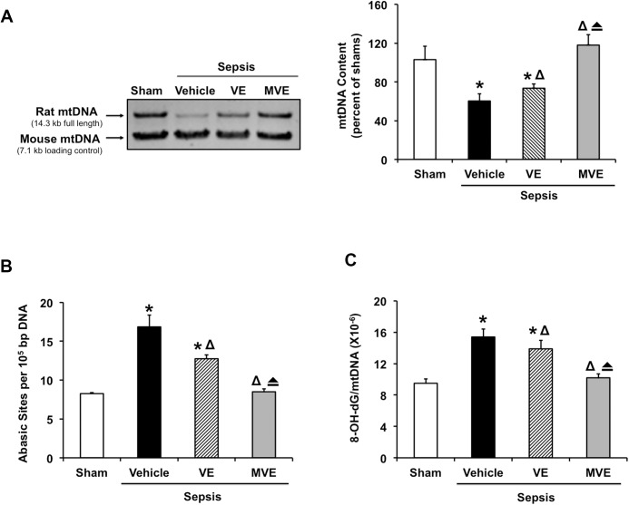
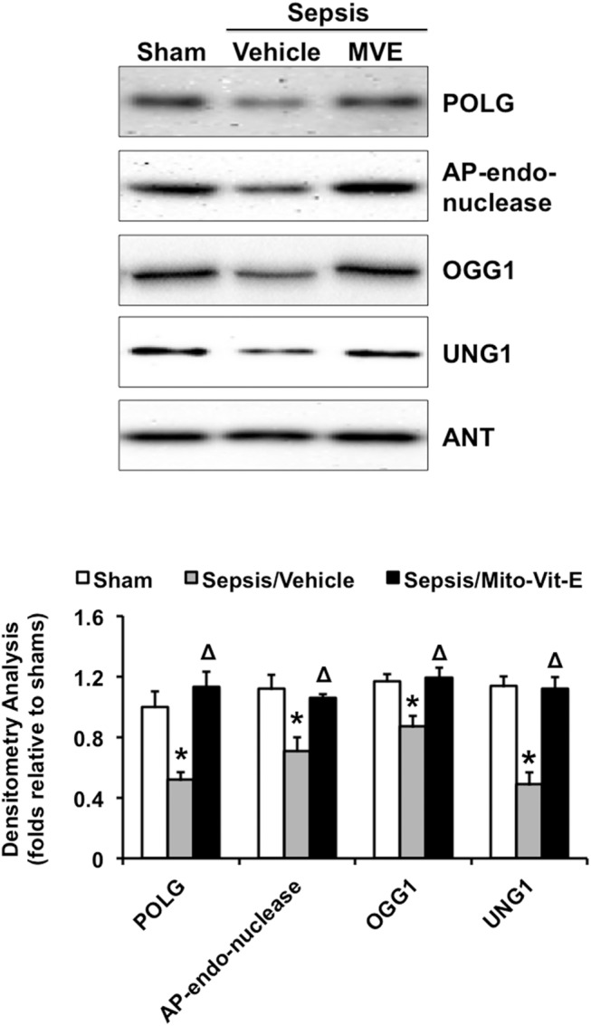
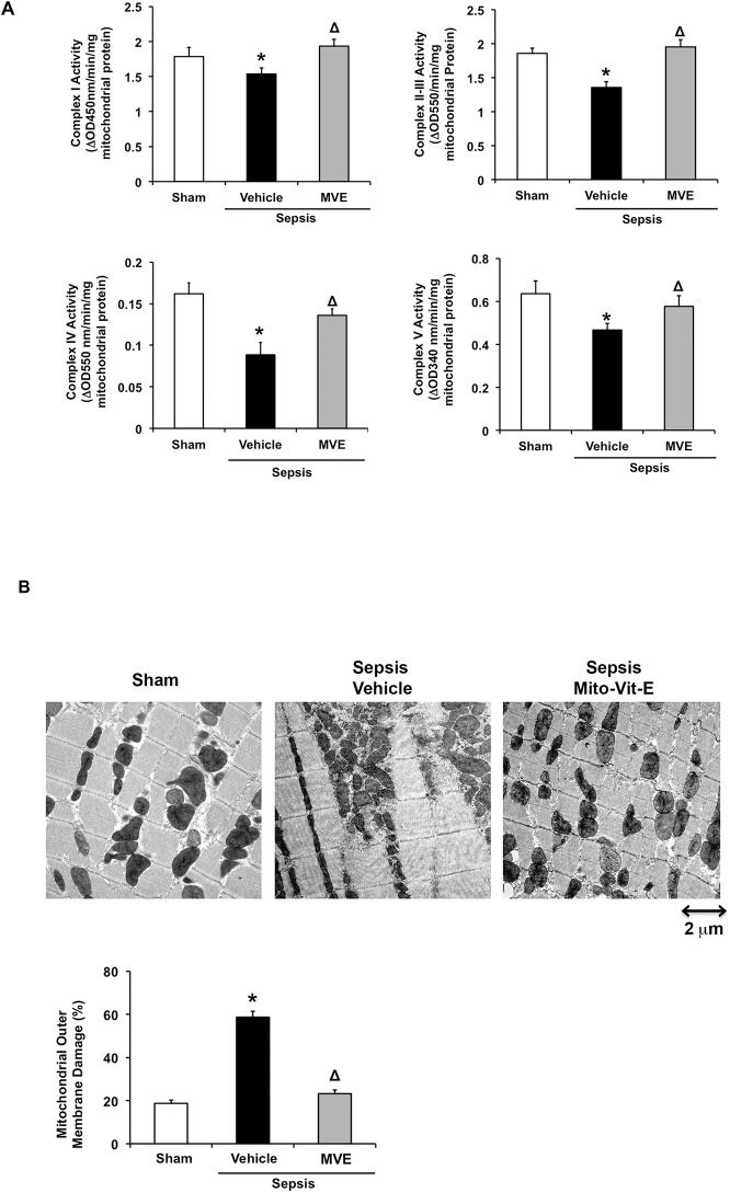
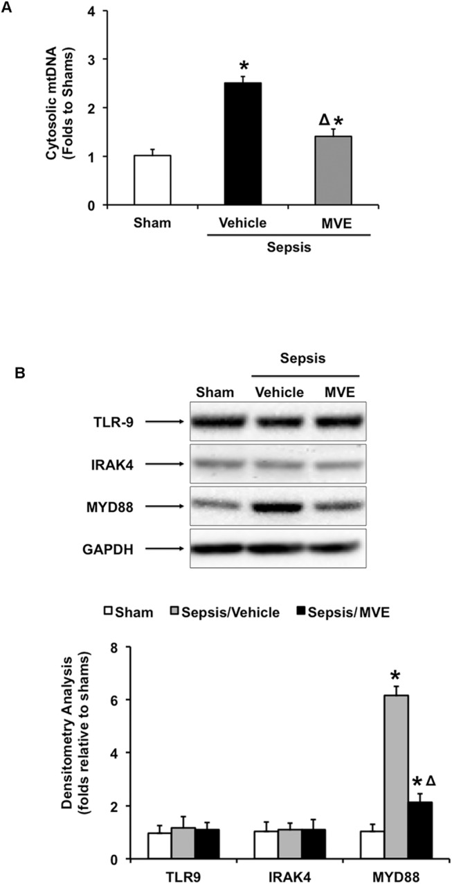
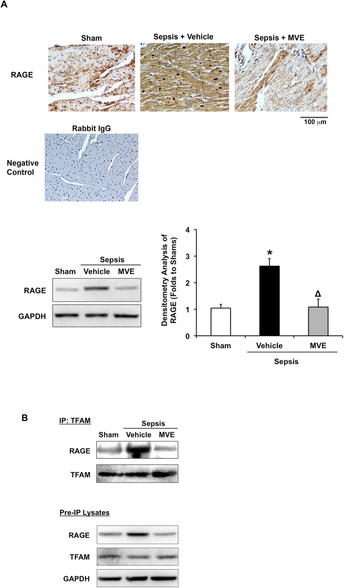
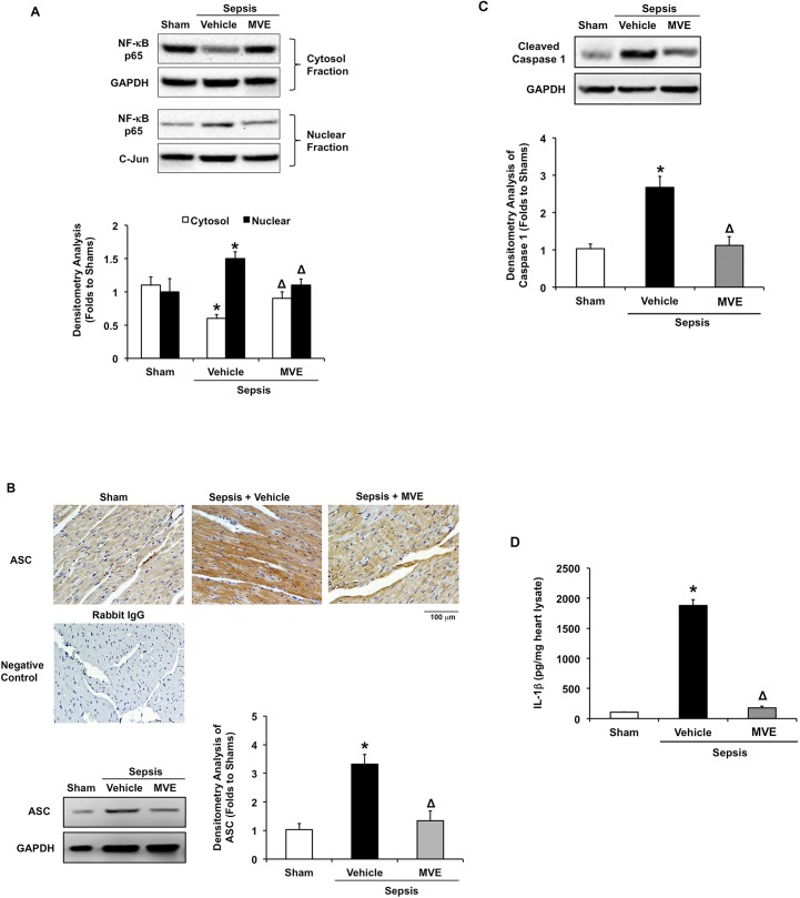
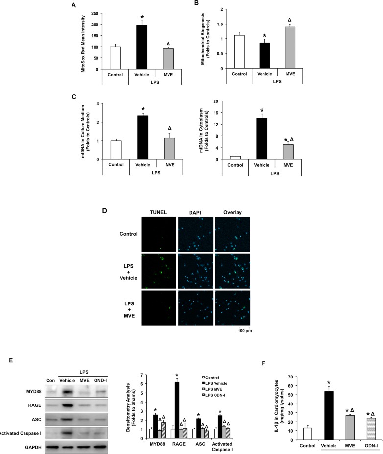
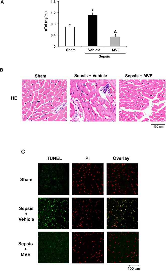
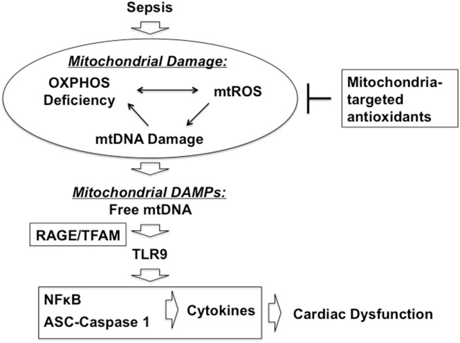
We have previously shown that mitochondria-targeted vitamin E (Mito-Vit-E), a mtROS specific antioxidant, improves cardiac performance and attenuates inflammation in a pneumonia-related sepsis model. In this study, we applied the same approaches to decipher the signaling pathway(s) of mtROS-dependent cardiac inflammation after sepsis. Sepsis was induced in Sprague Dawley rats by intratracheal injection of S. pneumoniae. Mito-Vit-E, vitamin E or vehicle was administered 30 minutes later. In myocardium 24 hours post-inoculation, Mito-Vit-E, but not vitamin E, significantly protected mtDNA integrity and decreased mtDNA damage. Mito-Vit-E alleviated sepsis-induced reduction in mitochondria-localized DNA repair enzymes including DNA polymerase γ, AP endonuclease, 8-oxoguanine glycosylase, and uracil-DNA glycosylase. Mito-Vit-E dramatically improved metabolism and membrane integrity in mitochondria, suppressed leakage of mtDNA into the cytoplasm, inhibited up-regulation of Toll-like receptor 9 (TLR9) pathway factors MYD88 and RAGE, and limited RAGE interaction with its ligand TFAM in septic hearts. Mito-Vit-E also deactivated NF-κB and caspase 1, reduced expression of the essential inflammasome component ASC, and decreased inflammatory cytokine IL-1β. In vitro, both Mito-Vit-E and TLR9 inhibitor OND-I suppressed LPS-induced up-regulation in MYD88, RAGE, ASC, active caspase 1, and IL-1β in cardiomyocytes. Since free mtDNA escaped from damaged mitochondria function as a type of DAMPs to stimulate inflammation through TLR9, these data together suggest that sepsis-induced cardiac inflammation is mediated, at least partially, through mtDNA-TLR9-RAGE. At last, Mito-Vit-E reduced the circulation of myocardial injury marker troponin-I, diminished apoptosis and amended morphology in septic hearts, suggesting that mitochondria-targeted antioxidants are a potential cardioprotective approach for sepsis.


Conflict of interest statement

Competing Interests: The authors have declared that no competing interests exist.

Figures

Fig 1
Fig 1. Mitochondrial ROS dependent mtDNA damage in the heart after sepsis.
Rats were infected by S. pneumoniae or given PBS sham control. 21.5 μmoles/kg Mito-Vit-E (MVE), vitamin E (VE) or vehicle was administered orally 30 minutes post-inoculation, and heart tissues were harvested 24 hours later. A. 0.4 ng genomic DNA from the heart tissue was amplified for 25 cycles by LPCR. Mouse DNA was included as an internal control. The PCR products were digested, separated on 1% agarose gel, and analyzed by densitometry. Levels of 8-hydroxy-2-deoxy guanosine (8-OH-dG) (B) and apurinic/apyrimidinic (AP) sites (C) were measured in DNA isolated from mitochondrial fractions. All values are means ±SE. Significant differences are shown as * between sham and sepsis, Δ between vehicle and drug-treated, and between vitamin E and Mito-Vit-E (p<0.01, n = 6).
Fig 2
Fig 2. DNA polymerase γ (POLG), AP endonuclease, 8-oxoguanine glycosylase (OGG1), and uracil-DNA glycosylase (UNG1) in mitochondria.
Rats were infected by S. pneumoniae or given PBS sham control. 21.5 μmoles/kg Mito-Vit-E (MVE) or vehicle was administered orally 30 minutes post-inoculation, and heart tissues were harvested 24 hours later. POLG, AP endonuclease, OGG1 and UNG1 were determined in mitochondrial fractions by Western blot using adenine nucleotide translocase (ANT) as a loading control. Results were quantified by densitometry and expressed as fold changes relative to shams. All values are means ±SE. Significant differences are shown as * between sham and sepsis and Δ between vehicle and Mito-Vit-E (p<0.02, n = 6).
Fig 3
Fig 3. Mitochondrial ROS dependent functional deficiency and structural impairment in cardiac mitochondria after sepsis.
Rats were infected by S. pneumoniae or given PBS sham control. 21.5 μmoles/kg Mito-Vit-E (MVE) or vehicle was administered orally 30 minutes post-inoculation, and heart tissues were harvested 24 hours later. A. Mitochondrial fractions were subjected to the measurements of complex I-V activities. B. Ultrastructure of myocardial mitochondria was observed by transmission electron microscope (TEM). Biochemically, mitochondrial outer membrane damage was measured using the mitochondrial fractions. All values are means ±SE. Significant differences are shown as * between sham and sepsis and Δ between vehicle and Mito-Vit-E (p<0.02, n = 6).
Fig 4
Fig 4. Mitochondrial ROS dependent activation of mtDNA-TLR9 pathway in the heart after sepsis.
Rats were infected by S. pneumoniae or given PBS sham control. 21.5 μmoles/kg Mito-Vit-E (MVE) or vehicle was administered orally 30 minutes post-inoculation, and heart tissues were harvested 24 hours later. A. Levels of mtDNA in cytosol fractions were measured by real-time PCR. B. Expression of TLR9, IRAK4 and MyD88 were determined in total tissue lysates by western blot using GAPDH as a loading control, and the results were quantified by densitometry. All values are means ±SE. Significant differences are shown as * between sham and sepsis and Δ between vehicle and Mito-Vit-E (p<0.02, n = 6).
Fig 5
Fig 5. Mitochondrial ROS dependent activation of RAGE pathway in the heart after sepsis.
Rats were infected by S. pneumoniae or given PBS sham control. 21.5 μmoles/kg Mito-Vit-E (MVE) or vehicle was administered orally 30 minutes post-inoculation, and heart tissues were harvested 24 hours later. A. Heart sections were stained with anti-RAGE (brown) and haematoxylin (blue). Negative control was stained with secondary antibody alone. Images are representative of a random selection of at least 3 sections of N = 6. B. RAGE-TFAM interaction was determined by co-immunoprecipitation in total heart lysate. Shown result was reprehensive of three independent experiments (N = 6).
Fig 6
Fig 6. Mitochondrial ROS-dependent activation of myocardial inflammation after sepsis.
Rats were infected by S. pneumoniae or given PBS sham control. 21.5 μmoles/kg Mito-Vit-E (MVE) or vehicle was administered orally 30 minutes post-inoculation, and heart tissues were harvested 24 hours later. A. NF-κB p65 subunit was detected in cytosolic and nuclear fractions by Western blot using GAPDH and c-Jun as a loading control respectively. Results were quantified by densitometry. B. Heart sections were co-stained with anti-ASC (brown) and haematoxylin (blue). Negative control was stained with secondary antibody alone. Images are representative of a random selection of at least 3 sections of N = 6. C. Activated form of caspase 1 was determined in heart tissue lysates by Western blot using GAPDH as a loading control, and results were quantified by densitometry. D. Production of IL–1β was measured in heart tissue lysates by ELISA. All values are means ±SE, and statistical significances are shown as * between sham and sepsis and Δ between vehicle and Mito-Vit-E (p<0.02, n = 6).
Fig 7
Fig 7. Effects of Mito-Vit-E and TLR9 inhibitor OND-I in LPS-challenged cardiomyocytes.
Cultured neonatal cardiomyocytes from rats were treated with ±LPS (100 ng/ml), ±Mito-Vit-E (MVE) (1μM), or ±ODN-I (0.5 μM) 4 hours prior to harvesting. A. Mitochondrial superoxide was labeled with MitoSox Red and quantified by flow cytometry. B. Mitochondrial biogenesis was quantified in live cells using MitoBiogenesis In-Cell ELISA assay. C. Levels of mtDNA in cell medium and in cytoplasm were measured by real-time PCR. D. Cells apoptosis was evaluated by TUNEL assay (green). Cell nucleuses were identified by DAPI staining (blue). E. Expression of MyD88, RAGE, ASC and activated form of caspase 1 were determined in cell lysates by western blot using GAPDH as a loading control, and results were quantified by densitometry. F. Cellular production of IL–1β was measured by ELISA. All the measurements were normalized by cell numbers and obtained in triplicate. All values are means ±SE. Significant differences are shown as * between control and LPS and Δ between vehicle and drug-treated groups (p<0.02 for A-C and p<0.01 for E-F, n = 4).
Fig 8
Fig 8. Mito-Vit-E attenuates myocardial damage after sepsis.
Rats were infected by S. pneumoniae or given PBS sham control. 21.5 μmoles/kg Mito-Vit-E (MVE) or vehicle was administered orally 30 minutes post-inoculation, and heart tissues were harvested 24 hours later. A. Serum levels of troponin I (cTnI) were measured by ELISA assay. All values are means ±SE. Significant differences are shown as * between sham and sepsis and Δ between vehicle and Mito-Vit-E (p<0.05, n = 6). In addition, heart tissue sections were applied to H&E staining (B) and TUNEL assay (green) (C). In C, cell nucleuses were identified by propidium iodide (PI) staining (Red). The original magnification is 40 fold, and images are representative of a random selection of at least 3 sections of N = 6.
Fig 9
Fig 9. Mitochondrial ROS-induced cardiac damage during sepsis and the mechanism of action of mitochondria-targeted antioxidants (MTAs).

References

    1. Angus DC, Pereira CA, Silva E. Epidemiology of severe sepsis around the world. Endocr Metab Immune Disord Drug Targets. 2006;6(2):207–12. Epub 2006/06/22. . - PubMed
    1. Bone RC, Balk RA, Cerra FB, Dellinger RP, Fein AM, Knaus WA, et al. Definitions for sepsis and organ failure and guidelines for the use of innovative therapies in sepsis. The ACCP/SCCM Consensus Conference Committee. American College of Chest Physicians/Society of Critical Care Medicine. Chest. 1992;101(6):1644–55. Epub 1992/06/01. . - PubMed
    1. Levy MM, Dellinger RP, Townsend SR, Linde-Zwirble WT, Marshall JC, Bion J, et al. The Surviving Sepsis Campaign: results of an international guideline-based performance improvement program targeting severe sepsis. Crit Care Med. 38(2):367–74. Epub 2009/12/26. 10.1097/CCM.0b013e3181cb0cdc . - DOI - PubMed
    1. O'Brien JM Jr., Ali NA, Aberegg SK, Abraham E. Sepsis. The American journal of medicine. 2007;120(12):1012–22. Epub 2007/12/07. 10.1016/j.amjmed.2007.01.035 . - DOI - PubMed
    1. Brealey D, Brand M, Hargreaves I, Heales S, Land J, Smolenski R, et al. Association between mitochondrial dysfunction and severity and outcome of septic shock. Lancet. 2002;360(9328):219–23. Epub 2002/07/23. 10.1016/S0140-6736(02)09459-X . - DOI - PubMed

Publication types

MeSH terms