Review
. 2015:2015:208303.
doi: 10.1155/2015/208303. Epub 2015 Sep 10.

Regulated Hyaluronan Synthesis by Vascular Cells

Affiliations
Review

Regulated Hyaluronan Synthesis by Vascular Cells

Manuela Viola et al. Int J Cell Biol. 2015.

Abstract

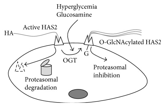
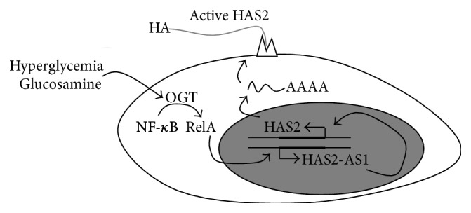
Cellular microenvironment plays a critical role in several pathologies including atherosclerosis. Hyaluronan (HA) content often reflects the progression of this disease in promoting vessel thickening and cell migration. HA synthesis is regulated by several factors, including the phosphorylation of HA synthase 2 (HAS2) and other covalent modifications including ubiquitination and O-GlcNAcylation. Substrate availability is important in HA synthesis control. Specific drugs reducing the UDP precursors are able to reduce HA synthesis whereas the hexosamine biosynthetic pathway (HBP) increases the concentration of HA precursor UDP-N-acetylglucosamine (UDP-GlcNAc) leading to an increase of HA synthesis. The flux through the HBP in the regulation of HA biosynthesis in human aortic vascular smooth muscle cells (VSMCs) was reported as a critical aspect. In fact, inhibiting O-GlcNAcylation reduced HA production whereas increased O-GlcNAcylation augmented HA secretion. Additionally, O-GlcNAcylation regulates HAS2 gene expression resulting in accumulation of its mRNA after induction of O-GlcNAcylation with glucosamine treatments. The oxidized LDLs, the most common molecules related to atherosclerosis outcome and progression, are also able to induce a strong HA synthesis when they are in contact with vascular cells. In this review, we present recent described mechanisms involved in HA synthesis regulation and their role in atherosclerosis outcome and development.


Figures

Figure 1
Figure 1
Schematic representation of the regulation of HA synthesis in ECs and the effects on immune cell-EC adhesion. Through their receptors, proinflammatory signals (i.e., cytokines) trigger NF-κB pathway that regulates both HAS2 and CD44 (and other adhesive molecules such as ICAM-1, E-selectin, VCAM-1, and MHC class I genes) [38]. HAS2 synthesizes high molecular weight HA that interacts with CD44 present on both ECs and immune cells (i.e., leukocytes) in the “sandwich model,” which drives immune cells to adhere to ECs contributing to inflammation. Gray circle represents the nucleus.
Figure 2
Figure 2
Schematic representation of the regulation of HA synthesis by AMPK in SMCs. Through the action of compounds such as AICAR, metformin, and resveratrol or by sensing ATP : AMP ratio or by the action of AMPK upstream kinases (AMPKK), AMPK phosphorylates HAS2 threonine 110 residue inhibiting HAS2 activity and reducing the HA production.
Figure 3
Figure 3
Schematic representation of the regulation of HA synthesis by OGT in SMCs. In normal conditions HAS2 in plasma membrane is active but can be rapidly degraded in a 26 S proteasome dependent manner. In hyperglycemic condition or after glucosamine treatments, OGT catalyzes the O-GlcNAcylation of HAS2 serine 221 residue, which greatly stabilizes HAS2 favoring HA production.
Figure 4
Figure 4
Schematic representation of the regulation of HAS2 expression by OGT in SMCs. In normal conditions, basal HAS2 and HAS2-AS1 expression are allowed. After the induction of O-GlcNAcylation (due to hyperglycemia or after glucosamine treatments), the NF-κB subunit RelA can be modified with O-GlcNAc by OGT. In the cell nucleus, glycosylated RelA can activate HAS2-AS1 transcription, which, in turn, changes chromatin structure around the HAS2 promoter (probably altering chromatin signature) favoring HAS2 expression leading to HA accumulation.
Figure 5
Figure 5
Schematic representation of HA metabolism in SMCs loaded with oxidized LDL (oxLDL). Nontoxic concentrations of oxLDL are driven inside the SMCs by the upregulation of the scavenger receptor LOX-1. Accumulation of oxLDL leads to ER stress with overexpression of the UPR factors CHOP and GRP78 as well as activation of the LXR sterol sensor system. One or both systems induce the overexpression in the nucleus of several genes: LOX-1, HAS2, and HAS3. The HASs are active on the plasma membrane where they synthesize HA that interacts with CD44 present on immune cells, driving their adhesion, which contributes to the inflammatory status of the atherosclerotic lesion.

References

    1. Roger V. L., Go A. S., Lloyd-Jones D. M., et al. Heart disease and stroke statistics—2012 update: a report from the American Heart Association. Circulation. 2012;125(1):e2–e220. doi: 10.1161/cir.0b013e31823ac046. - DOI - PMC - PubMed
    1. Vanhoutte P. M. Endothelial dysfunction—the first step toward coronary arteriosclerosis. Circulation Journal. 2009;73(4):595–601. doi: 10.1253/circj.cj-08-1169. - DOI - PubMed
    1. Merrilees M. J., Beaumont B. W., Braun K. R., et al. Neointima formed by arterial smooth muscle cells expressing versican variant V3 is resistant to lipid and macrophage accumulation. Arteriosclerosis, Thrombosis, and Vascular Biology. 2011;31(6):1309–1316. doi: 10.1161/ATVBAHA.111.225573. - DOI - PMC - PubMed
    1. Messner B., Bernhard D. Smoking and cardiovascular disease: mechanisms of endothelial dysfunction and early atherogenesis. Arteriosclerosis, Thrombosis, and Vascular Biology. 2014;34(3):509–515. doi: 10.1161/atvbaha.113.300156. - DOI - PubMed
    1. Badimon L., Vilahur G. LDL-cholesterol versus HDL-cholesterol in the atherosclerotic plaque: inflammatory resolution versus thrombotic chaos. Annals of the New York Academy of Sciences. 2012;1254(1):18–32. doi: 10.1111/j.1749-6632.2012.06480.x. - DOI - PubMed