Review
. 2015:2015:834893.
doi: 10.1155/2015/834893. Epub 2015 Sep 10.

Roles of Proteoglycans and Glycosaminoglycans in Wound Healing and Fibrosis

Affiliations
Review

Roles of Proteoglycans and Glycosaminoglycans in Wound Healing and Fibrosis

Shibnath Ghatak et al. Int J Cell Biol. 2015.

Abstract

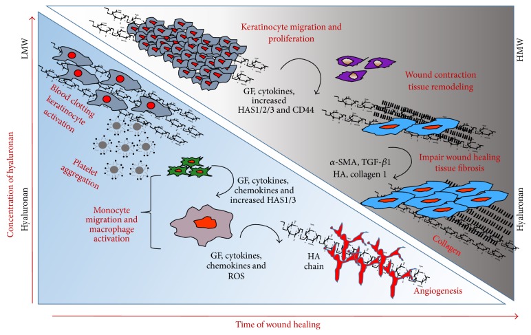
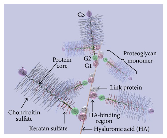
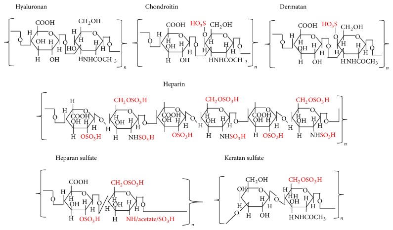
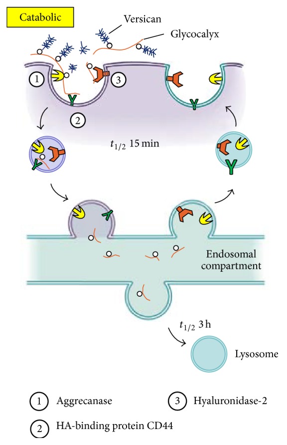
A wound is a type of injury that damages living tissues. In this review, we will be referring mainly to healing responses in the organs including skin and the lungs. Fibrosis is a process of dysregulated extracellular matrix (ECM) production that leads to a dense and functionally abnormal connective tissue compartment (dermis). In tissues such as the skin, the repair of the dermis after wounding requires not only the fibroblasts that produce the ECM molecules, but also the overlying epithelial layer (keratinocytes), the endothelial cells, and smooth muscle cells of the blood vessel and white blood cells such as neutrophils and macrophages, which together orchestrate the cytokine-mediated signaling and paracrine interactions that are required to regulate the proper extent and timing of the repair process. This review will focus on the importance of extracellular molecules in the microenvironment, primarily the proteoglycans and glycosaminoglycan hyaluronan, and their roles in wound healing. First, we will briefly summarize the physiological, cellular, and biochemical elements of wound healing, including the importance of cytokine cross-talk between cell types. Second, we will discuss the role of proteoglycans and hyaluronan in regulating these processes. Finally, approaches that utilize these concepts as potential therapies for fibrosis are discussed.


Figures

Figure 1
Figure 1
Schematic presentation of changes in hyaluronan synthesis/molecular size and cellular events and matrix events during the course of wound healing and fibrosis. Many of the biological processes mediated by HA are crucial for wound healing and fibrosis. After injury, wound healing follows a tightly regulated sequence of events. These phases are inflammation, granulation tissue formation, proliferation, reepithelization, and remodelling. In the early phases, high molecular HA is degraded by reactive oxygen species from activated granulocytes and by hyaluronidases secreted from platelets. Then monocytes secrete inflammatory mediators, which attract additional inflammatory cells. Keratinocytes become activated to migrate, proliferate, and to synthesize HA. As a result the LMW degradation products are active inducers of angiogenesis and inflammation. At later stages the interim matrix becomes supplemented with newly synthesized HMW HA, which contributes to tissue remodelling. During repetitive injury, the repairing processes are hindered, and the keratinocytes, the endothelial cells, and smooth muscle cells of the blood vessel, neutrophils, and macrophages together orchestrate the increased cytokine-mediated signaling and augment HA-CD44 signaling and excess collagen production that results in fibrosis.
Figure 2
Figure 2
Diagram of part of an aggrecan aggregate. G1, G2, and G3 are globular, folded regions of the central core protein. Proteoglycan aggrecan showing the noncovalent binding of proteoglycan to HA with the link proteins.
Figure 3
Figure 3
Structures of repeating disaccharides of glycosaminoglycans.
Figure 4
Figure 4
Model for catabolism of pericellular hyaluronan glycocalyx matrices (adapted from [86] with the permission from Dr. Hascall).
Figure 5
Figure 5
Structure, binding domains, and interactions of CD44 (Adapted from [7]). The ectodomain of CD44 contains HA-binding motifs and can contain chondroitin sulfate or heparan sulfate chains that can affect its HA-binding capacity and enable its interactions with growth factors and growth factor receptors, and its interaction with matrix metalloproteinases (MMPs). Transmembrane and cytoplasmic domains undergo multiple posttranslational modifications, including palmitoylation and phosphorylation on cysteine and serine residues, respectively, promoting the binding of proteins with crucial functions in cytoskeletal organization and signaling. ErbB2: epidermal growth factor receptor-2; ERM: ezrin–radixin–moesin; FGF: fibroblast growth factor; HGF: hepatocyte growth factor; IQGAP1: IQ motif containing GTPase activating protein 1; MAPK: mitogen-activated protein kinase; PDGFR: platelet-derived growth factor receptor; PI3K: phosphoinositide 3-kinase; TGFR: transforming growth factor receptor; VEGF: vascular endothelial growth factor.

References

    1. Menke N. B., Ward K. R., Witten T. M., Bonchev D. G., Diegelmann R. F. Impaired wound healing. Clinics in Dermatology. 2007;25(1):19–25. doi: 10.1016/j.clindermatol.2006.12.005. - DOI - PubMed
    1. Bissell M. J., Hall H. G., Parry G. How does the extracellular matrix direct gene expression? Journal of Theoretical Biology. 1982;99(1):31–68. doi: 10.1016/0022-5193(82)90388-5. - DOI - PubMed
    1. Hynes R. O. Integrins: bidirectional, allosteric signaling machines. Cell. 2002;110(6):673–687. doi: 10.1016/s0092-8674(02)00971-6. - DOI - PubMed
    1. Ponta H., Sherman L., Herrlich P. A. CD44: from adhesion molecules to signalling regulators. Nature Reviews Molecular Cell Biology. 2003;4(1):33–45. doi: 10.1038/nrm1004. - DOI - PubMed
    1. Sionov R. V., Naor D. Hyaluronan-independent lodgment of CD44+ lymphoma cells in lymphoid organs. International Journal of Cancer. 1997;71(3):462–469. doi: 10.1002/(sici)1097-0215(19970502)71:3lt;462::aid-ijc26>3.0.co;2-g. - DOI - PubMed