. 2015 Aug 31:5:733-40.
doi: 10.1016/j.fob.2015.08.010. eCollection 2015.

MicroRNAs downregulated in neuropathic pain regulate MeCP2 and BDNF related to pain sensitivity

Affiliations

MicroRNAs downregulated in neuropathic pain regulate MeCP2 and BDNF related to pain sensitivity

Melissa T Manners et al. FEBS Open Bio. .

Abstract

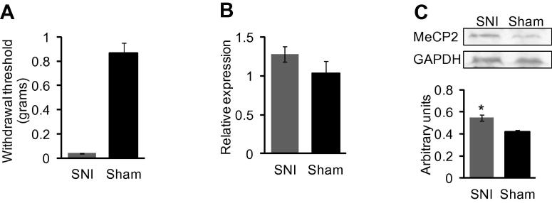
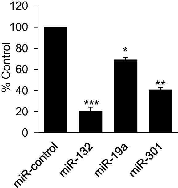
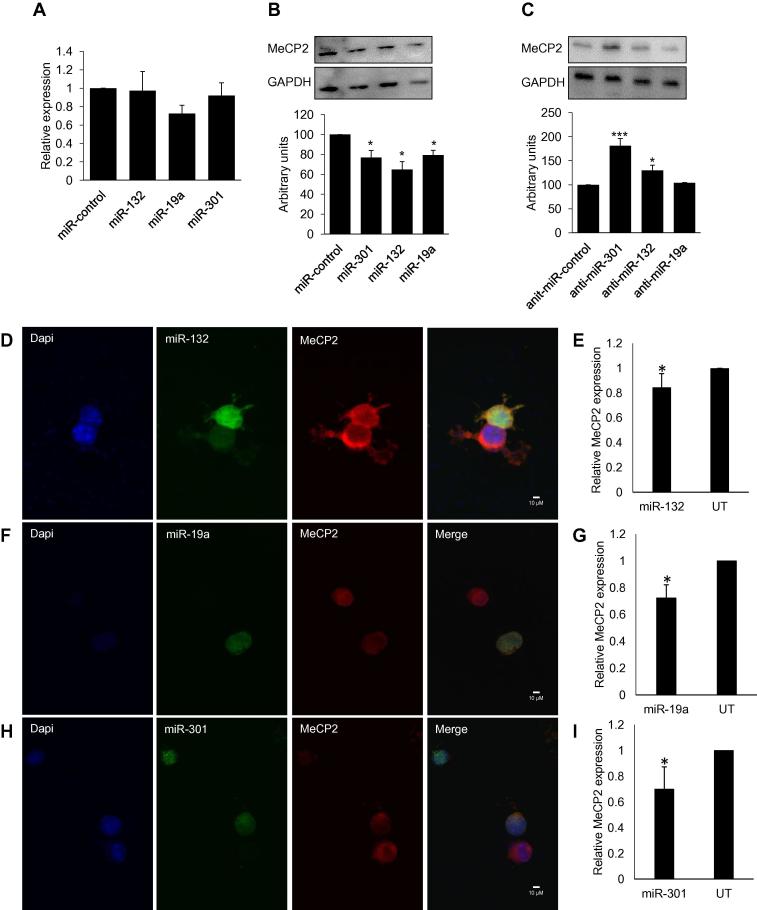
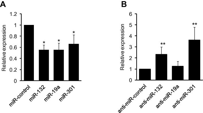
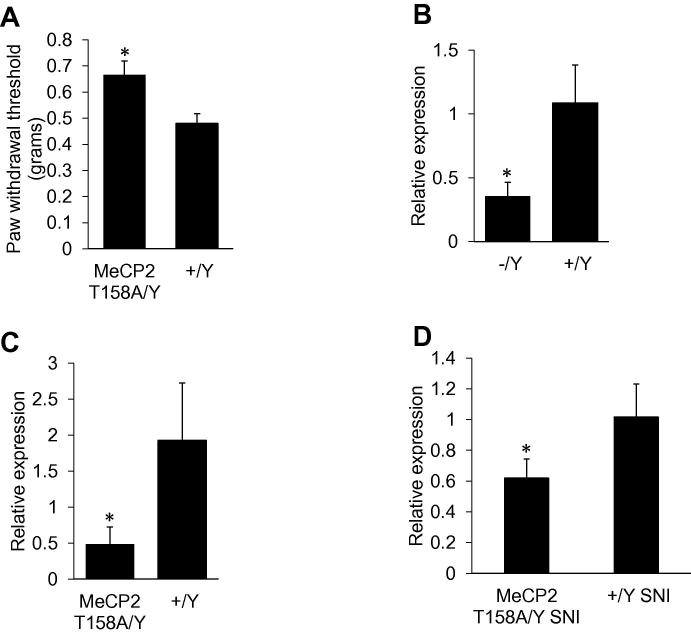
Nerve injury induces chronic pain and dysregulation of microRNAs in dorsal root ganglia (DRG). Several downregulated microRNAs are predicted to target Mecp2. MECP2 mutations cause Rett syndrome and these patients report decreased pain perception. We confirmed MeCP2 upregulation in DRG following nerve injury and repression of MeCP2 by miRNAs in vitro. MeCP2 regulates brain-derived neurotrophic factor (BDNF) and downregulation of MeCP2 by microRNAs decreased Bdnf in vitro. MeCP2 T158A mice exhibited reduced mechanical sensitivity and Mecp2-null and MeCP2 T158A mice have decreased Bdnf in DRG. MeCP2-mediated regulation of Bdnf in the DRG could contribute to altered pain sensitivity.

Keywords: +/Y, male wild-type littermate control for either MeCP2 T158A knock in mouse or Mecp2-null mouse; 3′UTR, three prime untranslated region; ATF3, activating transcription factor 3; BDNF; BDNF, brain derived neurotrophic factor; CFA, complete Freund’s adjuvant; DRG, dorsal root ganglia; L4/L5, 4th or 5th lumbar vertebra; MeCP2; MeCP2 T158A/Y, male MeCP2 T158A knock in mouse; MeCP2, methyl-CpG-binding protein 2; Neuropathic pain; RTT, Rett syndrome; SNI, spared nerve injury; T158A, threonine 158 conversion to alanine; TrkB, tropomyosin receptor kinase B; miRNA; −/Y, male Mecp2-null mouse.


Figures

Fig. 1
Fig. 1
MeCP2 expression in DRG 4 weeks after SNI or sham surgery in 12 weeks old mice. (A) von Frey filaments used to assess paw withdrawal threshold confirmed that mice were hypersensitive 4 weeks post SNI surgery (n = 10). Significance was determined using Mann–Whitney U test p value <0.001. (B) Taqman analysis of Mecp2 mRNA showed a trend toward increased expression that was not significant in SNI model compared to sham control (n = 7). Gapdh was used as the normalizer. (C) Western blot analysis of MeCP2 expression in DRG showed a significant increase in SNI model, determined using Student t-test, p value *<0.05. Tissue from 9 animals was pooled into 3 samples (n = 3). Protein expression was normalized to GAPDH.
Fig. 2
Fig. 2
miRNA binding to MeCP2 3′UTR. Luciferase assay showing miR-132, miR-19a, and miR-301 binding to the 3′UTR of Mecp2. The luciferase activity was measured 24 h after miRNA transfection. The data expressed as percentage of control is the average of 3 independent experiments. Statistically significant difference from control was calculated using one way ANOVA and Student t-test, p value **<0.01, ***<0.001.
Fig. 3
Fig. 3
Regulation of MeCP2 by miRNAs. (A) Taqman analysis of Mecp2 in Neuro-2a cells 48 h after miRNA transfection showed no significant reduction in mRNA (n = 3). (B) Western blot analysis after transfection of Neuro-2a cells with miR-301, miR-132 and miR-19a significantly reduced MeCP2 protein (n = 3). (C) MeCP2 expression increased when cells were transfected with anti-miR (miRNA inhibitors) for miR-301 and miR-132; (n = 3, representative blot shown). (D and E) Immunocytochemisty and quantification for MeCP2 in Neuro-2a cells transfected with miR-132. (F and G) Immunocytochemistry and quantification for MeCP2 in Neuro-2a cells transfected with miR-19a. (H and I) Immunocytochemistry and quantification for MeCP2 in Neuro-2a cells transfected with miR-301. MeCP2 levels were normalized to DAPI staining of the nucleus and the graph represents MeCP2 expression relative to untransfected cells. Representative images are shown, n = 3. Significance was determined using Student t-test, p value *<0.05, **<0.001 ***<0.001.
Fig. 4
Fig. 4
MeCP2 mediated changes in Bdnf transcripts. (A) miRNA-mediated reduction of MeCP2 expression decreased Bdnf mRNA in Neuro-2a cells 48 h after miRNA transfection as determined by Taqman analysis. (B) Antagomirs, inhibitors of endogenous miRNAs, increased Bdnf mRNA. Gapdh was used as the normalizer, n = 3. Significance determined using Student t-test, p value *<0.05, **<0.001.
Fig. 5
Fig. 5
MeCP2 T158A knockin mice have decreased mechanical sensitivity and lower Bdnf. (A) Mechanical sensitivity measured by von Frey filaments showed an increase in withdrawal threshold in 12 weeks old MeCP2 T158A mice, indicating hyposensitivity compared to wild-type littermate controls (n = 5 for MeCP2 T158A mice and n = 7 for wild-type littermate). Statistically significant difference was determined using the Mann–Whitney U test; there was a significant reduction in withdrawal threshold in the ipsilateral paw p value *<0.02. (B) DRG from Mecp2-null mice and (C) MeCP2 T158A mice has significantly lower Bdnf mRNA compared to wild-type littermates, (n = 6). (D) Bdnf was also lower in MeCP2 T158A mice compared to wild-type littermates after SNI surgery. Significance determined using Student t-test, p value *<0.05 (n = 3 for both MeCP2 T158A mice and wild-type littermate).

Similar articles

Cited by

References

    1. Gereau R.W., Sluka K.A., Maixner W., Savage S.R., Price T.J., Murinson B.B., Sullivan M.D., Fillingim R.B. A pain research agenda for the 21st century. J. Pain. 2014;15:1203–1214. - PMC - PubMed
    1. von Hehn C.A., Baron R., Woolf C.J. Deconstructing the neuropathic pain phenotype to reveal neural mechanisms. Neuron. 2012;73:638–652. - PMC - PubMed
    1. Basbaum A.I., Bautista D.M., Scherrer G., Julius D. Cellular and molecular mechanisms of pain. Cell. 2009;139:267–284. - PMC - PubMed
    1. Mogil J.S., Davis K.D., Derbyshire S.W. The necessity of animal models in pain research. Pain. 2010;151:12–17. - PubMed
    1. von Schack D. Dynamic changes in the microRNA expression profile reveal multiple regulatory mechanisms in the spinal nerve ligation model of neuropathic pain. PLoS ONE. 2011;6:e17670. - PMC - PubMed

LinkOut - more resources