. 2015:2015:305716.
doi: 10.1155/2015/305716. Epub 2015 Sep 10.

Efficient Mitochondrial Genome Editing by CRISPR/Cas9

Affiliations

Efficient Mitochondrial Genome Editing by CRISPR/Cas9

Areum Jo et al. Biomed Res Int. 2015.

Abstract

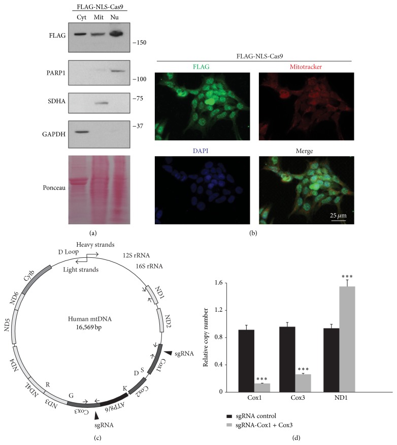
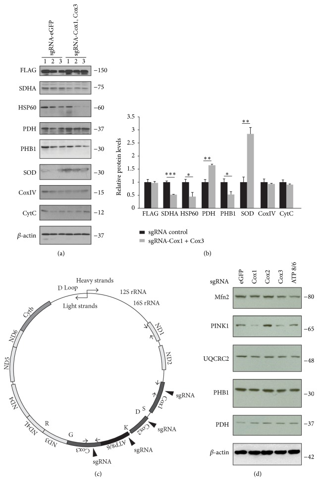
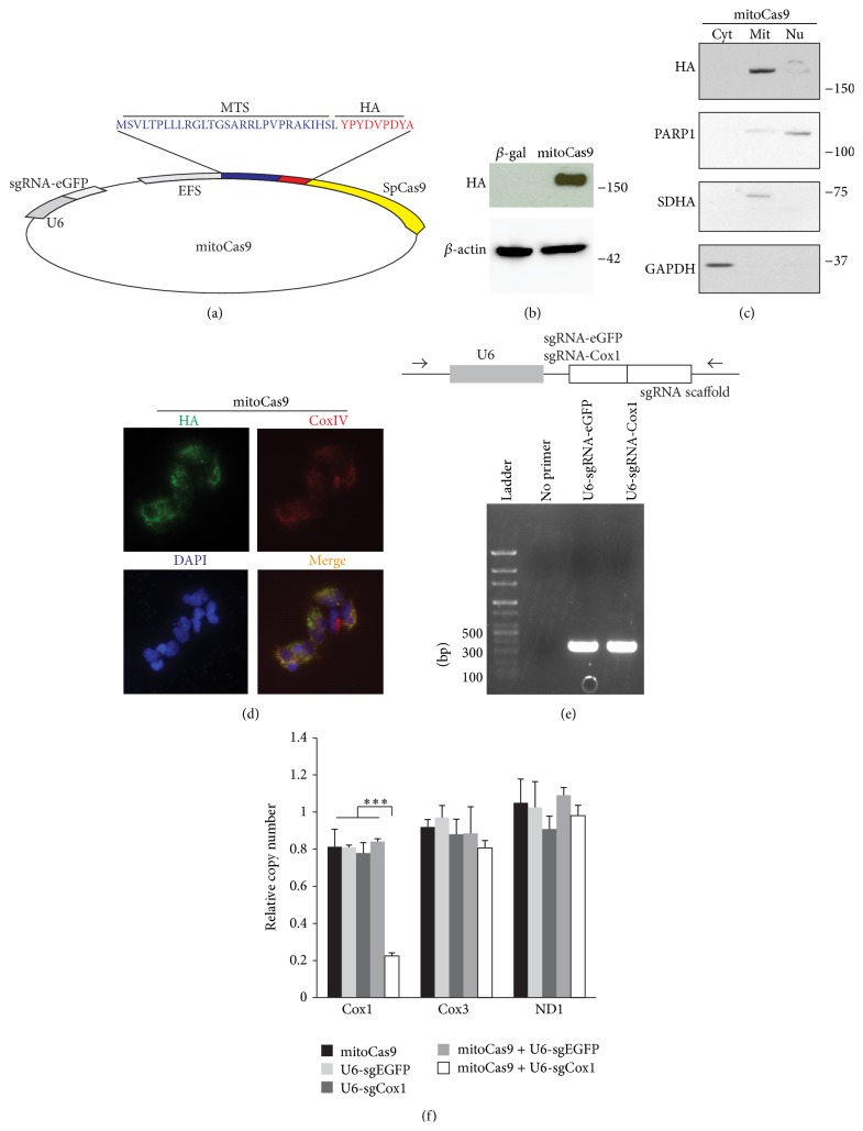
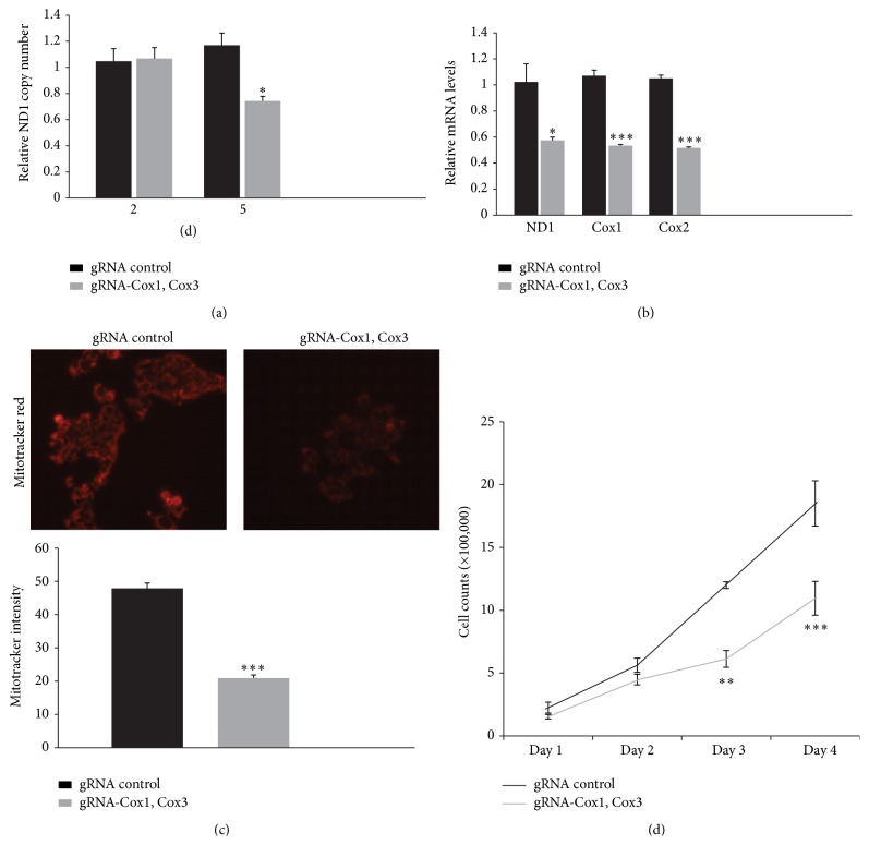
The Clustered Regularly Interspaced Short Palindromic Repeats (CRISPR)/Cas9 system has been widely used for nuclear DNA editing to generate mutations or correct specific disease alleles. Despite its flexible application, it has not been determined if CRISPR/Cas9, originally identified as a bacterial defense system against virus, can be targeted to mitochondria for mtDNA editing. Here, we show that regular FLAG-Cas9 can localize to mitochondria to edit mitochondrial DNA with sgRNAs targeting specific loci of the mitochondrial genome. Expression of FLAG-Cas9 together with gRNA targeting Cox1 and Cox3 leads to cleavage of the specific mtDNA loci. In addition, we observed disruption of mitochondrial protein homeostasis following mtDNA truncation or cleavage by CRISPR/Cas9. To overcome nonspecific distribution of FLAG-Cas9, we also created a mitochondria-targeted Cas9 (mitoCas9). This new version of Cas9 localizes only to mitochondria; together with expression of gRNA targeting mtDNA, there is specific cleavage of mtDNA. MitoCas9-induced reduction of mtDNA and its transcription leads to mitochondrial membrane potential disruption and cell growth inhibition. This mitoCas9 could be applied to edit mtDNA together with gRNA expression vectors without affecting genomic DNA. In this brief study, we demonstrate that mtDNA editing is possible using CRISPR/Cas9. Moreover, our development of mitoCas9 with specific localization to the mitochondria should facilitate its application for mitochondrial genome editing.


Figures

Figure 1
Figure 1
FLAG-NLS-Cas9 localizes to mitochondria. (a) Subcellular localization of FLAG-Cas9 assessed in the cytosolic (Cyt), mitochondrial (Mit), and nuclear (Nu) fractions of HEK-293T cells transfected with lentiCRISPR-sgRNA-eGFP#2 and monitored by Western blot. Glyceraldehyde 3-phosphate dehydrogenase (GAPDH) served as a cytosolic marker, poly(ADP-ribose) polymerase 1 (PARP1) served as a nuclear marker, and succinate dehydrogenase complex subunit A (SDHA) served as a mitochondrial marker. Ponceau staining of the blotted nitrocellulose membrane was presented in the bottom lane to visualize relative protein loading amounts. (b) Immunofluorescence images of FLAG-NLS-Cas9 demonstrating its localization to nucleus, cytoplasm, and mitochondria. HEK-293T cells were immunostained with mouse antibody to FLAG (green) 24 hours following transient transfection with FLAG-NLS-Cas9 construct. Mitochondria or nucleus was stained with mitotracker Red or DAPI, respectively. (c) Illustration of human mitochondrial DNA (mtDNA). Simplified view of mtDNA is presented to depict regions encoding peptides or tRNA for indicated amino acids. Filled triangles indicate sites targeted by gRNAs against Cox1 or Cox3. Arrows indicate primer annealing sites used for real-time PCR of Cox1 or Cox3 regions. (d) Quantification of copy numbers for Cox1, Cox3, and ND1 regions of mtDNA extracted from HEK-293T cells transiently transfected with lentiCRISPR-sgRNA-Cox1 and lentiCRISPR-sgRNA-Cox3 or lentiCRISPR-sgRNA-eGFP#2 as a control, determined by real-time quantitative PCR using primers listed in Table 2. GAPDH was used as an internal loading control (n = 3 per group). Quantified data (d) are expressed as mean ± s.e.m., ∗∗∗ P < 0.001, unpaired two-tailed Student's t-test.
Figure 2
Figure 2
Alterations in mitochondria-associated proteins following CRISPR/Cas9-mediated mtDNA editing. (a) Mitochondrial proteins in HEK-293T cells after CRISPR/Cas9-mediated cleavage of mtDNA at Cox1 and Cox3 loci as determined by Western blots using indicated antibodies. β-actin was used as a loading control. (b) Quantification of mitochondria proteins in HEK-293T cells transfected with lentiCRISPR-sgRNA-Cox1 + Cox3 or lentiCRISPR-sgRNA-eGFP#2 control as shown in panel (a) normalized to β-actin. (c) Illustration of human mtDNA. Specific loci targeted by lentiCRISPR-sgRNAs (Cox1, Cox2, Cox3, and ATP8/6) are indicated with filled triangles. (d) Representative Western blots showing differential alteration of mitochondrial proteins following cleavage of specific mtDNA loci mediated by indicated sgRNAs in HEK-293T cells. Quantified data (b) are expressed as mean ± s.e.m., P < 0.05, ∗∗ P < 0.01, and ∗∗∗ P < 0.001, unpaired two-tailed Student's t-test.
Figure 3
Figure 3
Construction of mitochondria-targeted MTS-HA-Cas9. (a) Schematic illustration of mitochondria-targeting Cas9 (mitoCas9). Mitochondria-targeting sequence (MTS) and HA tag information are presented. (b) Expression of mitoCas9 in HEK-293T cells transiently transfected with MTS-HA-Cas9 construct determined by Western blots using HA antibody. β-actin was used as a loading control. (c) Subcellular localization of MTS-HA-Cas9 assessed in the cytosolic (Cyt), mitochondrial (Mit), and nuclear (Nu) fractions of HEK-293T cells transfected with lentiCRISPR-sgRNA-eGFP#2 and monitored by Western blot. GAPDH served as a cytosolic marker, PARP1 served as a nuclear marker, and SDHA served as a mitochondrial marker. (d) Representative immunofluorescence microscopic image of MTS-HA-Cas9 (HA, green) and CoxIV (red) subcellular distributions in HEK-293T cells transfected with MTS-HA-Cas9 construct. The merged image (yellow, right panel) shows colocalization of mitoCas9 and CoxIV. (e) Representative gel images of the PCR product of hU6-sgRMA-eGFP and hU6-sgRNA-Cox1 which were purified by gel extraction (bottom panel). Schematics of primers and lentiCRISPR-sgRNA templates used to amplify U6 promoter and respective sgRNA components for transfection (upper panel). (f) Quantification of copy numbers for Cox1, Cox3, and ND1 regions of mtDNA extracted from HEK-293T cells transiently transfected with indicated constructs (mitoCas9 is a plasmid, while U6-sgRNAs are PCR product.), determined by real-time quantitative PCR using primers listed in Table 2. GAPDH was used as an internal loading control (n = 3 per group). Quantified data (b) are expressed as mean ± s.e.m., ∗∗∗ P < 0.001, analysis of variance (ANOVA) test followed by Student-Newman-Keuls post hoc analysis.
Figure 4
Figure 4
MitoCas9-induced mtDNA damage leads to mitochondria dysfunction. (a) Quantification of copy numbers for ND1 region of mtDNA extracted from HEK-293T cells transiently transfected with indicated constructs (gRNA control, mitoCas9 and U6-sgRNA to eGFP; gRNA Cox1, Cox3, mitoCas9, and U6-sgRNA to Cox1 and Cox3), determined by real-time quantitative PCR using primers listed in Table 2 at the indicated time points (2 days and 5 days after transfection). GAPDH was used as an internal loading control (n = 3 per group). (b) Quantification of messenger RNAs for ND1, Cox1, and Cox2 genes in HEK-293T cells 5 days following transient transfection with lentiCRISPR-sgRNA-Cox1 + Cox3 or lentiCRISPR-sgRNA-eGFP#2 control determined by real-time PCR and normalized to GAPDH (n = 3 per group). (c) Representative images of mitotracker Red staining for functional mitochondria in HEK-293T cells transfected with the indicated constructs (upper panel). Quantification of relative mitotracker Red staining intensities in two groups was shown in the bottom panel (n = 50 mitochondria from three independent cultures per group). (d) Cell counts demonstrating proliferation rate of the HEK-293T cells following cleavage of specific mtDNA loci (n = 6 individual measurements per group). Days indicate the time intervals proceeded after cell seeding following 5 days of transient transfectin. Quantified data are expressed as mean ± s.e.m., P < 0.05, ∗∗ P < 0.01, and ∗∗∗ P < 0.001, unpaired two-tailed Student's t-test.

Similar articles

Cited by

References

    1. Bratic I., Trifunovic A. Mitochondrial energy metabolism and ageing. Biochimica et Biophysica Acta. 2010;1797(6-7):961–967. doi: 10.1016/j.bbabio.2010.01.004. - DOI - PubMed
    1. Carafoli E. Historical review: mitochondria and calcium: ups and downs of an unusual relationship. Trends in Biochemical Sciences. 2003;28(4):175–181. doi: 10.1016/s0968-0004(03)00053-7. - DOI - PubMed
    1. Tait S. W. G., Green D. R. Mitochondria and cell death: outer membrane permeabilization and beyond. Nature Reviews Molecular Cell Biology. 2010;11(9):621–632. doi: 10.1038/nrm2952. - DOI - PubMed
    1. Taanman J.-W. The mitochondrial genome: structure, transcription, translation and replication. Biochimica et Biophysica Acta—Bioenergetics. 1999;1410(2):103–123. doi: 10.1016/s0005-2728(98)00161-3. - DOI - PubMed
    1. Filosto M., Scarpelli M., Cotelli M. S., et al. The role of mitochondria in neurodegenerative diseases. Journal of Neurology. 2011;258(10):1763–1774. doi: 10.1007/s00415-011-6104-z. - DOI - PubMed

Publication types