. 2015 Dec;8(12):1643-50.
doi: 10.1242/dmm.021394. Epub 2015 Oct 8.

CLARITY and PACT-based imaging of adult zebrafish and mouse for whole-animal analysis of infections

Affiliations

CLARITY and PACT-based imaging of adult zebrafish and mouse for whole-animal analysis of infections

Mark R Cronan et al. Dis Model Mech. 2015 Dec.

Abstract

Visualization of infection and the associated host response has been challenging in adult vertebrates. Owing to their transparency, zebrafish larvae have been used to directly observe infection in vivo; however, such larvae have not yet developed a functional adaptive immune system. Cells involved in adaptive immunity mature later and have therefore been difficult to access optically in intact animals. Thus, the study of many aspects of vertebrate infection requires dissection of adult organs or ex vivo isolation of immune cells. Recently, CLARITY and PACT (passive clarity technique) methodologies have enabled clearing and direct visualization of dissected organs. Here, we show that these techniques can be applied to image host-pathogen interactions directly in whole animals. CLARITY and PACT-based clearing of whole adult zebrafish and Mycobacterium tuberculosis-infected mouse lungs enables imaging of mycobacterial granulomas deep within tissue to a depth of more than 1 mm. Using established transgenic lines, we were able to image normal and pathogenic structures and their surrounding host context at high resolution. We identified the three-dimensional organization of granuloma-associated angiogenesis, an important feature of mycobacterial infection, and characterized the induction of the cytokine tumor necrosis factor (TNF) within the granuloma using an established fluorescent reporter line. We observed heterogeneity in TNF induction within granuloma macrophages, consistent with an evolving view of the tuberculous granuloma as a non-uniform, heterogeneous structure. Broad application of this technique will enable new understanding of host-pathogen interactions in situ.

Keywords: CLARITY; Imaging; Infection; Mouse; Mycobacteria; PACT; Tuberculosis; Zebrafish.


Conflict of interest statement

Competing interests

The authors declare no competing or financial interests.

Figures
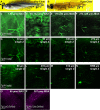
Fig. 1.
Fig. 1.
CLARITY protocol adapted for imaging intact zebrafish adults. (A,B) Zebrafish adult pre- and post-clearing. (C-N) Whole-body CLARITY allows imaging of fluorescent vasculature deep within the adult body. Blood vessels labeled by Tg(flk1:egfp) are imaged from the surface to deep within while maintaining fluorescence intensity and resolution. (C-J) Individual images obtained using an SP8 confocal microscope, ranging from the animal's scales (surface=1 µm) to 335 µm deep. (C-F) Z-stack is split into ∼50 µm maximum projection images to allow for clear views of vascular structures. (G-J) Individual Z planes from stack. (K-N) Individual images from two-photon microscopy ranging from the animal's scales (surface=1 µm) to >1 mm deep. 1-µm optical sections are shown. Scale bars: 100 µm. Single Z frames were exported and gamma adjusted in FIJI/ImageJ for increased visibility, with all gamma adjustments applied uniformly across all images. (O,P) CLARITY techniques are compatible with red fluorescent proteins. (O) Neutrophils within the epidermis were imaged using the transgenic line Tg(LysC:DsRed). 90-μm maximum projection image. (P) Neuronal cell bodies within the eye of cleared zebrafish in a 381-μm maximum projection from the transgenic line Tg(Xla.Tubb:DsRed).
Fig. 2.
Fig. 2.
PACT protocol maintains integrity of blood vessels. (A-D) Blood vessels in gills labeled by Tg(flk1:egfp) in a zebrafish adult pre-clearing (immediately post-euthanasia) and the same animal (E-H) post-clearing. Increasing numbers of gill blood vessels are visible deeper within the body following clearing (G compared to C). Single Z frames (D,H, and insets) demonstrate that fine structures are unaffected by the clearing process. (I,J) Blood vessels in the tail of the same animal pre- and post-clearing. (K,L) Large blood vessels in the mid-trunk of the same animal pre- and post-clearing. Scale bars: 120 µm. Single Z frames were exported and gamma adjusted in FIJI/ImageJ for increased visibility, with all gamma adjustments applied uniformly across all images.
Fig. 3.
Fig. 3.
Granuloma-induced angiogenesis and mycobacterial granulomas within intact adult zebrafish. (A-G) Tg(flk1:egfp) labels blood vessels in green; magenta labels cerulean-tagged Mycobacterium marinum (Mm-cerulean), which lies within a granuloma. Imaging commences at ∼400 µm below the scales; stack is ∼104 µm deep. Scale bar: 100 µm. Single Z frames were exported and gamma adjusted in FIJI/ImageJ for increased visibility, with all gamma adjustments applied uniformly across all images.
Fig. 4.
Fig. 4.
Fluorescent mycobacteria and cytokine induction can be imaged deep within intact adult zebrafish. (A) The TNF reporter (green) is expressed throughout a large granuloma (tdTomato-expressing M. marinum: magenta) in the TgBAC(tnf:GFP) line. Imaging begins 256 µm below the scales and the stack (A) is ∼100 µm deep; individual Z planes from the stack (B-D) reveal TNF reporter intensity throughout the granuloma. (E-H) TNF reporter expression in the granuloma is not dependent on infection status of individual cells: (F) an infected cell that does not express the TNF reporter; (G) an infected cell expressing the TNF reporter; (H) an uninfected cell expressing the TNF reporter. Scale bars: 50 µm (A-E); 5 µm (F-H). Single Z frames were exported and gamma adjusted in FIJI/ImageJ for increased visibility, with all gamma adjustments applied uniformly across all images.
Fig. 5.
Fig. 5.
Infection with fluorescent M.tuberculosis is visible throughout PACT-cleared mouse lungs. (A,B) Mouse lungs pre- and post-clearing. (C-F) Stack through lung infected with approximately 350 CFU tdTomato-expressing M. tuberculosis at 28 dpi taken on Spinning Disc Confocal ranges from the top to bottom surface of a lung lobe (surface=1 µm) to 665 µm deep with 20× objective. (C) 665 µm maximum projection image. (D-F) Individual Z planes from stack. (G) 411 µm maximum projection image taken with 10× objective; (H) detail of boxed region from G taken with 20× objective; 251 µm maximum projection image. Scale bars: 100 µm. Single Z frames were exported and gamma adjusted in FIJI/ImageJ for increased visibility, with all gamma adjustments applied uniformly across all images.

References

    1. Adams K. N., Takaki K., Connolly L. E., Wiedenhoft H., Winglee K., Humbert O., Edelstein P. H., Cosma C. L. and Ramakrishnan L. (2011). Drug tolerance in replicating mycobacteria mediated by a macrophage-induced efflux mechanism. Cell 145, 39-53. 10.1016/j.cell.2011.02.022 - DOI - PMC - PubMed
    1. Auer T. O., Duroure K., De Cian A., Concordet J.-P. and Del Bene F. (2014). Highly efficient CRISPR/Cas9-mediated knock-in in zebrafish by homology-independent DNA repair. Genome Res. 24, 142-153. 10.1101/gr.161638.113 - DOI - PMC - PubMed
    1. Brothers K. M., Newman Z. R. and Wheeler R. T. (2011). Live imaging of disseminated candidiasis in zebrafish reveals role of phagocyte oxidase in limiting filamentous growth. Eukaryot. Cell 10, 932-944. 10.1128/EC.05005-11 - DOI - PMC - PubMed
    1. Chang N., Sun C., Gao L., Zhu D., Xu X., Zhu X., Xiong J.-W. and Xi J. J. (2013). Genome editing with RNA-guided Cas9 nuclease in zebrafish embryos. Cell Res. 23, 465-472. 10.1038/cr.2013.45 - DOI - PMC - PubMed
    1. Chung K., Wallace J., Kim S.-Y., Kalyanasundaram S., Andalman A. S., Davidson T. J., Mirzabekov J. J., Zalocusky K. A., Mattis J., Denisin A. K. et al. (2013). Structural and molecular interrogation of intact biological systems. Nature 497, 332-337. 10.1038/nature12107 - DOI - PMC - PubMed

Publication types

Substances

LinkOut - more resources