. 2015 Dec;8(12):1579-89.
doi: 10.1242/dmm.019968. Epub 2015 Oct 8.

Regulation of mitochondrial oxidative stress by β-arrestins in cultured human cardiac fibroblasts

Affiliations

Regulation of mitochondrial oxidative stress by β-arrestins in cultured human cardiac fibroblasts

Jennifer L Philip et al. Dis Model Mech. 2015 Dec.

Abstract

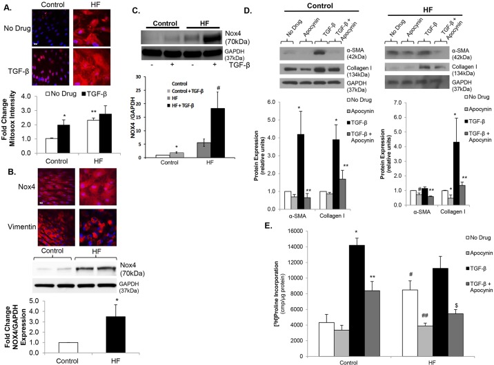
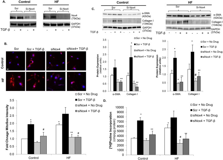
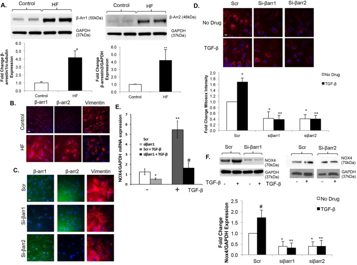
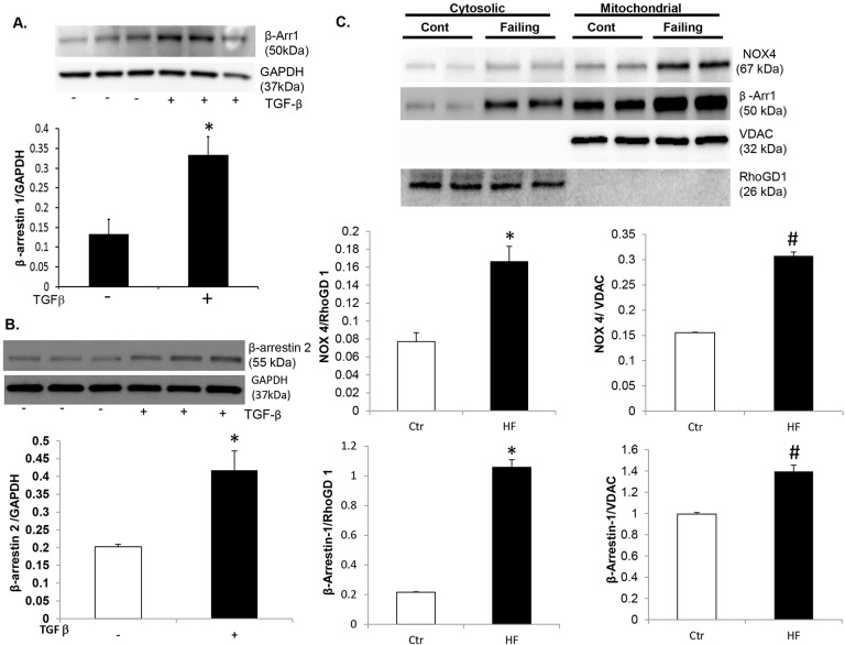
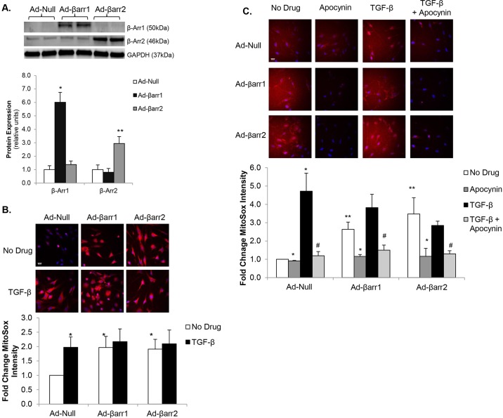
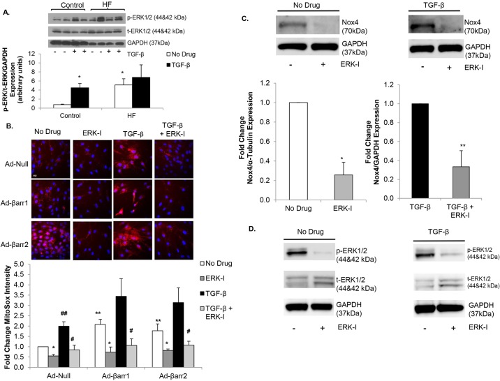
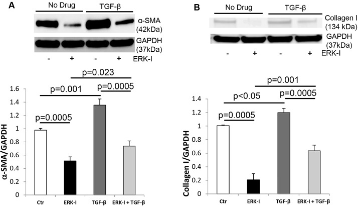
Oxidative stress in cardiac fibroblasts (CFs) promotes transformation to myofibroblasts and collagen synthesis leading to myocardial fibrosis, a precursor to heart failure (HF). NADPH oxidase 4 (Nox4) is a major source of cardiac reactive oxygen species (ROS); however, mechanisms of Nox4 regulation are unclear. β-arrestins are scaffold proteins that signal in G-protein-dependent and -independent pathways; for example, in ERK activation. We hypothesize that β-arrestins regulate oxidative stress in a Nox4-dependent manner and increase fibrosis in HF. CFs were isolated from normal and failing adult human left ventricles. Mitochondrial ROS/superoxide production was quantitated using MitoSox. β-arrestin and Nox4 expressions were manipulated using adenoviral overexpression or short interfering RNA (siRNA)-mediated knockdown. Mitochondrial oxidative stress and Nox4 expression in CFs were significantly increased in HF. Nox4 knockdown resulted in inhibition of mitochondrial superoxide production and decreased basal and TGF-β-stimulated collagen and α-SMA expression. CF β-arrestin expression was upregulated fourfold in HF. β-arrestin knockdown in failing CFs decreased ROS and Nox4 expression by 50%. β-arrestin overexpression in normal CFs increased mitochondrial superoxide production twofold. These effects were prevented by inhibition of either Nox or ERK. Upregulation of Nox4 seemed to be a primary mechanism for increased ROS production in failing CFs, which stimulates collagen deposition. β-arrestin expression was upregulated in HF and plays an important and newly identified role in regulating mitochondrial superoxide production via Nox4. The mechanism for this effect seems to be ERK-mediated. Targeted inhibition of β-arrestins in CFs might decrease oxidative stress as well as pathological cardiac fibrosis.

Keywords: Cardiac fibroblast; Collagen; Heart failure; Myocardial fibrosis; NADPH oxidase; Oxidative stress; β-arrestin.


Conflict of interest statement

Competing interests

The authors declare no competing or financial interests.

Figures

Fig. 1.
Fig. 1.
Mitochondrial superoxide production and Nox4 are upregulated in failing cardiac fibroblasts. (A) Confocal images (upper panel) of control and heart failure (HF) cardiac fibroblasts (CFs) stained with MitoSOX (red) under basal conditions (No Drug) vs TGF-β stimulation. Nuclei are stained blue with Hoechst 33342. Fluorescence quantitation shown below demonstrates an over twofold increase in mitochondrial oxidative stress in control CFs in response to TGF-β. *P<0.03 vs Control+No Drug; **P<0.005 vs Control+No Drug; n=3-4 in all groups. Scale bar: 20 μm. (B) Confocal images (upper panel) of Nox4 stained red with Alexa-Fluor-594 dye are show in in HF versus control CFs. Nuclei are stained blue with DAPI. Representative immunoblot (middle panel) showing Nox4 expression in HF versus control CFs. GAPDH was used as a loading control. Densitometric analysis (lower panel) demonstrates an over fourfold increase in Nox4 expression in HF. *P<0.05 vs Control; n=4 in all groups. This membrane was stripped, re-probed and quantitated in Fig. 3A. Scale bar: 20 μm. (C) Representative immunoblot (upper panel) showing Nox4 expression under basal conditions and following TGF-β stimulation in control and HF CFs. GAPDH was used as a loading control. Densitometric analysis (lower panel) demonstrates increased Nox4 expression in response to TGF-β in both control and HF. *P<0.05 vs No Drug; #P<0.05 vs No Drug; n=5 in all groups. (D) Representative immunoblots (upper panels) showing the effects of apocynin on basal and TGF-β-stimulated α-SMA and collagen I expression in both control (left) and HF (right) CFs. GAPDH was used as a loading control. Densitometric analysis shown below. *P<0.02 vs No Drug, **P<0.03 vs TGF-β, #P<0.05 vs No Drug; n=3-4 for all groups. (E) Collagen synthesis in HF versus control CFs under basal conditions, TGF-β stimulation, apocynin treatment, and pre-treatment with apocynin prior to stimulation with TGF-β. *P<0.001 vs Control+No Drug; **P<0.002 vs Control+TGF-β; #P<0.003 vs Control+No Drug; ##P<0.01 vs HF+No Drug; $P<0.002 vs HF+TGF-β; n=3 in all groups.
Fig. 2.
Fig. 2.
Nox4 mediates myofibroblast transformation and collagen synthesis via increased oxidative stress. (A) Representative immunoblots showing knockdown of Nox4 expression in control (left panel) and heart failure (HF) cardiac fibroblasts (CFs) (right panel) with Nox4 siRNA (siNox4) vs scrambled control (Scr). GAPDH was used as a loading control. (B) Confocal images of CFs stained with MitoSOX (red) showing inhibition of mitochondrial oxidative stress with siNox4 in control and HF CFs. Nuclei are stained blue with Hoechst 33342. Fluorescence quantitation is shown below those images. *P<0.003 vs Control Scr+No Drug, **P<0.005 vs Scr+No Drug, #P<0.02 vs Scr+TGF-β; n=3-4 in all groups. Scale bar: 20 μm. (C) Representative immunoblots (upper panels) showing the effects of Nox4 knockdown on basal and TGF-β-stimulated α-SMA and collagen I expression in control (left panel) and HF (right panel) CFs. GAPDH was used as a loading control. Densitometric analysis is shown below. *P<0.05 vs Scr+No Drug, **P<0.01 vs Scr+TGF-β, #P=0.05 vs Scr+No Drug; n=3-4 in all groups. (D) Basal and TGF-β-stimulated collagen synthesis in control and HF CFs following transfection with siNox4 or Scr control. *P<0.05 vs Control Scr+No Drug, **P<0.005 vs Scr+TGF-β; #P<0.01 vs HF Scr+No Drug; n=3-4 in all groups.
Fig. 3.
Fig. 3.
β-arrestins regulate mitochondrial superoxide production and Nox4 expression in cardiac fibroblasts. (A) The blot presented in Fig. 1B was stripped and re-probed (upper panels) for β-arrestin-1 (β-Arr1) (left) and β-arrestin-2 (β-Arr2) (right), showing expression in control vs heart failure (HF) cardiac fibroblasts (CFs) with the same GAPDH loading controls. Densitometric analysis (lower panels) demonstrates an over fourfold increase in β-arrestin-1 and β-arrestin-2 expression in HF. *P<0.01 vs Control; **P<0.04 vs Control; n=4 for β-arrestin-1 expression groups, n=5 for β-arrestin-2 expression groups. (B) Confocal images of β-arrestin-1, β-arrestin-2 and vimentin stained red with Alexa-Fluor-594 dye are shown in HF versus control CFs, demonstrating increased β-arrestin expression in HF. Nuclei are stained blue with DAPI. Scale bar: 20 μm. (C) Confocal images showing β-arrestin-1 and β-arrestin-2 stained green with FITC, and vimentin stained red with Alexa-Fluor-594 dye in HF CFs following siRNA knockdown of β-arrestin-1 (siβarr1), β-arrestin-2 (siβarr2) or scrambled control (Scr), demonstrating successful siRNA knockdown of β-arrestins. Nuclei are stained blue with DAPI. Scale bar: 20 μm. (D) Confocal images (upper panels) of failing CFs stained with MitoSOX (red) following siRNA knockdown of β-arrestin-1 (siβarr1), β-arrestin2 (siβarr2) or scrambled control (Scr). Nuclei are stained blue with Hoechst 33342. Fluorescence quantitation is shown below. *P<0.03 vs Scr+No Drug; **P<0.001 vs Scr+TGF-β; n=3 for all groups. Scale bar: 20 μm. (E) Quantitative PCR showing basal and TGF-β-stimulated Nox4 mRNA expression following siβarr1 vs Scr control. Values are normalized to GAPDH. *P<0.03 vs siβarr1, **P<0.05 vs Scr+TGF-β, #P<0.05 vs siβarr1+TGF-β; n=3 for all groups. (F) Representative immunoblot (upper panels) showing basal and TGF-β-stimulated Nox4 expression following siβarr1 or siβarr2 vs Scr control. GAPDH was used as a loading control. Densitometric analysis is shown below. *P<0.05 vs Scr+No Drug, **P<0.001 vs Scr+TGF-β, #P<0.05 vs Scr+No Drug; n=3 in each group.
Fig. 4.
Fig. 4.
β-arrestin is robustly expressed in fibroblast mitochondria. (A) Representative immunoblot (upper panel) showing β-arrestin-1 expression under basal conditions and following TGF-β stimulation in control human cardiac fibroblasts (CFs). GAPDH was used as a loading control. Densitometric analysis (lower panel) demonstrates increased β-arrestin-1 expression in response to TGF-β in CFs. *P<0.004 vs No Drug. n=3 in each group. (B) Representative immunoblot (upper panel) showing β-arrestin-2 expression under basal conditions and following TGF-β stimulation in control human CFs. GAPDH was used as a loading control. Densitometric analysis (lower panel) demonstrates increased β-arrestin-2 expression in response to TGF-β in CFs. *P<0.01 vs No Drug. n=3 in each group. (C) Cytosolic and mitochondrial fractionations were isolated from control and failing human CFs. Representative immunoblot (upper panel) showing Nox4 and β-arrestin-1 expression in cytosolic and mitochondrial fractions of normal (Cont) and failing human CFs. This blot was stripped and re-probed for RhoGD1 and VDAC, which were used as cytosolic and mitochondrial loading controls, respectively. Densitometric analysis (middle panels) demonstrates increased Nox4 expression in both cytosolic (left) and mitochondrial (right) fractions in the failing (HF) CFs. *P=0.02 vs control in the cytosolic fraction and #P=0.001 vs control in the mitochondrial fraction. Densitometric analysis (lower panels) demonstrates increased β-arrestin-1 expression in both cytosolic and mitochondrial fractions. *P=0.003 vs control in the cytosolic fraction and #P=0.01 vs control in the mitochondrial fraction.
Fig. 5.
Fig. 5.
β-arrestin overexpression increases mitochondrial oxidative stress. (A) Representative immunoblot (upper panel) showing β-arrestin-1 (β-Arr1) and β-arrestin2 (β-Arr2) expression following transfection with adenoviruses overexpressing β-arrestin-1 (Ad-βarr1), β-arrestin-2 (Ad-βarr2), or control null virus (Ad-Null). GAPDH was used as a loading control. Densitometric analysis of β-arrestin-1 and β-arrestin-2 normalized to GAPDH expression is shown below. *P<0.001 vs Ad-Null and vs Ad-βarr2, **P<0.03 vs Ad-Null and vs Ad-βarr1; n=3-4 in all groups. (B) Confocal images (upper panel) of control cardiac fibroblasts (CFs) stained with MitoSOX (red) following transfection with Ad-βarr1, Ad-βarr2 or Ad-Null. Nuclei are stained blue with Hoechst 33342. Fluorescence quantitation is shown below. *P<0.05 vs Ad-Null+No Drug; n=3-5 in all groups. (C) Confocal images of transfected CFs stained with MitoSOX (red) showing mitochondrial oxidative stress under basal conditions, TGF-β stimulation, apocynin treatment, and pre-treatment with apocynin prior to stimulation with TGF-β (TGF-β+Apocynin). Nuclei are stained blue with Hoechst 33342. Fluorescence quantitation is shown below. *P<0.02 vs No Drug; **P<0.03 vs Ad-Null+No Drug; #P<0.02 vs TGF-β; n=3 in each group. Scale bars: 20 μm.
Fig. 6.
Fig. 6.
β-arrestin-stimulated oxidative stress is mediated by ERK signaling. (A) Representative immunoblot (upper panel) showing phosphorylated ERK1/2 (p-ERK1/2) and total ERK1/2 (t-ERK1/2) expression under basal conditions versus TGF-β stimulation in control and heart failure (HF) cardiac fibroblasts (CFs). GAPDH was used as a loading control. Densitometric analysis is shown below. *P<0.05 vs Control+No Drug; n=3-6 in all groups. (B) Confocal images (upper panel) of control CFs stained with MitoSOX (red) following transfection with adenoviruses overexpressing β-arrestin-1 (Ad-βarr1), β-arrestin-2 (Ad-βarr2) or control null virus (Ad-Null) under basal conditions, treatment with ERK inhibitor PD98059 (ERK-I), TGF-β-stimulation, and pre-treatment with PD98059 prior to stimulation with TGF-β (TGF-β+ERK-I). Nuclei are stained blue with Hoechst 33342. Fluorescence quantitation is shown below. *P<0.05 vs No Drug; **P<0.04 vs Ad-Null+No Drug, #P<0.001 vs TGF-β, ##P=0.05 vs Ad-Null+No Drug; n=4 for all groups. Scale bar: 20 μm. (C) Representative immunoblots (upper panels) showing the effect of ERK inhibitor on basal (left panel) and TGF-β-stimulated (right panel) Nox4 expression in failing CFs. GAPDH was used as a loading control. Densitometric analysis is shown below. *P=0.01 vs No Drug, **P<0.03 vs TGF-β; n=3 in each group. (D) Representative immunoblots showing decreased basal (No Drug) (left panel) and TGF-β-stimulated (right panel) ERK1/2 phosphorylation following treatment with the ERK inhibitor (ERK-I) PD98059. GAPDH was used as a loading control.
Fig. 7.
Fig. 7.
NOX4/oxidative-stress-induced myofibroblast transformation is mediated by ERK signaling. (A) Representative immunoblot (upper panel) showing α-SMA expression in control cardiac fibroblasts (CFs) under basal conditions (Ctr), TGF-β-stimulation, treatment with the ERK inhibitor PD98059 (ERK-I), and pre-treatment with PD98059 prior to stimulation with TGF-β (TGF-β+ERK-I). GAPDH was used as a loading control. Densitometric analysis is shown below. n=3 for all groups. (B) Representative immunoblot (upper panel) showing collagen I expression in control CFs under basal conditions, TGF-β-stimulation, treatment with the ERK inhibitor PD98059 (ERK-I), and pre-treatment with PD98059 prior to stimulation with TGF-β (TGF-β+ERK-I). GAPDH was used as a loading control. Densitometric analysis is shown below. n=3 for all groups.

Similar articles

Cited by

References

    1. Ago T., Kuroda J., Pain J., Fu C., Li H. and Sadoshima J. (2010). Upregulation of Nox4 by hypertrophic stimuli promotes apoptosis and mitochondrial dysfunction in cardiac myocytes. Circ. Res. 106, 1253-1264. 10.1161/CIRCRESAHA.109.213116 - DOI - PMC - PubMed
    1. Al Ghouleh I., Khoo N. K. H., Knaus U. G., Griendling K. K., Touyz R. M., Thannickal V. J., Barchowsky A., Nauseef W. M., Kelley E. E., Bauer P. M. et al. (2011). Oxidases and peroxidases in cardiovascular and lung disease: new concepts in reactive oxygen species signaling. Free Radic. Biol. Med. 51, 1271-1288. 10.1016/j.freeradbiomed.2011.06.011 - DOI - PMC - PubMed
    1. Baudino T. A., Carver W., Giles W. and Borg T. K. (2006). Cardiac fibroblasts: friend or foe? Am. J. Physiol. Heart Circ. Physiol. 291, H1015-H1026. 10.1152/ajpheart.00023.2006 - DOI - PubMed
    1. Blanchette F., Rivard N., Rudd P., Grondin F., Attisano L. and Dubois C. M. (2001). Cross-talk between the p42/p44 MAP kinase and Smad pathways in transforming growth factor beta 1-induced furin gene transactivation. J. Biol. Chem. 276, 33986-33994. 10.1074/jbc.M100093200 - DOI - PubMed
    1. Bristow M. R., Ginsburg R., Minobe W., Cubicciotti R. S., Sageman W. S., Lurie K., Billingham M. E., Harrison D. C. and Stinson E. B. (1982). Decreased catecholamine sensitivity and beta-adrenergic-receptor density in failing human hearts. N. Engl. J. Med. 307, 205-211. 10.1056/NEJM198207223070401 - DOI - PubMed

Publication types

MeSH terms