. 2015 Oct 9:6:8546.
doi: 10.1038/ncomms9546.

Substrate specificity and transport mechanism of amino-acid transceptor Slimfast from Aedes aegypti

Affiliations

Substrate specificity and transport mechanism of amino-acid transceptor Slimfast from Aedes aegypti

Dmitri Y Boudko et al. Nat Commun. .

Abstract

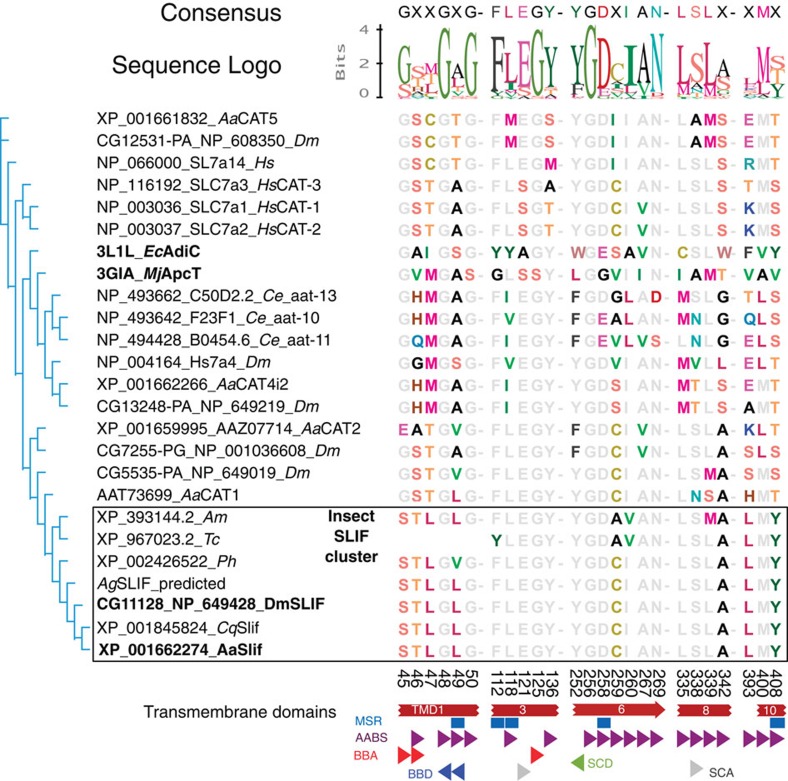
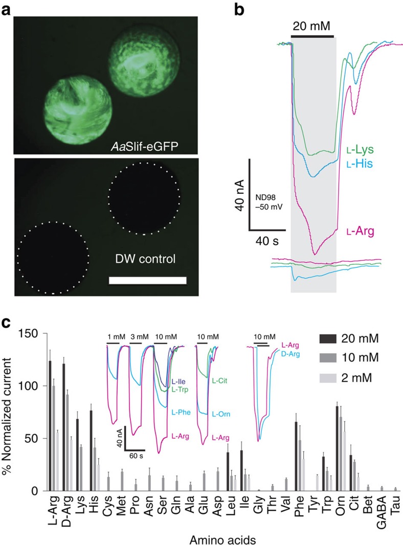
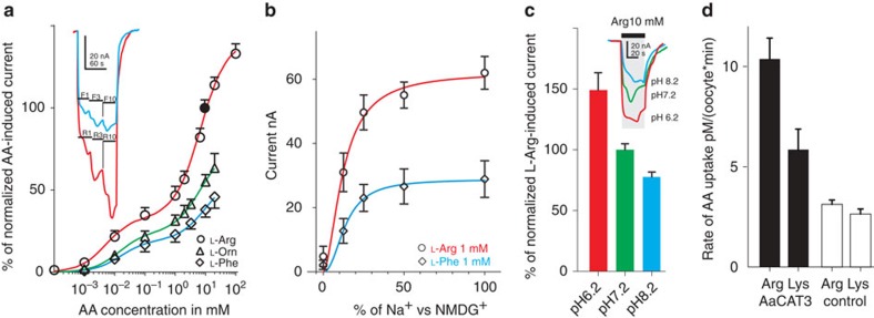
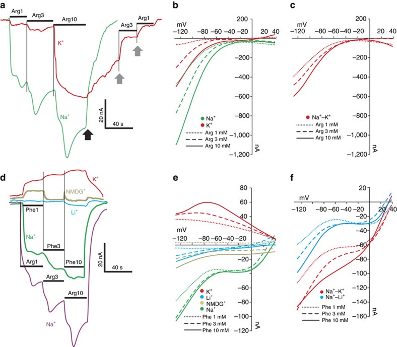
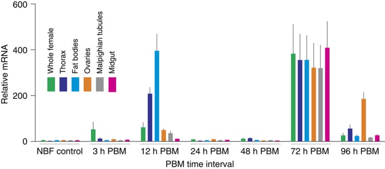
Anautogenous mosquitoes depend on vertebrate blood as nutrient source for their eggs. A highly efficient set of membrane transporters mediates the massive movement of nutrient amino acids between mosquito tissues after a blood meal. Here we report the characterization of the amino-acid transporter Slimfast (Slif) from the yellow-fever mosquito Aedes aegypti using codon-optimized heterologous expression. Slif is a well-known component of the target-of-rapamycin signalling pathway and fat body nutrient sensor, but its substrate specificity and transport mechanism were unknown. We found that Slif transports essential cationic and neutral amino acids with preference for arginine. It has an unusual dual-affinity mechanism with only the high affinity being Na(+) dependent. Tissue-specific expression and blood meal-dependent regulation of Slif are consistent with conveyance of essential amino acids from gut to fat body. Slif represents a novel transport system and type of transceptor for sensing and transporting essential amino acids during mosquito reproduction.


Figures

Figure 1
Figure 1. Bioinformatics analysis of the AaSlif substrate-binding motif (SBM).
Shown are a neighbour-joining consensus tree and an alignment of the putative SBM residues of selected CAT-SLC7 members. Putative Slif orthologues (outlined box) form a phylogenetic cluster with exactly one orthologue per selected insect genome and also show strong conservation of the SBM. The names of Drosophila and Aedes slif orthologues and their prokaryotic homologues used for the identification and alignment of SBMs are highlighted by bold font. Coloured and grey fonts depict variable and significantly conserved residues, respectively. The aligned numbers indicate the amino-acid positions in the AaSlif protein sequence. The partial SBM of the Tribolium Slif is due to its incomplete annotation. The coloured shapes depict specific putative components of the SBMs abbreviated as: AABS, amino acid-binding sites; BBA, backbone acceptor; BBD, backbone donor; MSR, mutation sensitive residues; SCA, side chain acceptor; SCD, side chain donor. Species abbreviations are: Aa, Aedes aegypti; Ce, Caenorhabditis elegans; Cq, Culex quinquefasciatus; Hs, Homo sapiens; Ph, Pediculus humanus; Tc, Tribolium castaneum.
Figure 2
Figure 2. Functional expression and AA-specificity profile of AaSlif.
(a) Functional expression of codon-optimized AaSlif-eGFP fusion protein showed bright fluorescence compared with control deionized water-injected oocytes (bottom panel, white dashed outlines indicate position of control oocytes that are almost invisible with identical settings of the GFP-specific filter cube (Nikon 49583; EN GFP LP: Excitation Hq 470/40, Dichroic 495EM, Emission 500LP) and camera sensitivity); scale bar represents 1 mm. (b) The expression of AaSlif correlates with large cationic AA-induced current responses. The coloured lines represent currents induced in the AaSlif-eGFP-expressing oocytes (top traces) versus control oocytes (bottom traces) after application of 20 mM of L-enantiomer of Lysine (K), Histidine (H) and Arginine (R). (c) AA specificity profile reconstructed from AA-induced currents. The bars are mean of percent normalized current+s.d. (n>2 oocytes/samples per point) at 20, 10 and 2 mM concentrations of AAs. Currents were recorded using two-electrode voltage clamp. The coloured traces show representative recordings of currents induced by three different concentration of L-Arg and L-Phe, as well as 10 mM of L-Trp, L-Ile, L-Arg, L-Orn and L-Cit. Oocytes were clamped to –50 mV holding voltage in a constant flow micro-chamber perfused at ∼2-chamber volumes per second with ND98 or indicated solutions of AAs in 98 Na+ buffer. Currents were measured considering a steady-state current value and normalized to the mean of 10 mM L-Arg (=100%) response in each oocyte for cross-oocyte comparison. Cit, citrulline; DW, deionized water; GABA, γ-aminobutyric acid; Orn, ornithine; Tau, taurine.
Figure 3
Figure 3. AaSlif substrate saturation kinetics, pH dependency and substrate specificity.
(a) Saturation kinetic graphs for L-Arg-, L-Phe- and L-Orn-induced currents. The insert shows the relative amplitudes of L-Arg and L-Phe-induced currents in AaSlif-expressing oocytes stimulated with three different concentrations of L-Arg and L-Phe. The data were normalized between different oocytes using mean value responses to 10 mM L-Arg (Filled circle point). Data are mean±s.d. for n>3. (b) Sodium saturation kinetics for 1 mM L-Arg- and L-Phe-induced currents. Data are mean±s.d. for n=3. (c) pH dependency of 10 mM L-Arg-induced currents. Bars are mean+s.d. for n=3 oocyte/samples, that are significantly different P>0.001; t-test. Insert shows a scaled superposition of typical L-Arg-induced currents induced upon sequential application of L-Arg at specified pH values. (d) Uptake of radiolabelled cationic AAs in AaSlif-expressing oocytes (black) versus control oocytes (white). Data are mean+s.d. for n=5 individual oocyte assays.
Figure 4
Figure 4. Cation dependency of AA-induced currents.
(a) Example of substrate-induced currents in an AaSlif-expressing oocyte after application of 1, 3 and 10 mM of L-Arg in 98 Na+ (green line) and 98 K+ (red line) buffers (containing identical concentrations of Cl and other ions). Black arrow indicates washing after L-Arg application with initial buffer saline. Grey arrows indicate accelerated recovery after re-application of 3 or 1 mM of L-Arg (for 98 K+ buffer only). (b) Background-subtracted I/V plots of AaSlif-expressing oocytes in 98 Na+ and 98 K+ buffers with three different concentrations of L-Arg (insert). (c) I/V plot calculated by subtraction of the current values measured in 98 K+ from those in 98 Na+. Results show no significant dependency of current on L-Arg concentration. (d) L-Phe-induced currents in four different buffers. Results show that substitution of Na+ with Li+ and NMDG+ ions reduced substrate-induced currents, whereas the substitution with K+ reversed the current. (e) I/V plots acquired from the same oocyte as in d with different cation compositions, as colour-coded in insert. (f) I/V plots calculated by subtraction of current values measured in 98 K+ and 98 Li+ buffers from those in 98 Na+ buffer.
Figure 5
Figure 5. AaSlif expression and regulation in selected mosquito tissues and organs.
The transcript levels were determined using quantitative PCR (qPCR). The data represent relative quantities of AaSlif transcript that were normalized with qPCR levels of ribosomal protein S7 (rpS7) mRNA in the same tissue sample set. Bars are means+s.e.m. for n=3 replicates collected from different sets of mosquitoes (analysis of variance followed by Tukey's HSD post hoc test). The RNA samples were isolated from whole body and various body parts and organs of adult female mosquitoes grouped as shown by the colour-coding insert. The horizontal scale indicates group-specific conditions: non-blood fed (NBF, control), 3, 12, 24, 48, 72 and 96 h post blood meal (PBM).

References

    1. Guedes R. et al.. Amino acids biosynthesis and nitrogen assimilation pathways: a great genomic deletion during eukaryotes evolution. BMC Genomics 12, S2 (2011). - PMC - PubMed
    1. Boudko D. Y. Molecular basis of essential amino acid transport from studies of insect nutrient amino acid transporters of the SLC6 family (NAT-SLC6). J. Insect. Physiol. 58, 433–449 (2012). - PMC - PubMed
    1. Boudko D. Y. In: Epithelial Transport Physiology Springer (2010).
    1. Attardo G. M., Hansen I. A. & Raikhel A. S. Nutritional regulation of vitellogenesis in mosquitoes: implications for anautogeny. Insect. Biochem. Mol. Biol. 35, 661–675 (2005). - PubMed
    1. Kokoza V. A. et al.. Transcriptional regulation of the mosquito vitellogenin gene via a blood meal-triggered cascade. Gene 274, 47–65 (2001). - PubMed

Publication types