. 2016 Jun;27(6):1678-88.
doi: 10.1681/ASN.2014090937. Epub 2015 Oct 8.

Cystinosin is a Component of the Vacuolar H+-ATPase-Ragulator-Rag Complex Controlling Mammalian Target of Rapamycin Complex 1 Signaling

Affiliations

Cystinosin is a Component of the Vacuolar H+-ATPase-Ragulator-Rag Complex Controlling Mammalian Target of Rapamycin Complex 1 Signaling

Zuzanna Andrzejewska et al. J Am Soc Nephrol. 2016 Jun.

Abstract

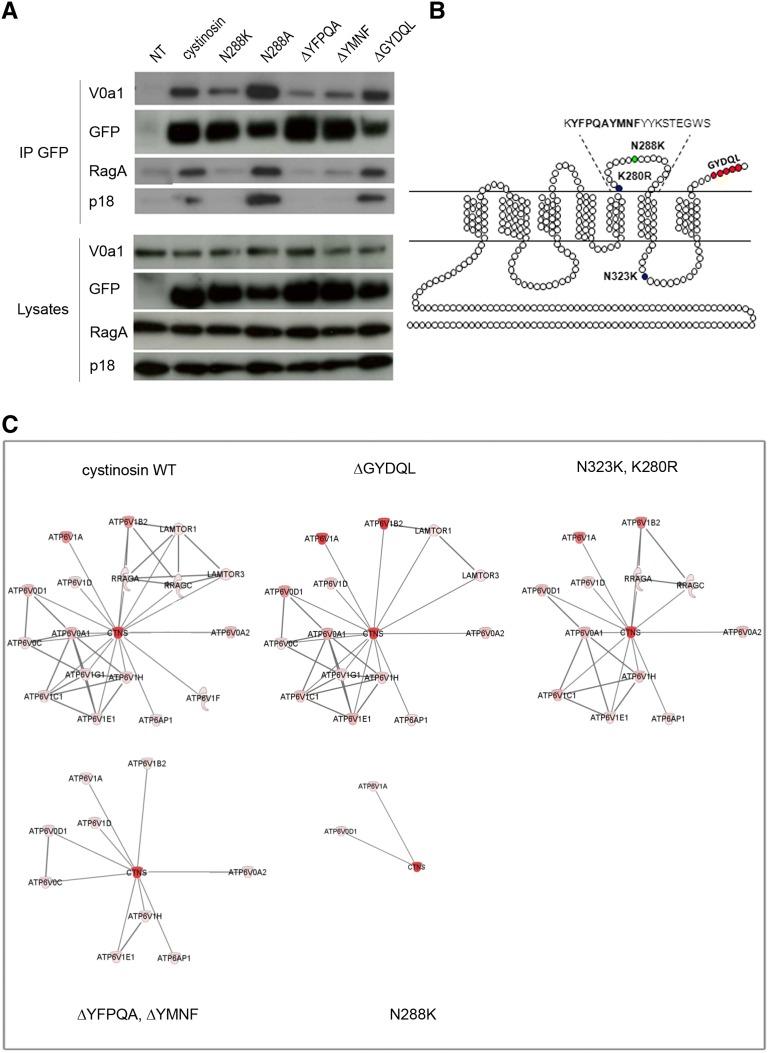
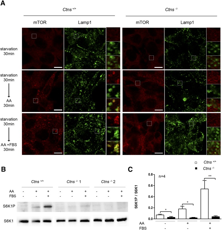
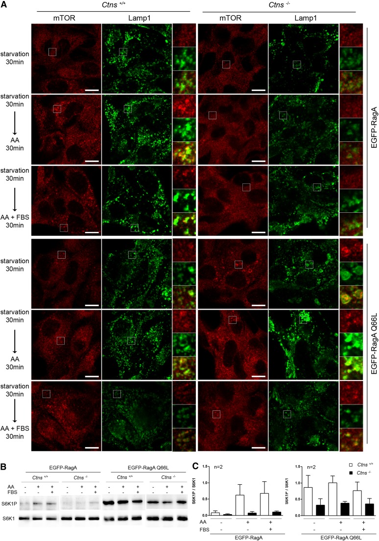
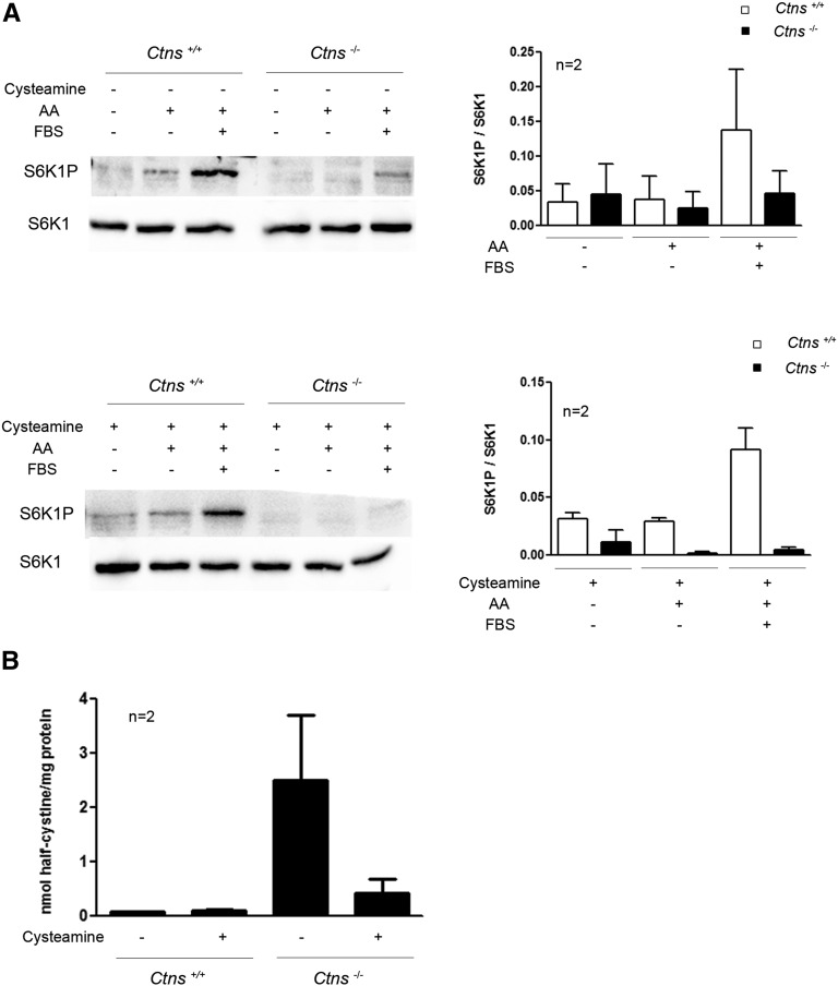
Cystinosis is a rare autosomal recessive storage disorder characterized by defective lysosomal efflux of cystine due to mutations in the CTNS gene encoding the lysosomal cystine transporter, cystinosin. Lysosomal cystine accumulation leads to crystal formation and functional impairment of multiple organs. Moreover, cystinosis is the most common inherited cause of renal Fanconi syndrome in children. Oral cysteamine therapy delays disease progression by reducing intracellular cystine levels. However, because cysteamine does not correct all complications of cystinosis, including Fanconi syndrome, we hypothesized that cystinosin could have novel roles in addition to transporting cystine out of the lysosome. By coimmunoprecipitation experiments and mass spectrometry, we found cystinosin interacts with almost all components of vacuolar H(+)-ATPase and the Ragulator complex and with the small GTPases Ras-related GTP-binding protein A (RagA) and RagC. Furthermore, the mammalian target of rapamycin complex 1 (mTORC1) pathway was downregulated in proximal tubular cell lines derived from Ctns(-/-) mice. Decrease of lysosomal cystine levels by cysteamine did not rescue mTORC1 activation in these cells, suggesting that the downregulation of mTORC1 is due to the absence of cystinosin rather than to the accumulation of cystine. Our results show a dual role for cystinosin as a cystine transporter and as a component of the mTORC1 pathway, and provide an explanation for the appearance of Fanconi syndrome in cystinosis. Furthermore, this study highlights the need to develop new treatments not dependent on lysosomal cystine depletion alone for this devastating disease.

Keywords: Fanconi syndrome; cystinosin; cystinosis; mTORC1.


Figures

Figure 1.
Figure 1.
Cystinosin interacts and colocalizes with components of the mTORC1 pathway. Lysates of 3T3 cells stably expressing cystinosin-EGFP or EGFP-CD63 were immunoprecipitated with (A) anti-GFP or (B) anti-RagC antibodies, and coimmunoprecipitated proteins were analyzed by Western blotting (n=1). IP, immunoprecipitates; NT, non transfected cells.
Figure 2.
Figure 2.
Mutations in the fifth inter-TM loop of cystinosin suppress interactions with the mTORC1 complex. (A) Lysates of 3T3 cells stably expressing WT cystinosin-EGFP and its mutated forms (N288K, N288A, ΔYFPQA, ΔYMNF, or ΔGYDQL) were immunoprecipitated with anti-GFP antibodies and coimmunoprecipitated proteins were analyzed by Western blotting. Representative blots from two independent experiments are shown. (B) Schematic representation of cystinosin indicating point mutations and deletions used in the study. (C) Representation of differential protein networks of WT or mutated cystinosin and its partners. The connections irradiating from cystinosin can represent both direct and indirect physical interactions between cystinosin and the proteins partners identified by co-IP. The connections between the proteins partners represent direct and indirect interactions established according to literature archived in Ingenuity Knowledge Database. The red color code reflects the abundance of the protein in the complex, estimated according to the number of identified peptides (brighter red = higher abundance). IP, immunoprecipitates; NT, non transfected cells.
Figure 3.
Figure 3.
The mTORC1 pathway is downregulated in Ctns−/− MPT cells. Ctns+/+ or Ctns−/− MPT cells were either AA/FBS-starved for 30 minutes, or starved and then allowed to recover in AA- or AA/FBS-containing medium for 30 minutes. (A) Cells were coimmunolabeled with mTOR and Lamp1 antibodies (confocal microscopy; scale bars = 10 µm); similar results were obtained for two Ctns−/− and two Ctns+/+ lines in three independent experiments. Zoomed areas and merge images are presented on the right of the panels to better appreciate the lysosomal or diffuse localization of mTOR in the various experimental conditions. (B) Total cell lysates were analyzed by Western blotting with anti-S6K1 and anti-S6K1P antibodies to evaluate the phosphorylation levels of the S6K1 protein. Ctns−/− 1 and 2 are two cell lines derived from different Ctns−/− mice. Representative blots from at least three independent experiments are shown. (C) Quantification of phosphorylation levels of S6K1 protein (each bar represents the mean±SEM from four independent experiments; *P<0.05; **P<0.01).
Figure 4.
Figure 4.
Cystinosin acts upstream of RagA in the mTORC1 signaling pathway. Ctns+/+ or Ctns−/− MPT cells stably expressing EGFP-RagA or its constitutively active form (EGFP-RagA Q66L) were either AA/FBS-starved for 30 minutes, or starved and then allowed to recover in medium containing AA or AA/FBS for 30 minutes. (A) Cells were coimmunolabeled for mTOR and Lamp1 (confocal microscopy; scale bars = 10 µm); similar results were obtained for two Ctns−/− and two Ctns+/+ lines in two independent experiments. Zoomed areas and merge images are presented on the right of the panels to better appreciate the lysosomal or diffuse localization of mTOR in the various experimental conditions. (B) Total cell lysates were analyzed by Western blotting with anti-S6K1 and anti-S6K1P antibodies. Representative blots from two independent experiments are shown. (C) Quantification of the phosphorylation levels of the S6K1 protein (each bar represents the mean±SEM from two independent experiments).
Figure 5.
Figure 5.
Cysteamine treatment has no effect on mTORC1 signaling in Ctns−/− MPT cells. (A) Ctns+/+ or Ctns−/− MPT cells were treated or not for 2 hours with 1 mM cysteamine hydrochloride and either AA/FBS-starved for 30 minutes, or starved and allowed to recover in medium containing AA or AA/FBS for 30 minutes. Total cell lysates were analyzed by Western blotting with anti-S6K1 and anti-S6K1P antibodies. Representative blots from two independent experiments and quantification of the phosphorylation levels of the S6K1 protein are shown (each bar represents the mean±SEM from two independent experiments). (B) Quantification of half-cystine levels in Ctns+/+ or Ctns−/− MPT cells before and after cysteamine treatment (each bar represents the mean±SEM from two independent experiments).

References

    1. Bar-Peled L, Schweitzer LD, Zoncu R, Sabatini DM: Ragulator is a GEF for the rag GTPases that signal amino acid levels to mTORC1. Cell 150: 1196–1208, 2012 - PMC - PubMed
    1. Sancak Y, Bar-Peled L, Zoncu R, Markhard AL, Nada S, Sabatini DM: Ragulator-Rag complex targets mTORC1 to the lysosomal surface and is necessary for its activation by amino acids. Cell 141: 290–303, 2010 - PMC - PubMed
    1. Laplante M, Sabatini DM: Regulation of mTORC1 and its impact on gene expression at a glance. J Cell Sci 126: 1713–1719, 2013 - PMC - PubMed
    1. Martina JA, Puertollano R: RRAG GTPases link nutrient availability to gene expression, autophagy and lysosomal biogenesis. Autophagy 9: 928–930, 2013 - PMC - PubMed
    1. Zoncu R, Bar-Peled L, Efeyan A, Wang S, Sancak Y, Sabatini DM: mTORC1 senses lysosomal amino acids through an inside-out mechanism that requires the vacuolar H(+)-ATPase. Science 334: 678–683, 2011 - PMC - PubMed

MeSH terms

Substances