Case Reports
. 2015 Oct 8:8:111.
doi: 10.1186/s13045-015-0213-6.

Increase of PD-L1 expressing B-precursor ALL cells in a patient resistant to the CD19/CD3-bispecific T cell engager antibody blinatumomab

Affiliations
Case Reports

Increase of PD-L1 expressing B-precursor ALL cells in a patient resistant to the CD19/CD3-bispecific T cell engager antibody blinatumomab

Thomas Köhnke et al. J Hematol Oncol. .

Abstract

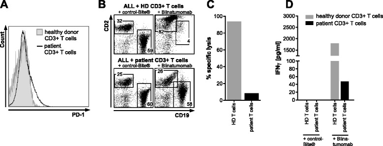
The bispecific T cell engager blinatumomab has shown encouraging clinical activity in B-precursor acute lymphoblastic leukemia (ALL). However, about half of relapsed/refractory patients do not respond to therapy. Here, we present the case of a 32-year-old male patient with refractory B-precursor ALL who was resistant to treatment with blinatumomab. Bone marrow immunohistochemistry revealed T cell infiltrates and an increase in programmed death-ligand 1 (PD-L1)-positive ALL cells as a potential immune escape mechanism. We were able to recapitulate the clinical observation in vitro by showing that blinatumomab was not able to mediate cytotoxicity of CD19-positive ALL cells using autologous T cells. In contrast, the addition of healthy donor T cells led to lysis of ALL cells.These results strongly encourage further systematic evaluation of checkpoint molecules in cases of blinatumomab treatment failure and might highlight a possible mechanism to overcome resistance to this otherwise highly effective treatment.


Figures

Fig. 1
Fig. 1
Increase of PD-1 and PD-L1 positivity after treatment with blinatumomab. a CD19 vs. CD34 expression of lymphoblasts detected by flow cytometry. Lymphoblasts showed homogenous expression of CD19 at baseline (pre-treatment) as well as after blinatumomab treatment (post-treatment). b Lymphocyte counts on peripheral blood during blinatumomab treatment. Lymphocyte counts decreased during blinatumomab treatment (1596/μl on day 0, 986/μl on day 7, 464/μl on day 14, 368/μl on day 21, and 217/μl on day 28). c Hematoxylin and eosin stain of paraffin embedded bone marrow core biopsy showing diffuse infiltration of immature progenitors at both time points (pre-treatment blast count 30 %, post-treatment 60 %). d Immunohistochemistry of paraffin embedded bone marrow core biopsy stained for CD3 showing spotted infiltration of CD3-positive T cells at baseline (5–10 %, pre-treatment) and diffuse infiltration after blinatumomab treatment (20–30 %, post-treatment). e Immunohistochemistry of paraffin embedded bone marrow core biopsy stained for PD-1 showing 5 % PD-1-positive cells at baseline (pre-treatment) vs. 15 % after blinatumomab treatment (post-treatment). f Immunohistochemistry of paraffin embedded bone marrow core biopsy stained for PD-L1 showing 2 % PD-L1-positive blasts at baseline (pre-treatment) vs. 40 % after blinatumomab treatment (post-treatment)
Fig. 2
Fig. 2
Decreased in vitro blinatumomab-mediated lysis of ALL blasts by patient CD3-positive T cells. a PD-1 expression on peripheral CD3-positive T cells was compared to PD-1 expression on healthy donor CD3-positive T cells showing no detectable difference. b The patient’s ALL blasts were cocultured either with healthy donor CD3-positive T cells (upper panels) or the patient’s own CD3-positive T cells (lower panels) with either control-Bite® (left panels) or blinatumomab (right panels) and analyzed by flow cytometry after 3 days. c Specific lysis was calculated as one minus the ratio of CD19-positive cells treated with blinatumomab and CD19-positive cells treated with control-Bite®. Healthy donor CD3-positive T cells showed efficient lysis of our patient’s ALL blasts (specific lysis 93.6 %) whereas CD3-positive T cells from our patient showed inefficient lysis of autologous ALL blasts (specific lysis 8.5 %). d IFN-γ concentration in cell culture supernatants were considerably lower for our patient’s T cells cocultured with his ALL blasts, whereas coculture of healthy donor T cells and ALL blasts from our patient led to considerable IFN-γ production. Cell cultures with control-Bite® did not show any IFN-γ production

References

    1. Suresh T, Lee LX, Joshi J, Barta SK. New antibody approaches to lymphoma therapy. J Hematol Oncol. 2014;7(1):58. doi: 10.1186/s13045-014-0058-4. - DOI - PMC - PubMed
    1. Wu J, Fu J, Zhang M, Liu D. Blinatumomab: a bispecific T cell engager (BiTE) antibody against CD19/CD3 for refractory acute lymphoid leukemia. J Hematol Oncol. 2015;8(1):104. doi: 10.1186/s13045-015-0195-4. - DOI - PMC - PubMed
    1. Zugmaier G, Klinger M, Schmidt M, Subklewe M. Clinical overview of anti-CD19 BiTE(®) and ex vivo data from anti-CD33 BiTE(®) as examples for retargeting T cells in hematologic malignancies. Mol Immunol. 2015;67(2 Pt A):58–66. doi: 10.1016/j.molimm.2015.02.033. - DOI - PubMed
    1. Topp MS, Gökbuget N, Stein AS, Zugmaier G, O'Brien S, Bargou RC, et al. Safety and activity of blinatumomab for adult patients with relapsed or refractory B-precursor acute lymphoblastic leukaemia: a multicentre, single-arm, phase 2 study. Lancet Oncol. 2014;16(1):57–66. doi: 10.1016/S1470-2045(14)71170-2. - DOI - PubMed
    1. Portell CA, Wenzell CM, Advani AS. Clinical and pharmacologic aspects of blinatumomab in the treatment of B-cell acute lymphoblastic leukemia. Clin Pharmacol. 2013;5(Suppl 1):5–11. - PMC - PubMed

Publication types

MeSH terms