. 2015 Oct 9:5:14959.
doi: 10.1038/srep14959.

Paradoxical effect of rapamycin on inflammatory stress-induced insulin resistance in vitro and in vivo

Affiliations

Paradoxical effect of rapamycin on inflammatory stress-induced insulin resistance in vitro and in vivo

Ping Yang et al. Sci Rep. .

Abstract

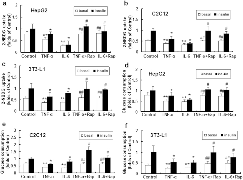
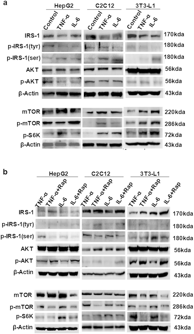
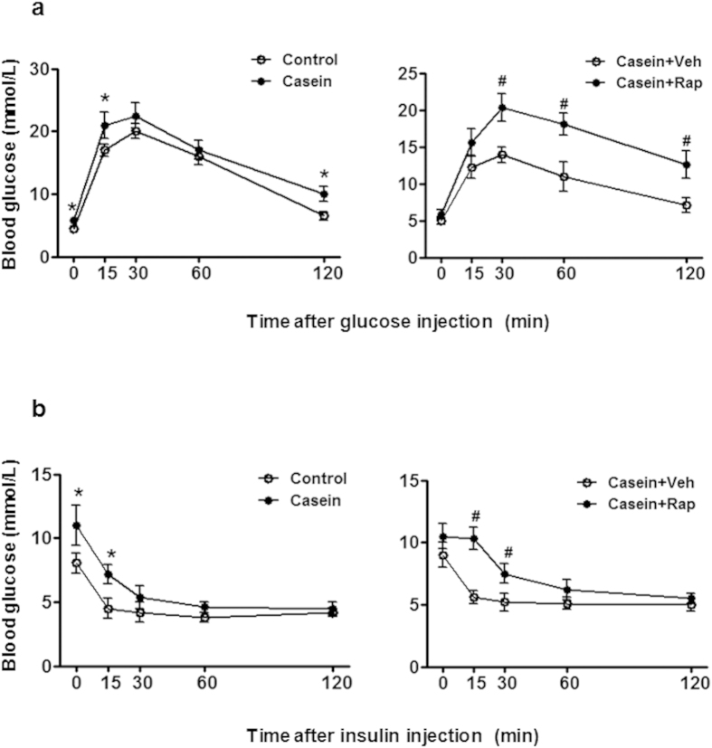
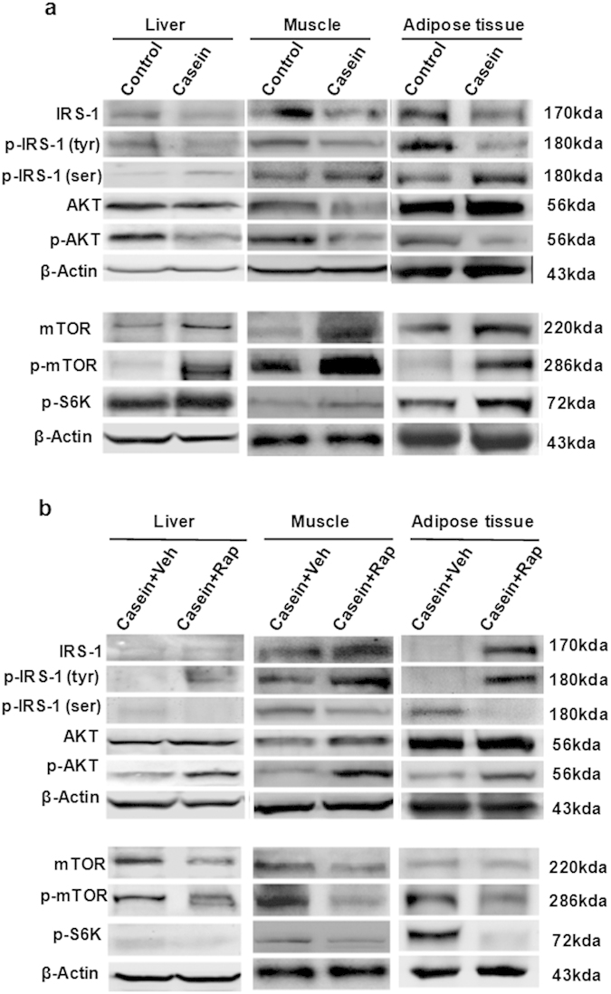
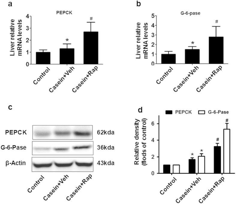
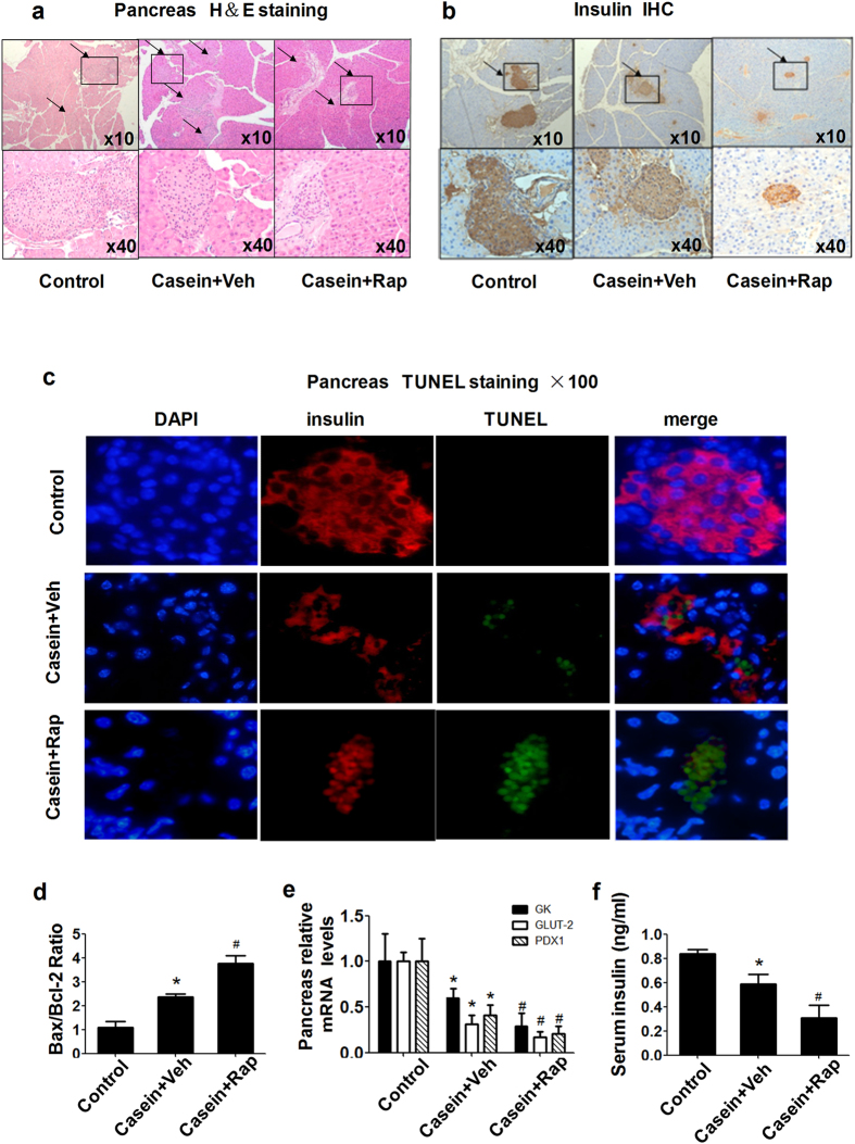
Insulin resistance is closely related to inflammatory stress and the mammalian target of rapamycin/S6 kinase (mTOR/S6K) pathway. The present study investigated whether rapamycin, a specific inhibitor of mTOR, ameliorates inflammatory stress-induced insulin resistance in vitro and in vivo. We used tumor necrosis factor-alpha (TNF-α) and interleukin-6 (IL-6) stimulation in HepG2 hepatocytes, C2C12 myoblasts and 3T3-L1 adipocytes and casein injection in C57BL/6J mice to induce inflammatory stress. Our results showed that inflammatory stress impairs insulin signaling by reducing the expression of total IRS-1, p-IRS-1 (tyr632), and p-AKT (ser473); it also activates the mTOR/S6K signaling pathway both in vitro and in vivo. In vitro, rapamycin treatment reversed inflammatory cytokine-stimulated IRS-1 serine phosphorylation, increased insulin signaling to AKT and enhanced glucose utilization. In vivo, rapamycin treatment also ameliorated the impaired insulin signaling induced by inflammatory stress, but it induced pancreatic β-cell apoptosis, reduced pancreatic β-cell function and enhanced hepatic gluconeogenesis, thereby resulting in hyperglycemia and glucose intolerance in casein-injected mice. Our results indicate a paradoxical effect of rapamycin on insulin resistance between the in vitro and in vivo environments under inflammatory stress and provide additional insight into the clinical application of rapamycin.


Figures

Figure 1
Figure 1. Effects of inflammatory stress and rapamycin on glucose utilization in vitro.
HepG2 cells, differentiated C2C12 myoblasts and 3T3-L1 adipocytes were incubated in serum-free medium for 24 h. The medium was then replaced by fresh serum-free medium containing 0.04 mmol/L palmitic acid alone or with TNF-α (25 ng/ml) or IL-6 (20 ng/ml) in the absence or presence of rapamycin (10 ng/ml) for 24 h. Glucose uptake (ac) was determined with a fluorescent D-glucose analog, 2NBDG, after treatment with or without insulin. Glucose consumption (df) was measured as described in the Materials and Methods section and normalized by cell protein. The results are shown as the mean ± SD (n = 6). **P < 0.05 versus control (basal), *P < 0.05 versus control (insulin), ##P < 0.05 versus inflammatory cytokine treated group (basal), #P < 0.05 versus inflammatory cytokine treated group (insulin).
Figure 2
Figure 2. Effects of inflammatory stress and rapamycin on protein expression of insulin signaling pathway in vitro.
HepG2 cells, C2C12 myoblasts and 3T3-L1 adipocytes were treated with insulin prior to harvest. (a) IRS1, p-IRS1 (tyr), and p-AKT, proteins of insulin signaling, were downregulated in the TNF-α- or IL-6-treated group. The proteins involved in the mTOR/S6K pathway were upregulated in the presence of inflammatory cytokines. (b) Under inflammatory stress, rapamycin inhibited the protein expression of mTOR, p-mTOR and p-S6K, while increasing the proteins involved in insulin signaling.
Figure 3
Figure 3. Casein injection induced chronic systemic inflammation in vivo.
The C57BL/6J mice fed with high-fat diet (HFD) were randomly assigned to receive a subcutaneous injection of normal saline (control group) or casein (casein group). (a) The levels of SAA and TNF-α in the serum of casein-injected mice were significant higher. The mRNA (b) and protein (c) levels of TNF-α and MCP-1 in the liver, muscle and adipose tissues increased in the casein group, which was parallel to the upregulation of SR-A and NF-kB protein in the liver, muscle and adipose tissue. The histogram represents the mean ± SD (n = 3) of the densitometric scans for protein bands, normalized by comparison with β-actin, and expressed as folds of control. *P < 0.05 versus control.
Figure 4
Figure 4. Effects of chronic inflammation and rapamycin on glucose and insulin tolerance in vivo.
The C57BL/6J mice fed with a high-fat diet (HFD) were randomly assigned to receive a subcutaneous injection of normal saline, casein, casein plus vehicle and casein plus rapamycin. (a) Glucose tolerance tests (glucose-stimulated blood glucose concentrations, IGTT) performed after 14 weeks of treatment. (b) Insulin tolerance tests (insulin-stimulated blood glucose concentrations, IITT) performed at the end of the experiments. The results represent the mean ± SD (n = 6). * P < 0.05 versus control. #P < 0.05 versus casein plus vehicle group.
Figure 5
Figure 5. Effects of chronic inflammatory and rapamycin on protein expression of insulin signaling in vivo.
(a) IRS1, p-IRS1 (tyr), and p-AKT, proteins of insulin signaling, were downregulated in casein group. The proteins involved in the mTOR/S6K pathway were upregulated in the presence of casein. (b) Under inflammatory stress, rapamycin inhibited the protein expression of mTOR, p-mTOR and p-S6K, while increasing the proteins involved in insulin signaling.
Figure 6
Figure 6. Rapamycin treatment enhanced hepatic gluconeogenesis.
(a) PEPCK and (b) G-6-Pase mRNA expression in the liver of C57BL/6J mice. The graphs depict mRNA expression in the liver of target genes corrected for the expression of β-actin as the housekeeping gene. (c) Representative western blotting of PEPCK and G-6-pase proteins in the liver. (d)The histogram represents the mean ± SD (n = 3) of the densitometric scans for protein bands from three experiments, normalized by comparison with β-actin, and expressed as folds of control. *P < 0.05 versus control, #P < 0.05 versus casein plus vehicle.
Figure 7
Figure 7. Rapamycin induced pancreatic β-cell dysfunction and apoptosis in vivo.
(a) Hematoxylin–eosin staining in the pancreas. (b) Insulin immunohistochemistry. Magnification, 10× (top panels); 40× (bottom panels). (c) Apoptosis in situ. Apoptosis of insulin-expressing cells on islet sections was determined by the TUNEL assay. Representative examples of pancreatic islets stained by immunofluorescence for insulin (red), marker of cell apoptosis TUNEL (green), and nuclear stain DAPI (blue) imaged at 100× in casein plus vehicle (upper) and casein plus rapamycin (lower). (d) Effects of rapamycin on the mRNA expression of Bax/Bcl2 in the pancreas of C57BL/6J mice. (e) Effects of rapamycin on β-cell gene expression. The expression of GK, GLUT2 and PDX-1 in islets was determined by real-time quantitative RT-PCR using β-actin as an internal standard. (f) Serum insulin concentrations in C57BL/6J mice were analyzed using a mouse-specific insulin ELISA kit. The results represent the mean ± SD (n = 6). *P < 0.05 versus control, #P < 0.05 versus casein plus vehicle.

References

    1. Biddinger S. B. & Kahn C. R. From mice to men: insights into the insulin resistance syndromes. Annu Rev Physiol 68, 123–158 (2006). - PubMed
    1. Lillioja S. et al. Insulin resistance and insulin secretory dysfunction as precursors of non-insulin-dependent diabetes mellitus. Prospective studies of Pima Indians. N Engl J Med 329, 1988–1992 (1993). - PubMed
    1. Michael M. D. et al. Loss of insulin signaling in hepatocytes leads to severe insulin resistance and progressive hepatic dysfunction. Mol Cell 6, 87–97 (2000). - PubMed
    1. Miyake K. et al. Hyperinsulinemia, glucose intolerance, and dyslipidemia induced by acute inhibition of phosphoinositide 3-kinase signaling in the liver. J Clin Invest 110, 1483–1491 (2002). - PMC - PubMed
    1. Morino K. et al. Reduced mitochondrial density and increased IRS-1 serine phosphorylation in muscle of insulin-resistant offspring of type 2 diabetic parents. J Clin Invest 115, 3587–3593 (2005). - PMC - PubMed

Publication types

MeSH terms