. 2016 Mar;24(3):564-9.
doi: 10.1038/mt.2015.192. Epub 2015 Oct 9.

CRISPR-mediated Genome Editing Restores Dystrophin Expression and Function in mdx Mice

Affiliations

CRISPR-mediated Genome Editing Restores Dystrophin Expression and Function in mdx Mice

Li Xu et al. Mol Ther. 2016 Mar.

Abstract

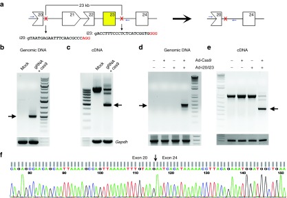
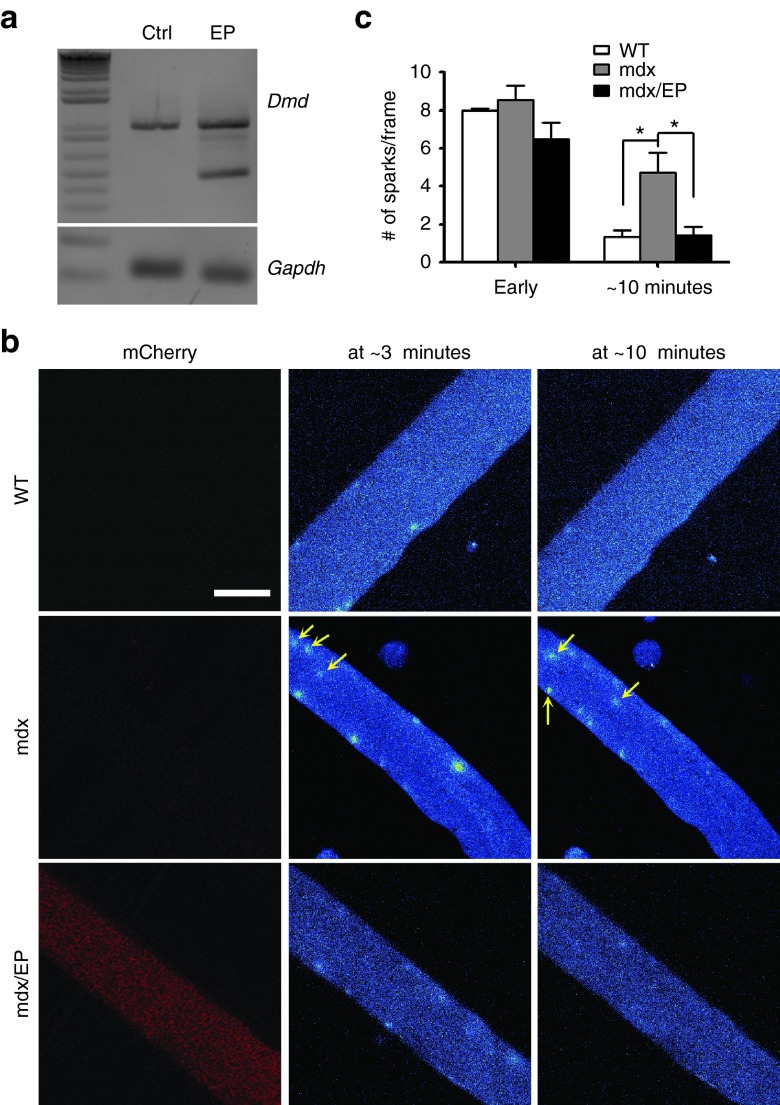
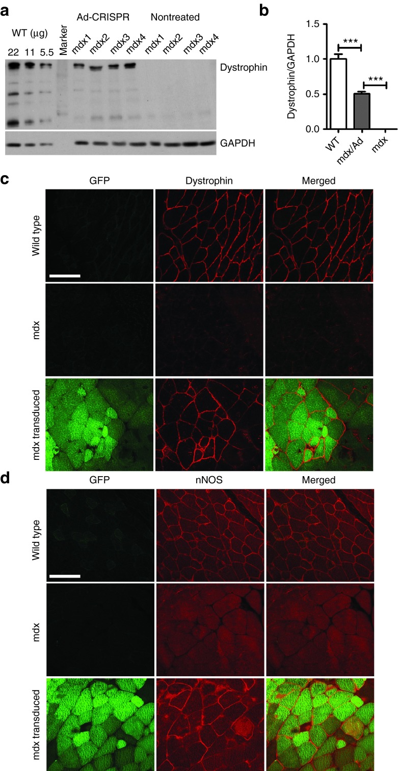
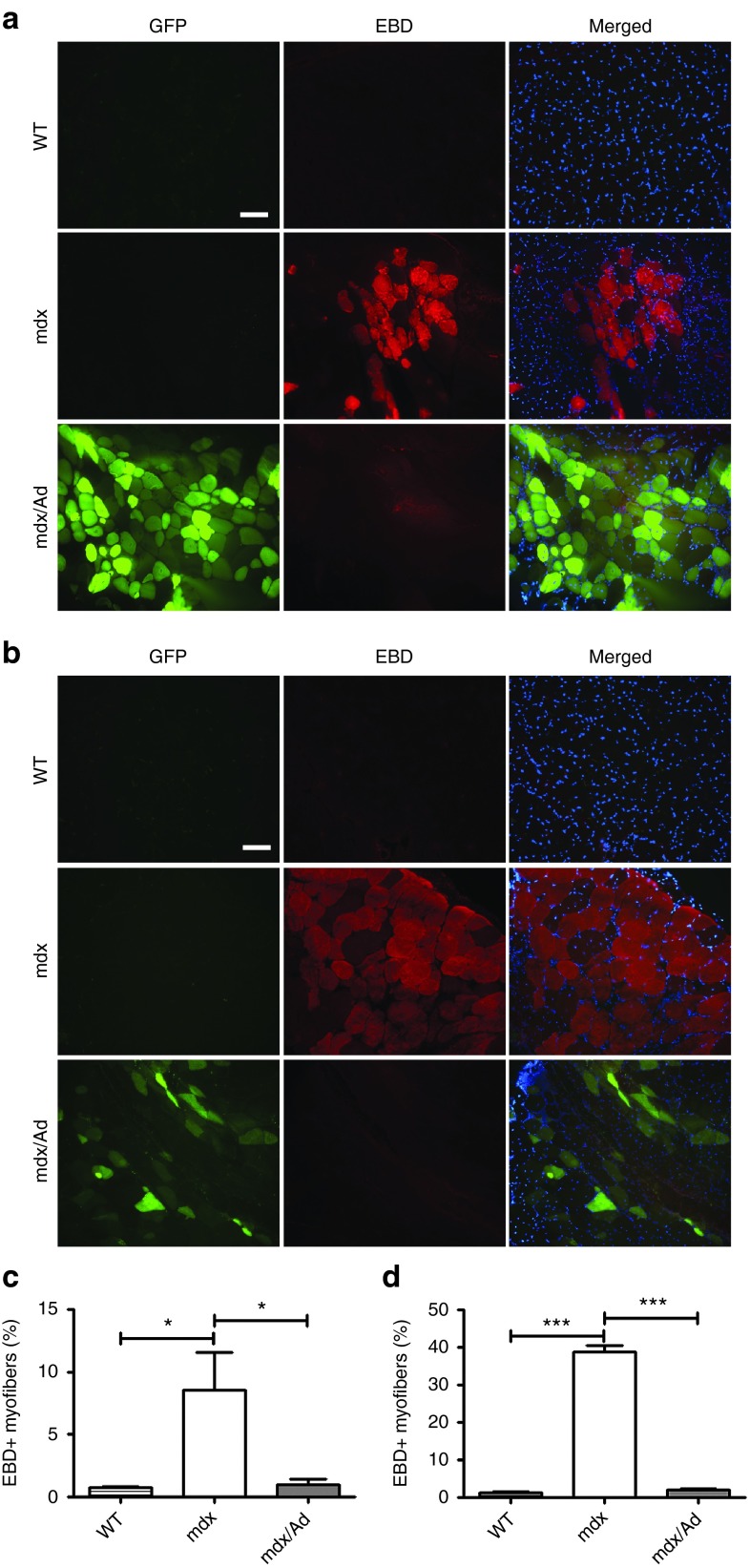
Duchenne muscular dystrophy (DMD) is a degenerative muscle disease caused by genetic mutations that lead to the disruption of dystrophin in muscle fibers. There is no curative treatment for this devastating disease. Clustered regularly interspaced short palindromic repeat/Cas9 (CRISPR/Cas9) has emerged as a powerful tool for genetic manipulation and potential therapy. Here we demonstrate that CRIPSR-mediated genome editing efficiently excised a 23-kb genomic region on the X-chromosome covering the mutant exon 23 in a mouse model of DMD, and restored dystrophin expression and the dystrophin-glycoprotein complex at the sarcolemma of skeletal muscles in live mdx mice. Electroporation-mediated transfection of the Cas9/gRNA constructs in the skeletal muscles of mdx mice normalized the calcium sparks in response to osmotic shock. Adenovirus-mediated transduction of Cas9/gRNA greatly reduced the Evans blue dye uptake of skeletal muscles at rest and after downhill treadmill running. This study provides proof evidence for permanent gene correction in DMD.


Figures

Figure 1
Figure 1
CRISPR-mediated deletion of a large region in mouse Dmd gene in vitro. (a) Diagram showing the genomic locus of mouse X-chromosome and the gRNA targeting sites. The mutant exon 23 is highlighted in yellow. (b) Polymerase chain reaction (PCR) analysis of genomic DNA extracted from C2C12 cells treated with or without gRNA and Cas9 constructs. (c) RT-PCR analysis of the dystrophin transcript expression in C2C12 cells. (d) PCR analysis of genomic DNA extracted from mdx myoblasts transduced with or without gRNA (Ad-i20/i23) and Cas9 expressing adenovirus. (e) RT-PCR analysis of the dystrophin transcript expression in mdx myoblasts as treated in (d). (f) DNA sequencing analysis of the smaller RT-PCR product (475 bp) in (c) and (e). Arrows indicate the expected bands after gene editing. All data are representative of a minimum of three experiments.
Figure 2
Figure 2
Osmotic shock-induced calcium sparks in mdx muscle fibers treated with or without mCherry-2A-Cas9/gRNA. (a) RT-PCR analysis of the total RNA extracted from the control flexor digitorum longus (FDB) muscles (Ctrl) or those electroporated with mCherry-2A-Cas9/gRNA plasmids (EP). (b) Osmotic shock-induced calcium sparks at ~3 minutes and ~10 minutes in isolated FDB muscle fibers from mdx mice electroporated with (left) or without (right) mCherry-2A-Cas9/gRNA plasmids. Arrows point to the calcium sparks located at the center of the muscle fibers. Scale bar: 50 µm. (c) Statistical analysis of calcium sparks in mdx muscle fibers electroporated with (male mdx/EP, n = 3) or without (male mdx, n = 4) mCherry-2A-Cas9/gRNA plasmids immediately (Early) or 10 minutes after osmotic shock and male WT controls (n = 4). *P < 0.05.
Figure 3
Figure 3
Restoration of dystrophin and its associated proteins at the sarcolemma of mdx muscles by genome-editing. (a) Western blotting of gastrocnemius muscle homogenates from wild-type (WT), mdx, and mdx with adenoviral vectors carrying GFP-2A-cas9 and gRNA using anti-dystrophin and anti-glyceraldehyde 3-phosphate dehydrogenase (GAPDH) antibodies. 22, 11, and 5.5 µg total proteins from the WT muscles were loaded per lane. (b) Quantitative analysis of western blotting in WT, mdx, and mdx injected with adenoviral vectors. (c,d) Confocal immunofluorescence images of dystrophin (c, red) and neuronal nitric oxide synthase (nNOS) (d, red) in muscle cryosections treated with or without EGFP-2A-cas9/gRNA adenovirus. The images are representative of five experiments. Scale bar: 100 µm. ***P < 0.001.
Figure 4
Figure 4
Functional rescue of dystrophin in mdx mice by CRISPR-mediated gene surgery. (a,b) Evans blue dye (EBD) uptake in the gastrocnemius muscles from male WT (n = 4), mdx (n = 4) and mdx (n = 4) mice injected with cas9/gRNA adenovirus at rest (a) or after downhill treadmill running exercise (b). The images are representative of four experiments. Red: EBD, green: GFP, blue: 4′,6-diamidino-2-phenylindole (DAPI). Scale bar: 100 µm. (c,d) Statistical analysis of EBD-positive muscle fiber percentage in WT, mdx and mdx/Ad preparations at rest (c) and after downhill treadmill running exercise (d). *P < 0.05; ***P < 0.001.

Comment in

References

    1. Cohn, RD and Campbell, KP (2000). Molecular basis of muscular dystrophies. Muscle Nerve 23: 1456–1471. - PubMed
    1. Wallace, GQ and McNally, EM (2009). Mechanisms of muscle degeneration, regeneration, and repair in the muscular dystrophies. Annu Rev Physiol 71: 37–57. - PubMed
    1. Koenig, M, Hoffman, EP, Bertelson, CJ, Monaco, AP, Feener, C and Kunkel, LM (1987). Complete cloning of the Duchenne muscular dystrophy (DMD) cDNA and preliminary genomic organization of the DMD gene in normal and affected individuals. Cell 50: 509–517. - PubMed
    1. Emery, AE (1991). Population frequencies of inherited neuromuscular diseases–a world survey. Neuromuscul Disord 1: 19–29. - PubMed
    1. Campbell, KP (1995). Three muscular dystrophies: loss of cytoskeleton-extracellular matrix linkage. Cell 80: 675–679. - PubMed

Publication types