. 2015 Dec 1;408(1):151-63.
doi: 10.1016/j.ydbio.2015.10.001. Epub 2015 Oct 21.

Neural crest-mediated bone resorption is a determinant of species-specific jaw length

Affiliations

Neural crest-mediated bone resorption is a determinant of species-specific jaw length

Erin L Ealba et al. Dev Biol. .

Abstract

Precise control of jaw length during development is crucial for proper form and function. Previously we have shown that in birds, neural crest mesenchyme (NCM) confers species-specific size and shape to the beak by regulating molecular and histological programs for the induction and deposition of cartilage and bone. Here we reveal that a hitherto unrecognized but similarly essential mechanism for establishing jaw length is the ability of NCM to mediate bone resorption. Osteoclasts are considered the predominant cells that resorb bone, although osteocytes have also been shown to participate in this process. In adults, bone resorption is tightly coupled to bone deposition as a means to maintain skeletal homeostasis. Yet, the role and regulation of bone resorption during growth of the embryonic skeleton have remained relatively unexplored. We compare jaw development in short-beaked quail versus long-billed duck and find that quail have substantially higher levels of enzymes expressed by bone-resorbing cells including tartrate-resistant acid phosphatase (TRAP), Matrix metalloproteinase 13 (Mmp13), and Mmp9. Then, we transplant NCM destined to form the jaw skeleton from quail to duck and generate chimeras in which osteocytes arise from quail donor NCM and osteoclasts come exclusively from the duck host. Chimeras develop quail-like jaw skeletons coincident with dramatically elevated expression of TRAP, Mmp13, and Mmp9. To test for a link between bone resorption and jaw length, we block resorption using a bisphosphonate, osteoprotegerin protein, or an MMP13 inhibitor, and this significantly lengthens the jaw. Conversely, activating resorption with RANKL protein shortens the jaw. Finally, we find that higher resorption in quail presages their relatively lower adult jaw bone mineral density (BMD) and that BMD is also NCM-mediated. Thus, our experiments suggest that NCM not only controls bone resorption by its own derivatives but also modulates the activity of mesoderm-derived osteoclasts, and in so doing enlists bone resorption as a key patterning mechanism underlying the functional morphology and evolution of the jaw.

Keywords: Avian beak development and evolution; Bisphosphonates; Bone mineral density; Bone remodeling; Bone resorption; Cranial neural crest; Evolutionary developmental biology; Jaw length; Mmp13; Mmp9; OPG; Osteoclasts; Osteocytes; Osteocytic osteolysis; Quail-duck chimeras; Quck; RANKL; TRAP staining.


Figures

Fig. 1
Fig. 1
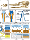
Species-specific jaw length, the quail-duck chimeric system, and lineages that resorb bone. Adult (A) quail and (B) duck skulls exhibit species-specific differences in jaw length. (C) Neural crest mesenchyme (NCM) that gives rise to the jaw skeleton is excised unilaterally from the boundary between the forebrain (fb) and midbrain (mb) to the rostral hindbrain (hb) of a quail donor (light blue) and transplanted orthotopically into a duck host (dark blue) at Hamburger and Hamilton (HH) stage 9.5 to make chimeric quck. (D) Schematic of a chimeric quck lower jaw in dorsal view at HH39 showing quail donor-derived bones and cartilages on one side (light blue) and duck host-derived bones and cartilages on the other (dark blue). Almost all of the lower jaw bones form through intramembranous ossification and these are the dentary, splenial, surangular, and angular. The articular cartilage undergoes endochondral ossification whereas Meckel’s cartilage remains unossified. (E) Schematic of an HH10 chimeric quck embryo in a transverse section plane through the caudal midbrain showing embryonic precursor populations that participate in the deposition and resorption of bone in the jaw skeleton. Specifically, osteoblasts and osteocytes are derived from quail donor NCM (light blue), while osteoclasts are derived from duck host mesoderm (orange). Osteoblasts begin depositing bone by HH33 whereas osteocytes and osteoclasts participate in bone resorption starting around HH37. In the lower jaw of quck chimeras, tartrate-resistant acid phosphatase (TRAP) is expressed by both quail donor-derived osteocytes and duck host-derived osteoclasts whereas Mmp13 is expressed by osteocytes, Mmp9 is expressed by osteoclasts, and neither is expressed by chondrocytes in Meckel’s cartilage since there is no endochondral ossification. Based on this timeline, treatments to inhibit or activate bone resorption were administered at HH33.
Fig. 2
Fig. 2
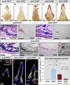
Species-specific differences in TRAP activity are mediated by NCM. (A) Whole mount TRAP staining in the head skeleton of quail (n=4) versus, (B) duck (n=4) reveals species-specific differences in levels and spatial domains of bone resorption at HH37, especially at the distal and proximal regions of the jaw. (C) Quck demonstrate that NCM controls bone resorption as indicated by higher quail-like levels and spatial patterns of TRAP activity on the donor side (arrows) of duck hosts (n=8). (D) By HH40, TRAP staining is more robust and widespread in quail (n=4) versus (E) duck (n=6) demonstrating both species- and stage-specific regulation of bone resorption. (F) Sections through the distal lower jaw skeleton at HH37 demonstrate that in the osteoid matrix of developing bone (stained blue), quail (n=6) have (G) high levels of TRAP-positive cells (stained red). (H) In equivalent bony regions of the duck jaw (I) less TRAP-positive staining can be observed (n=6). (J) Sections through the lower jaw of chimeric quck reveal that (K) coincident with the presence of quail-derived bones (Q¢PN-positive donor cells stained black) (L) there are quail-like levels of TRAP-positive staining (n=6). (M) Based on their small size and location within the bone matrix, osteocytes (ocy) can be recognized as being both TRAP-positive and Q¢PN-positive (arrow). Based on their large and irregular morphology, osteoclasts (ocl) can be identified as TRAP-positive but Q¢PN-negative. (N) 3D reconstruction of a quail mandible at HH37 showing the distribution of osteoid matrix (yellow) and TRAP staining (red) down the length of Meckel’s cartilage (blue). (O) Substantially lower levels of TRAP staining can be seen in the duck mandible. (P) The donor side of the chimeric quck mandible has higher levels of TRAP (arrows) like that observed in quail whereas the host side has lower duck-like levels. (Q) Quantification of the ratio of TRAP to osteoid volume in duck (n=4), quail (n=4), and quck (n=2) demonstrates that there are statistically significant differences between duck and quail in the amount of bone resorption, and that the donor side of quck is more quail-like whereas the host side is more duck-like.
Fig. 3
Fig. 3
NCM controls expression of genes involved in bone resorption. (A) Using RT-qPCR to assay for Col1α1 mRNA as a marker for bone deposition shows a 2.5-fold increase in duck (dark blue) from HH34 (n=5) to HH37 (n=5) and 4-fold increase in quail (light blue) from HH34 (n=5) to HH37 (n=5). Levels of Col1α1 expression in chimeric quck (red) at HH34 (n=2) are like that observed in quail controls at HH37. (B) Levels of Mmp13 in duck show a 3-fold increase from HH34 to HH37, whereas quail have an approximately 35-fold increase in expression from HH34 to HH37. In chimeric quck at HH34, quail donor cells maintain their higher stage-specific and species-specific levels of expression with an approximately 37-fold increase in Mmp13. (C) There is no significant increase in Mmp9 expression from HH34 to HH37 in duck whereas quail have a 6-fold increase. In quck, there is a 4-fold increase in Mmp9 at HH34 like that observed in quail at HH37. P-values: quail at HH37 compared to HH34; duck at HH37 to HH34; and quck at HH34 to HH34. (D) Sagittal sections showing the dentary bone and Meckel’s cartilage in the lower jaw of quail at HH37. (E) Through in situ hybridization on adjacent sections and coincident with the intramembranous ossification of bone, Mmp13 transcripts can be observed in the osteoid matrix (yellow dashed line), but not in Meckel’s cartilage. (F) Mmp9 expression is localized to discrete domains in bone presumably coincident with the distribution of osteoclasts. Mmp9 is not detected in Meckel’s cartilage. (G) Endochondral ossification of cartilage is restricted to the proximal-most region of the duck lower jaw at HH39. (H, I) Mmp13 and Mmp9 are expressed in cartilage undergoing endochondral ossification. (J, K, N and O) Osteoid in quail and duck at HH36 and HH39. (L, M) In quck, duck host bone is Q¢PN-negative while quail donor bone is Q¢PN-positive (black cells). (P, Q, T and U) Mmp13 is expressed at higher levels in quail versus duck at HH36 and HH39. (R) Mmp13 is not detected on the duck host side of quck (S) but is highly expressed on the quail donor side coincident with Q¢PN-positive cells.
Fig. 4
Fig. 4
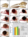
Bone resorption regulates jaw length. Differences in upper versus lower beak length occur naturally in some adult birds such as the (A) kea (Nestor notabilis), which has a shorter lower jaw, and (B) black skimmer (Rynchops niger) which has a longer lower jaw. (C and D) Quail and duck beak skeletons at HH39 stained with alizarin red showing differences in jaw length. Note the normal relations of the upper and lower portions that approximate one another at the distal tip, with the lower jaw being slightly shorter than the upper (arrows). (E) The beak is lengthened in duail when lower jaw NCM comes from a duck (n=4), and (F) shortened in quck when lower jaw NCM comes from a quail (n=7), demonstrating that NCM regulates size. (G) Quail treated at HH33 with a bisphosphonate (n=14), (H) recombinant OPG (rOPG) protein (n=8), and (I) an MMP13 inhibitor (iMMP13) have longer beaks, especially the lower jaw (n=6). (J) rRANKL protein treatments at HH33 decreases jaw length (n=8). (K) Quantifying jaw size reveals significant treatment-dependent increases and decreases in length relative to control embryos (n=12).
Fig. 5
Fig. 5
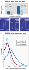
NCM regulates bone mineral density. (A) Duck jaw bones (n=3) have higher BMD than do quail (n=3). (B) µCT reconstructions of mineralized bone in lower jaws for duck at HH38, (C) quck at HH35, and (D) quail at HH38. (E) µCT histogram of BMD in lower jaws of HH38 quail (n=2, light blue), donor-side HH35 quck (n=3, red) and HH38 duck (n=3, dark blue). Duck have a higher average BMD than quail, and quck are quail-like.

Similar articles

Cited by

References

    1. Abzhanov A, Kuo WP, Hartmann C, Grant BR, Grant PR, Tabin CJ. The calmodulin pathway and evolution of elongated beak morphology in Darwin’s finches. Nature. 2006;442:563–567. - PubMed
    1. Abzhanov A, Protas M, Grant BR, Grant PR, Tabin CJ. Bmp4 and morphological variation of beaks in Darwin’s finches. Science. 2004;305:1462–1465. - PubMed
    1. Abzhanov A, Tabin CJ. Shh and Fgf8 act synergistically to drive cartilage outgrowth during cranial development. Dev. Biol. 2004;273:134–148. - PubMed
    1. Ainsworth SJ, Stanley RL, Evans DJ. Developmental stages of the Japanese quail. J. Anat. 2010;216:3–15. - PMC - PubMed
    1. Albrecht UEG, Helms JA, Lin H. Visualization of gene expression patterns by in situ hybridization. In: Daston GP, editor. Molecular and Cellular Methods in Developmental Toxicology. Boca Raton, FL: CRC Press; 1997. pp. 23–48.

Publication types

LinkOut - more resources