. 2015 Dec;14(12):3196-210.
doi: 10.1074/mcp.M115.052795. Epub 2015 Oct 8.

Dithiothreitol (DTT) Acts as a Specific, UV-inducible Cross-linker in Elucidation of Protein-RNA Interactions

Affiliations

Dithiothreitol (DTT) Acts as a Specific, UV-inducible Cross-linker in Elucidation of Protein-RNA Interactions

Uzma Zaman et al. Mol Cell Proteomics. 2015 Dec.

Abstract

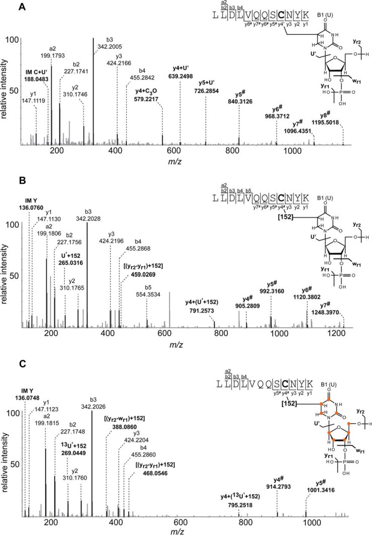
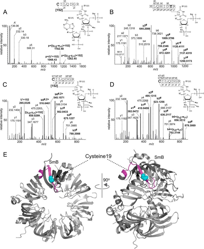
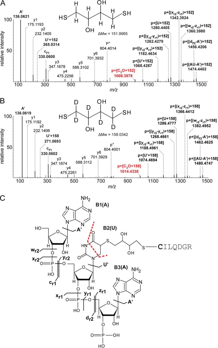
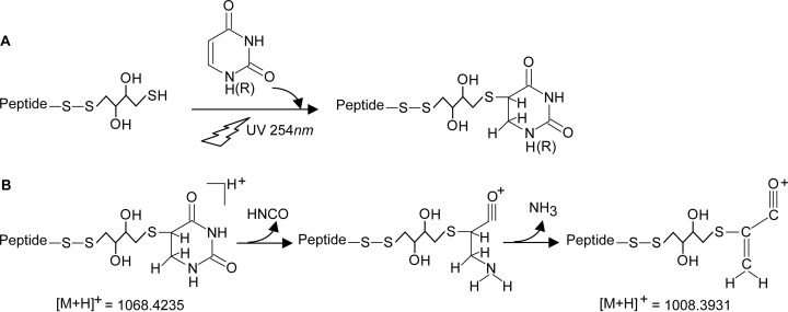
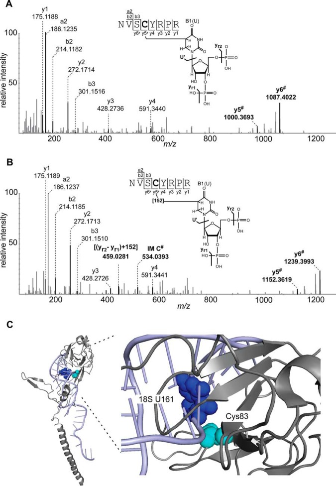
Protein-RNA cross-linking by UV irradiation at 254 nm wavelength has been established as an unbiased method to identify proteins in direct contact with RNA, and has been successfully applied to investigate the spatial arrangement of protein and RNA in large macromolecular assemblies, e.g. ribonucleoprotein-complex particles (RNPs). The mass spectrometric analysis of such peptide-RNA cross-links provides high resolution structural data to the point of mapping protein-RNA interactions to specific peptides or even amino acids. However, the approach suffers from the low yield of cross-linking products, which can be addressed by improving enrichment and analysis methods. In the present article, we introduce dithiothreitol (DTT) as a potent protein-RNA cross-linker. In order to evaluate the efficiency and specificity of DTT, we used two systems, a small synthetic peptide from smB protein incubated with U1 snRNA oligonucleotide and native ribonucleoprotein complexes from S. cerevisiae. Our results unambiguously show that DTT covalently participates in cysteine-uracil crosslinks, which is observable as a mass increment of 151.9966 Da (C(4)H(8)S(2)O(2)) upon mass spectrometric analysis. DTT presents advantages for cross-linking of cysteine containing regions of proteins. This is evidenced by comparison to experiments where (tris(2-carboxyethyl)phosphine) is used as reducing agent, and significantly less cross-links encompassing cysteine residues are found. We further propose insertion of DTT between the cysteine and uracil reactive sites as the most probable structure of the cross-linking products.


Figures

Fig. 1.
Fig. 1.
Sequence analysis of peptide-RNA (oligo)nucleotide cross-links derived from human spliceosomal complex U4 specific 15.5K protein cross-linked to U4 snRNA. A, MS/MS spectrum of m/z 874.38562+ annotated as LLDLVQQSCNYK (positions 22–33) cross-linked to nucleotide U. The peptide fragment signals shifted by (U-H3PO4) or (226 Da) are indicated by #. B, MS/MS spectrum of m/z 663.92343+ annotated as LLDLVQQSCNYK (positions 22–33) cross-linked to nucleotide U with the additional mass of 152 Da. The peptide fragment signals shifted by [(U-H3PO4)+152 Da] or (378 Da) are indicated by #. C, MS/MS spectrum of m/z 954.89762+ annotated as LLDLVQQSCNYK (positions 22–33) cross-linked to nucleotide 13U with the additional mass of 152 Da. Fragment ions shifted by an additional mass of 387.0858 Da are indicated by #. Signals corresponding to uracil fragments with the 152 Da increment are annotated as U', (yr2-yr1) and (yr2-wr1). All signals corresponding to RNA and peptide-RNA adducts are highlighted by bold letter. RNA oligonucleotide fragmentation nomenclature is according to McLuckey et al. (61). A proposed schematic illustration of the peptide-RNA cross-link along with cross-link product of +152 Da is shown on the right. Cysteine as the cross-linked amino acid identified by shift in the y ion series (starting from the y4 ion), is highlighted by bold letter in the peptide sequence. y-type fragmentation of RNA is referred to as U', wr1, yr1, yr2. Positions of 13C on UTP are marked by orange circles in lower panel.
Fig. 2.
Fig. 2.
Sequence analysis of peptide-RNA (oligo)nucleotide cross-links derived from human spliceosomal complex U1 SnRNP specific SmB/B′ peptide. A, MS/MS spectrum of m/z 805.24682+ showing the peptide CILQDGR (positions 19–25) cross-linked to AU with an additional mass of 152 Da in the presence of DTT. Spectrum obtained from LTQ-Orbitrap XL (HCD fragmentation). All signals corresponding to peptide-RNA adducts are highlighted by bold letter. B, MS/MS spectrum of m/z 691.28582+ shows the peptide MLQHIDYR (positions 9–16) cross-linked to U-H2O in the presence of DTT. A shifting of y ions by 306 Da corresponding to (yr2-yr1) is indicated by #. Tyrosine as the cross-linked amino acid, identified by a shift in y ion series from y2 to y7, is highlighted by bold letter. C, MS/MS spectrum of m/z 640.72022+ in the presence of DTT shows the peptide cross-linked to U with an additional mass of 152 Da. Shifting of a and b fragment ions of the peptide by different RNA fragments identified cysteine as cross-linked amino acid. D, MS/MS spectrum of m/z 564.72522+ shows the peptide cross-linked to U in the absence of DTT. In all spectra signal correspond to RNA and RNA fragments and shifting of the peptide signals by different RNA fragments masses are annotated and highlighted as bold letter. Proposed schematic illustration structures for peptide-RNA cross-links along with and without cross-link product of 152 Da mass are shown on the right side of each corresponding spectrum. Fragmentation of RNA refers as yr1, xr1, wr1 and yr2 by McLuckey et al. (61). E, Mapping of MLQHIDYRMRCILQDGR peptide (positions 9–25) on the crystal structure of Sm ring of U1 SnRNP (PDB ID: 4PJO, (62)). In SmB/B′ protein (both front and side view), the peptide is highlighted in pink color whereas the cross-linked cysteine is highlighted in cyan color as a sphere.
Fig. 3.
Fig. 3.
Sequence analysis of peptide-RNA (oligo)nucleotide cross-links derived from human spliceosomal complex U1 SnRNP specific SmB/B' peptide CILQDGR (positions 19–25). A, MS/MS spectrum of m/z 646.85103+ in the presence of DTT shows the peptide cross-linked to the trinucleotide AAU with an additional mass of 152 Da. The fragment nominally corresponding to the [M+H]+1 of the unmodified peptide is annotated as p+. All signals corresponding to RNA and RNA fragments and shifting of peptide signal fragment signals by different RNA fragments via 152 Da mass are annotated and highlighted by bold letters according to the nomenclature by McLuckey et al. (61). Marker ions of the adenine base and nucleotide with loss of water are annotated as A' and [yr1-(yr-1)] respectively. Similarly, uracil base and uracil nucleotide with the loss of water are annotated as U' and (yr2-yr1) respectively. Shifting in signal of uracil nucleobase (U') with 152 Da is also depicted. B, MS/MS spectrum of m/z 972.79572+ in the presence of d10-DTT shows the peptide cross-linked to the trinucleotide AAU with the additional mass of 158 Da. Signals in the high m/z range correspond to shifting of mass of peptide shifted peptide fragment masses by different RNA fragments along with 158 Da mass, and are annotated as in spectrum A. The difference of 6 Da is clearly observed between the two spectra in the high m/z region. C, Proposed structure for peptide-DTT-uracil cross-link formation. Upon UV irradiation at 254 nm, DTT can form covalent bonds with uracil and the thiol group of cysteine.
Fig. 4.
Fig. 4.
Proposed scheme for the cross-linking reaction of uridine/uracil to cysteine/DTT. Panel (A) shows UV induced peptide-DTT-uridine cross-link formation. Panel (B) shows CID fragmentation of cross-linked uracil according to Nelson and McCloskey (53). Sequential loss of HNCO and NH3 from uracil result in formation of C3H2O+ covalently linked to DTT-peptide. Abbreviations used in the figure are “P” for peptide and “R” for ribose.
Fig. 5.
Fig. 5.
Comparison of MS/MS fragment spectra of a peptide derived from S. cerevisiae mRNPs in two cross-linking experiments. A, MS/MS spectrum of m/z 650.76092+ shows the peptide NVSCYRPR (positions 80–87) cross-linked to U-H2O without the additional mass of 152 Da observed in the presence of TCEP. Shifts of the y5 and y6 ions by 306 Da corresponding to (yr2-yr1) are indicated by #. B, MS/MS spectrum of m/z 726.76052+ shows peptide cross-linked to U-H2O with the increment of 152 Da, obtained in the presence of DTT. Signals corresponding to shifts of the y5 and y6 ions by 458 Da [(yr2-yr1) + 152 Da] are highlighted by bold letters and indicated by # according to the nomenclature by McLuckey et al. (61). Proposed schematic structures for peptide-RNA (oligo)nucleotide cross-links with and without the 152 Da increment are also shown. Cysteine is identified as the cross-linked amino acid by the shifting of the y4 ion, highlighted as bold letter in the sequence. C, Crystal structure of S6–40S ribosomal protein in complex with 18S rRNA (PDB ID: 3J78, (54)). The cross-linked peptide NVSCYRPR (positions 80–87) is mapped on the crystal structure of S6–40S ribosomal protein. Cross-linked cysteine (position 83) and uracil161 (18S-rRNA), which are in close spatial proximity, are shown as cyan and blue spheres within the structure, respectively.

References

    1. Sinz A. (2006) Chemical cross-linking and mass spectrometry to map three-dimensional protein structures and protein-protein interactions. Mass Spectrom. Rev. 25, 663–682 - PubMed
    1. Leitner A., Walzthoeni T., Kahraman A., Herzog F., Rinner O., Beck M., and Aebersold R. (2010) Probing native protein structures by chemical cross-linking, mass spectrometry, and bioinformatics. Mol. Cell. Proteomics 9, 1634–1649 - PMC - PubMed
    1. Rappsilber J. (2011) The beginning of a beautiful friendship: cross-linking/mass spectrometry and modelling of proteins and multi-protein complexes. J. Struct. Biol. 173, 530–540 - PMC - PubMed
    1. Petrotchenko E. V., and Borchers C. H. (2010) Crosslinking combined with mass spectrometry for structural proteomics. Mass Spectrom. Rev. 29, 862–876 - PubMed
    1. Paramelle D., Miralles G., Subra G., and Martinez J. (2013) Chemical cross-linkers for protein structure studies by mass spectrometry. Proteomics 13, 438–456 - PubMed

Publication types