

Transcrestal Sinus Lift Procedure Approaching Atrophic Maxillary Ridge: A 60-Month Clinical and Radiological Follow-Up Evaluation
- PMID: 26451145
- PMCID: PMC4588341
- DOI: 10.1155/2015/261652
Transcrestal Sinus Lift Procedure Approaching Atrophic Maxillary Ridge: A 60-Month Clinical and Radiological Follow-Up Evaluation
Abstract
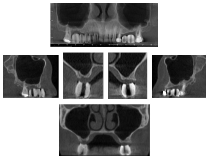
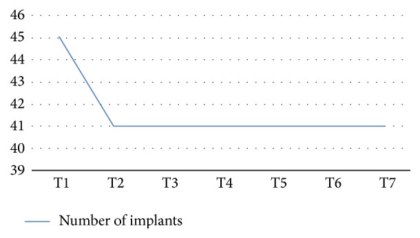
Aim. The aim of this study was to assess the success and the survival rate of dental implants placed in augmented bone after sinus lifting procedures. Material and Methods. 31 patients were mainly enrolled for a residual upper jaw crest thickness of 3 mm. CBCT scans were performed before and after the augmentation technique and at the follow-up appointments, at 3, 6, 12, 24, and up to 60 months. The follow-up examination included cumulative survival rate of implants, peri-implant marginal bone loss, and the height of sinus floor augmentation. Results. This retrospective study on 31 patients and 45 implants later inserted in a less than 3 mm crest showed excellent survival rates (99.5%), one implant was lost before loading due to an acute infection after 24 days, and two implants did not osteointegrate and were removed after 3 months. The radiological evaluation showed an average bone loss of 0.25 mm (±0.78 mm) at the first follow-up appointment (3 months) up to 0.30 mm (±1.28 mm) after 60-month follow-up. Conclusion. In this study it was reported how even in less than 3 mm thick crest a transcrestal technique can predictably be used with a long-term clinical and radiological outcome, giving patients excellent stability of the grafted material and healthy clinical results.
Figures


Similar articles
-
Minimally invasive transcrestal guided sinus lift (TGSL): a clinical prospective proof-of-concept cohort study up to 52 months.Clin Implant Dent Relat Res. 2014 Aug;16(4):582-93. doi: 10.1111/cid.12034. Epub 2013 Jan 28. Clin Implant Dent Relat Res. 2014. PMID: 23356732
-
Flapless, CBCT-guided osteotome sinus floor elevation with simultaneous implant installation. I: radiographic examination and surgical technique. A prospective 1-year follow-up.Clin Oral Implants Res. 2012 Jan;23(1):28-34. doi: 10.1111/j.1600-0501.2010.02151.x. Epub 2011 Mar 28. Clin Oral Implants Res. 2012. PMID: 21443611
-
A 10-year clinical and radiographic study of implants placed after maxillary sinus floor augmentation with an 80:20 mixture of deproteinized bovine bone and autogenous bone.Clin Implant Dent Relat Res. 2014 Jun;16(3):435-46. doi: 10.1111/cid.12008. Epub 2012 Oct 15. Clin Implant Dent Relat Res. 2014. PMID: 23066860
-
Transcrestal Sinus Lift Using Platelet Concentrates in Association to Short Implant Placement: A Retrospective Study of Augmented Bone Height Remodeling.Clin Implant Dent Relat Res. 2016 Oct;18(5):993-1002. doi: 10.1111/cid.12383. Epub 2015 Oct 20. Clin Implant Dent Relat Res. 2016. PMID: 26482330
-
Clinical and Radiological Long-Term Outcome of a Tapered Implant System with Special Emphasis on the Influence of Augmentation Procedures.Clin Implant Dent Relat Res. 2016 Aug;18(4):810-20. doi: 10.1111/cid.12338. Epub 2015 Mar 25. Clin Implant Dent Relat Res. 2016. PMID: 25810365
Cited by
-
Odontogenic Sinusitis Caused by an Inflammation of a Dentigerous Cyst and Subsequent Finding of a Fibrous Dysplasia. A Case Report.Open Dent J. 2016 Nov 30;10:647-655. doi: 10.2174/1874210601610010647. eCollection 2016. Open Dent J. 2016. PMID: 28077969 Free PMC article.
-
Evaluation of graft osteogenesis using fractal dimension analysis on cone-beam computed tomography images following maxillary sinus lift surgery.BMC Oral Health. 2025 Aug 21;25(1):1346. doi: 10.1186/s12903-025-06695-8. BMC Oral Health. 2025. PMID: 40841897 Free PMC article.
-
Platelet-Rich Fibrin (PRF) in Implants Dentistry in Combination with New Bone Regenerative Flapless Technique: Evolution of the Technique and Final Results.Open Med (Wars). 2017 Mar 9;12:24-32. doi: 10.1515/med-2017-0005. eCollection 2017 Jan. Open Med (Wars). 2017. PMID: 28401197 Free PMC article.
-
Accuracy of Periapical Radiography and CBCT in Endodontic Evaluation.Int J Dent. 2018 Oct 16;2018:2514243. doi: 10.1155/2018/2514243. eCollection 2018. Int J Dent. 2018. PMID: 30410540 Free PMC article.
-
Stress Distribution Pattern in Zygomatic Implants Supporting Different Superstructure Materials.Materials (Basel). 2022 Jul 16;15(14):4953. doi: 10.3390/ma15144953. Materials (Basel). 2022. PMID: 35888420 Free PMC article.
References
-
- Bernardello F., Righi D., Cosci F., Bozzoli P., Soardi Carlo M., Spinato S. Crestal sinus lift with sequential drills and simultaneous implant placement in sites with <5 mm of native bone: a multicenter retrospective study. Implant Dentistry. 2011;20(6):439–444. doi: 10.1097/id.0b013e3182342052. - DOI - PubMed
-
- Boyne P. J., James R. A. Grafting of the maxillary sinus floor with autogenous marrow and bone. Journal of Oral Surgery. 1980;38(8):613–616. - PubMed
-
- Tatum H., Jr. Maxillary and sinus implant reconstructions. Dental clinics of North America. 1986;30(2):207–229. - PubMed
-
- Summers R. B. A new concept in maxillary implant surgery: the osteotome technique. Compendium. 1994;15:154–156. - PubMed
-
- Summers R. B. The osteotome technique: part 3—less invasive methods of elevating the sinus floor. Compendium. 1994;15(6):698–700. - PubMed
LinkOut - more resources
Full Text Sources
Other Literature Sources
Medical
