. 2015:2015:814089.
doi: 10.1155/2015/814089. Epub 2015 Sep 14.

Protective Effect of Triptolide against Glomerular Mesangial Cell Proliferation and Glomerular Fibrosis in Rats Involves the TGF- β 1/Smad Signaling Pathway

Affiliations

Protective Effect of Triptolide against Glomerular Mesangial Cell Proliferation and Glomerular Fibrosis in Rats Involves the TGF- β 1/Smad Signaling Pathway

Yingjie Cao et al. Evid Based Complement Alternat Med. 2015.

Abstract

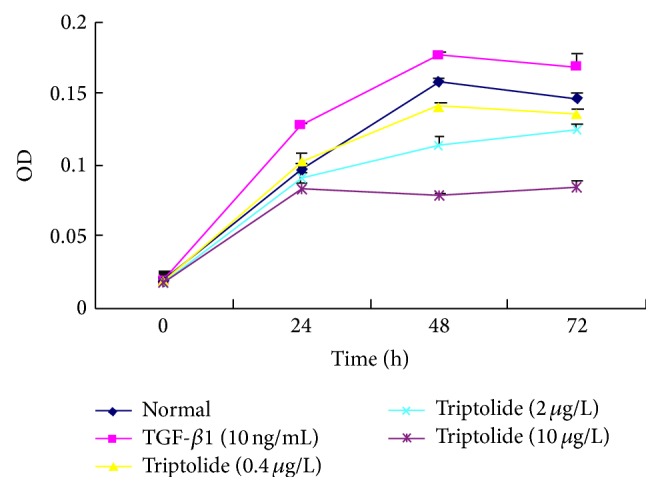
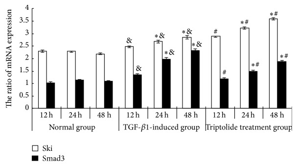
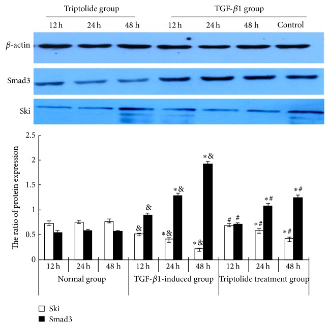
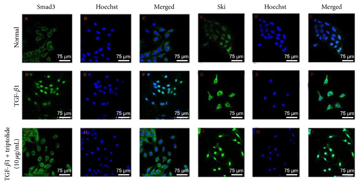
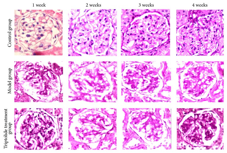
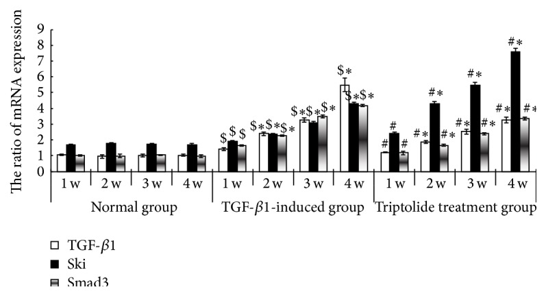
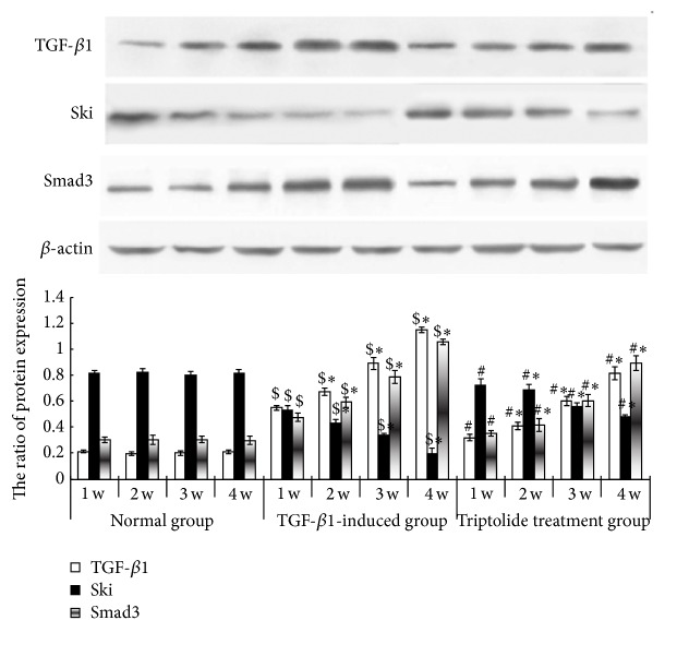
Triptolide as a main active ingredient of Tripterygium wilfordii is known to be exerting anti-inflammatory, marked immunosuppressive, and podocyte-protective effects. In this study, we investigated the protective effect of triptolide in kidney disease. Rat glomerular mesangial cells were randomly divided into three groups: (1) control group, (2) TGF-β1 (10 μg/mL) group, and (3) triptolide group (triptolide 10 μg/L + TGF-β1 10 μg/L). Sixty male Sprague-Dawley rats were randomly divided into three groups: (1) control group, (2) chronic serum sickness glomerulonephritis model group, and (3) triptolide (0.2 mg/kg·d) group. Reverse transcription PCR was used to assess Ski and Smad3 mRNA expression in the mesangial cells and renal tissues. Western blotting was used to determine Ski and Smad3 protein expressions. Laser confocal fluorescence microscopy was used to observe the subcellular localization of Smad3 and Ski proteins in the mesangial cells. Triptolide inhibited the TGF-β1-induced proliferation of mesangial cells. It significantly upregulated Ski protein expression and downregulated Smad3 mRNA and protein expressions in a time-dependent manner. Laser confocal fluorescence microscopy detected high Smad3 fluorescence intensity in the cytoplasm and low Smad3 and high Ski fluorescence intensity in the nucleus. By upregulating Ski protein expression triptolide decreased the extent of fibrosis by affecting the TGF-β1/Smad3 signaling pathway.


Figures

Figure 1
Figure 1
The effect of triptolide on the TGF-β1-induced RMC proliferation. Triptolide inhibited the TGF-β1-induced proliferation of mesangial cell. The triptolide effect enhanced gradually over a period of 48 h reaching its peak at 48 h and eventually decreasing after 48 h. When the concentration of the triptolide was gradually increased from 0.4 to 10 μg/L, the efficiency of the drug simultaneously increased (P < 0.05, n = 6).
Figure 2
Figure 2
Effect of triptolide on the mRNA expression of Ski and Smad3 of the TGF-β1-induced mesangial cells. Triptolide downregulated Smad3 mRNA expression but upregulated Ski mRNA expression in the TGF-β1-treated mesangial cells. Compared to the TGF-β1-induced group, under the effect of triptolide, the level of expression of Smad3 mRNA decreased but the level of expression of Ski mRNA had significantly increased in a time-dependent manner (& P < 0.05 versus the control group, P < 0.05 versus the different time points in the same group, and # P < 0.05 versus the TGF-β1-induced group, n = 6).
Figure 3
Figure 3
Effect of triptolide on the Ski and Smad3 protein expression of the TGF-β1-induced mesangial cells. Triptolide downregulated Smad3 protein expression but upregulated Ski protein expression in the TGF-β1-treated cells. Western blot analysis: the protein levels of Smad3 and Ski of the glomerular mesangial cells at different time points in control, TGF-β1-stimulated, and triptolide treatment groups. Compared to the TGF-β1-induced group, under the effect of triptolide, the level of expression of Smad3 protein decreased but the level of expression of Ski protein had significantly increased in a time-dependent manner (& P < 0.05 versus the control group, P < 0.05 versus the different time points in the same group, and # P < 0.05 versus the TGF-β1-induced group, n = 6).
Figure 4
Figure 4
The effect of triptolide on the subcellular localization of the Smad3 and Ski proteins of the TGF-β1-induced RMC. In control group, Smad3 protein was mainly expressed in the cytoplasm but, in the TGF-β1-induced group, the level of expression of Smad3 protein was significantly higher and was mainly localized in the nucleus. Ski protein of the control group was expressed mainly in the nucleus. The level of expression of Ski in and out of the nucleus of the TGF-β1-induced group was significantly higher and was mainly localized in the cytoplasm (bar = 75 μm).
Figure 5
Figure 5
Renal pathological changes of the different groups at different time points. Triptolide alleviated pathological damage of rat renal tissues in chronic serum sickness glomerulonephritis model group. There was no significant hyperplasia in the glomerular mesangial cells and cellular matrix, with no obvious broadening of the mesangial region in the renal tissues from the normal control group. However, there was significant hyperplasia in the glomerular mesangial cells and cellular matrix, with significant broadening of the mesangial region in the renal tissues obtained from the model group, increasing with respect to time, with the tendency to glomerular fibrosis. The lesions of the triptolide treatment group were, however, alleviated.
Figure 6
Figure 6
The expression of TGF-β1 mRNA, Smad3, and Ski in the renal tissues of the different groups. Triptolide downregulated Smad3 mRNA expression but upregulated Ski mRNA expression in the renal tissues of the model rats. In the model group, the level of expression of TGF-β1 and Smad3 mRNA in the renal tissues of the rats of the model group at the different time points was high and time-dependent but the expression of Ski mRNA was lower than that in the control group. Compared to the triptolide treatment group, the level of expression of TGF-β1 and Smad3 mRNA in the renal tissues of the rats of the model group at the different time points was lower and time-dependent but the expression of Ski mRNA was higher ($ P < 0.05 versus the control group, P < 0.05 versus the different time points in the same group, and # P < 0.05 versus the TGF-β1-induced group, n = 18–20).
Figure 7
Figure 7
Expressions of TGF-β1, Smad3, and Ski proteins in the renal tissues of the different groups. Triptolide downregulated Smad3 protein expression but upregulated Ski protein expression in the renal tissues of the model rats. Western blot analysis of Ski, TGF-β1, and Smad3 protein expressions in the renal tissues at different time points in the control, model, and triptolide treatment groups. In the model group, the level of expression of TGF-β1 and Smad3 proteins in the renal tissues of the rats of the model group at the different time points was higher and time-dependent but the expression of Ski protein was lower than that in the control group. Compared to the triptolide treatment group, the level of expression of TGF-β1 and Smad3 protein in the renal tissues of the rats of the model group at the different time points was lower and time-dependent but the expression of Ski protein was higher ($ P < 0.05 versus the control group, P < 0.05 versus the different time points in the same group, and # P < 0.05 versus the TGF-β1-induced group, n = 18–20).

References

    1. Stavnezer D., Barkas A. E., Brennan L. A., Brodeur D., Li Y. Transforming Sloan-Kettering viruses generated from the cloned v-ski oncogene by in vitro and in vivo recombinations. Journal of Virology. 1986;57(3):1073–1083. - PMC - PubMed
    1. Phillips R. L., Ernst R. E., Brunk B., et al. The genetic program of hematopoietic stem cells. Science. 2000;288(5471):1635–1640. doi: 10.1126/science.288.5471.1635. - DOI - PubMed
    1. Mauviel A. Transforming growth factor-beta: a key mediator of fibrosis. Methods in molecular medicine. 2005;117:69–80. - PubMed
    1. Attisano L., Wrana J. L. Signal transduction by the TGF-beta superfamily. Science. 2002;296(5573):1646–1647. doi: 10.1126/science.1071809. - DOI - PubMed
    1. Yang J., Dai C., Liu Y. Hepatocyte growth factor suppresses renal interstitial myofibroblast activation and intercepts Smad signal transduction. The American Journal of Pathology. 2003;163(2):621–632. doi: 10.1016/s0002-9440(10)63689-9. - DOI - PMC - PubMed

LinkOut - more resources