. 2015:2015:689240.
doi: 10.1155/2015/689240. Epub 2015 Sep 15.

Actinomycotic Osteomyelitis of Maxilla Presenting as Oroantral Fistula: A Rare Case Report

Affiliations

Actinomycotic Osteomyelitis of Maxilla Presenting as Oroantral Fistula: A Rare Case Report

Ashalata Gannepalli et al. Case Rep Dent. 2015.

Abstract

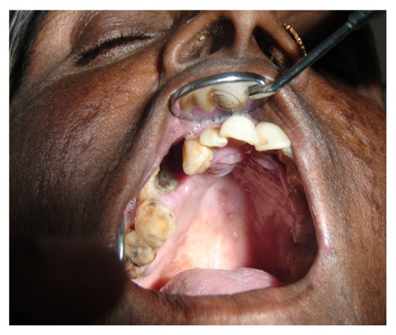
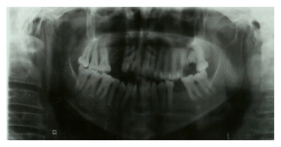
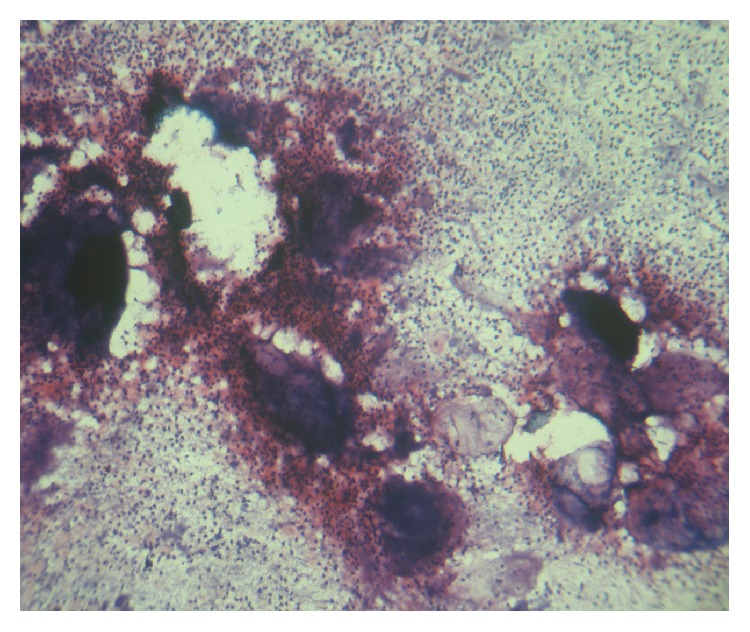
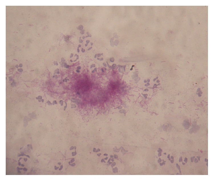
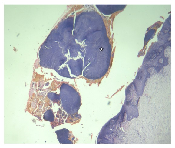
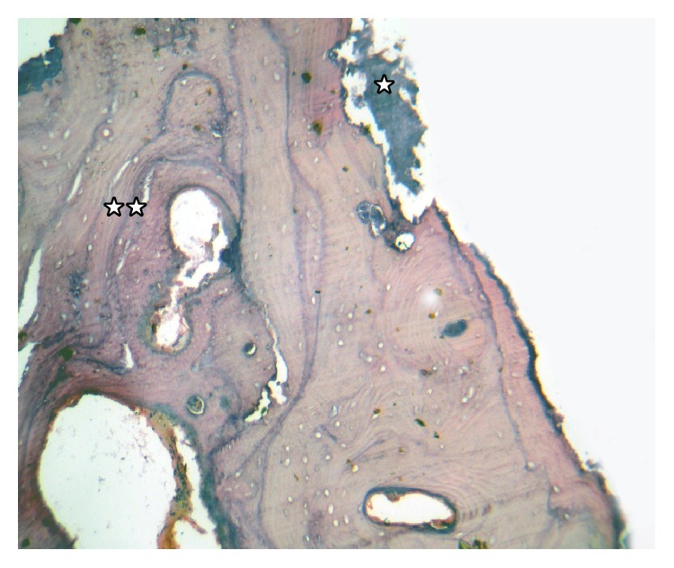
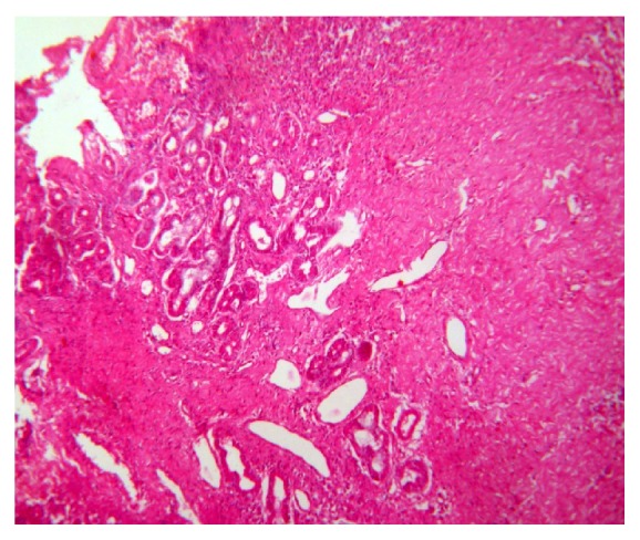
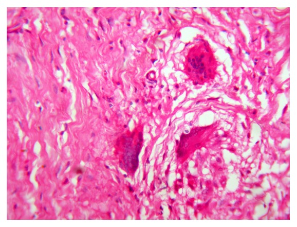
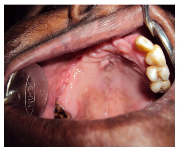
Actinomycosis is a chronic granulomatous infection caused by Actinomyces species which may involve only soft tissue or bone or the two together. Actinomycotic osteomyelitis of maxilla is relatively rare when compared to mandible. These are normal commensals and become pathogens when they gain entry into tissue layers and bone where they establish and maintain an anaerobic environment with extensive sclerosis and fibrosis. This infection spreads contiguously, frequently ignoring tissue planes and surrounding tissues or organ. The portal of entry may be pulpal, periodontal infection, and so forth which may lead to involvement of adjacent structures as pharynx, larynx, tonsils, and paranasal sinuses and has the propensity to damage extensively. Diagnosis is often delayed and is usually based on histopathology as they are cultured in fewer cases. The chronic clinical course without regional lymphadenopathy may be essential in diagnosis. The management of actinomycotic osteomyelitis is surgical debridement of necrotic tissue combined with antibiotics for 3-6 months. The primary actinomycosis arising within the maxilla with contiguous involvement of paranasal sinus with formation of oroantral fistula is rare. Hence, we present a 50-year-old female patient with chronic sclerosing osteomyelitis of maxilla which presented as oroantral fistula with suppurative and sclerotic features.


Figures

Figure 1
Figure 1
Clinical photograph showing oroantral fistula.
Figure 2
Figure 2
OPG showing destructive radiolucent lesion of right maxilla.
Figure 3
Figure 3
Photomicrograph of smear (10x) Gram's staining image of actinomycotic colony with peripheral neutrophilic aggregation.
Figure 4
Figure 4
Photomicrograph of smear (oil immersion). Tangled mass of Periodic acid-Schiff (PAS) positive branching filaments.
Figure 5
Figure 5
Photomicrograph of biopsy (10x) H&E oral epithelium with large actinomycotic colonies (☆).
Figure 6
Figure 6
Photomicrograph (10x) of H&E decalcified section. Necrotic thickened bony trabeculae with Actinomyces filaments on trabecular surface of bony spicules (☆), with extensive sclerosis of bone showing prominent resting and reversal lines (☆☆).
Figure 7
Figure 7
Photomicrograph (10x) H&E section. Extensive fibrosis with granulomas and degeneration of salivary gland acini.
Figure 8
Figure 8
Photomicrograph (40x) of H&E. Resolving granulomas with giant cells.
Figure 9
Figure 9
Postoperative photograph after 2 months of follow-up.

References

    1. Rubin M. M., Krost B. S. Actinomycosis presenting as a midline palatal defect. Journal of Oral and Maxillofacial Surgery. 1995;53(6):701–703. doi: 10.1016/0278-2391(95)90176-0. - DOI - PubMed
    1. Miller M., Haddad A. J. Cervicofacial actinomycosis—review. Oral Surgery, Oral Medicine, Oral Pathology, Oral Radiology, and Endodontics. 1998;85(5):496–508. doi: 10.1016/s1079-2104(98)90280-3. - DOI - PubMed
    1. Marx R. E., Stern D. Oral and Maxillofacial Pathology—A Rational for Diagnosis and Treatment. Quintessence Publishing; 2003. Inflammatory, Reactive, and infectious Diseases; pp. 57–67.
    1. Crossman T., Herold J. Actinomycosis of the maxilla—a case report of a rare oral infection presenting in general dental practice. British Dental Journal. 2009;206(4):201–202. doi: 10.1038/sj.bdj.2009.115. - DOI - PubMed
    1. Sezer B., Akdeniz B. G., Günbay S., Hilmioğlu-Polat S., Başdemir G. Actinomycosis osteomyelitis of the jaws: report of four cases and a review of the literature. Journal of Dental Sciences. 2013 doi: 10.1016/j.jds.2013.02.031. - DOI - PMC - PubMed

LinkOut - more resources