. 2015 Apr 1;4(10):e1022301.
doi: 10.1080/2162402X.2015.1022301. eCollection 2015 Oct.

Progressive loss of anti-HER2 CD4+ T-helper type 1 response in breast tumorigenesis and the potential for immune restoration

Affiliations

Progressive loss of anti-HER2 CD4+ T-helper type 1 response in breast tumorigenesis and the potential for immune restoration

Jashodeep Datta et al. Oncoimmunology. .

Abstract

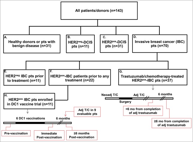
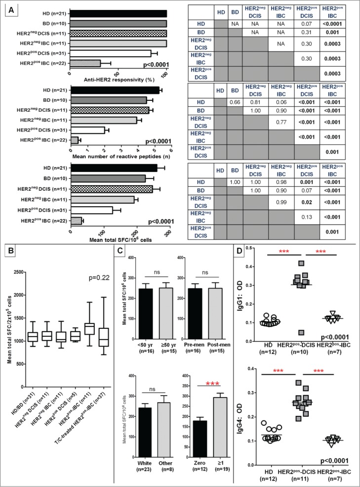
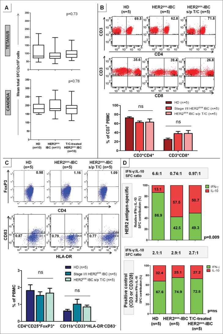
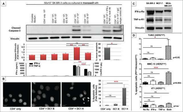
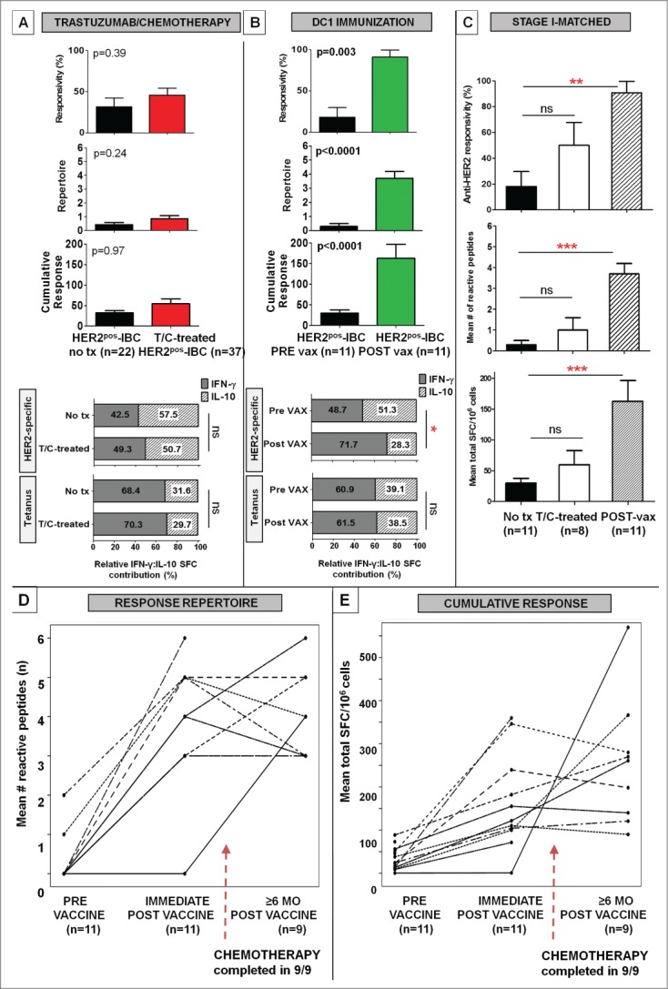
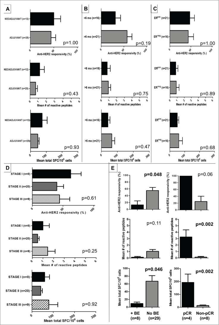
Genomic profiling has identified several molecular oncodrivers in breast tumorigenesis. A thorough understanding of endogenous immune responses to these oncodrivers may provide insights into immune interventions for breast cancer (BC). We investigated systemic anti-HER2/neu CD4+ T-helper type-1 (Th1) responses in HER2-driven breast tumorigenesis. A highly significant stepwise Th1 response loss extending from healthy donors (HD), through HER2pos-DCIS, and ultimately to early stage HER2pos-invasive BC patients was detected by IFNγ ELISPOT. The anti-HER2 Th1 deficit was not attributable to host-level T-cell anergy, loss of immune competence, or increase in immunosuppressive phenotypes (Treg/MDSCs), but rather associated with a functional shift in IFNγ:IL-10-producing phenotypes. HER2high, but not HER2low, BC cells expressing IFNγ/TNF-α receptors were susceptible to Th1 cytokine-mediated apoptosis in vitro, which could be significantly rescued by neutralizing IFNγ and TNF-α, suggesting that abrogation of HER2-specific Th1 may reflect a mechanism of immune evasion in HER2-driven tumorigenesis. While largely unaffected by cytotoxic or HER2-targeted (trastuzumab) therapies, depressed Th1 responses in HER2pos-BC patients were significantly restored following HER2-pulsed dendritic cell (DC) vaccinations, suggesting that this Th1 defect is not "fixed" and can be corrected by immunologic interventions. Importantly, preserved anti-HER2 Th1 responses were associated with pathologic complete response to neoadjuvant trastuzumab/chemotherapy, while depressed responses were observed in patients incurring locoregional/systemic recurrence following trastuzumab/chemotherapy. Monitoring anti-HER2 Th1 reactivity following HER2-directed therapies may identify vulnerable subgroups at risk of clinicopathologic failure. In such patients, combinations of existing HER2-targeted therapies with strategies to boost anti-HER2 CD4+ Th1 immunity may decrease the risk of recurrence and thus warrant further investigation.

Keywords: CD4+ T-helper immunity; HER2/neu; breast cancer; dendritic cell; immune monitoring; immune restoration; vaccination.


Figures

Figure 1.
Figure 1.
Study-eligible patient and donor cohorts. Hierarchy diagram representing patient/donor groups included in study. Cohorts are labeled A–H for ease of comparison (of immune responses), and are referred to in Results. Treatment schedules in cohorts G and H, as well as time-points at which blood was drawn are indicated in red callout boxes. Specifically, in the T/C-treated HER2pos-IBC cohort (G), patients received either neoadjuvant T/C, followed by surgery and completion of adjuvant trastuzumab; patients selected for a surgery-first approach completed adjuvant T/C. Blood was drawn either <6 mo or ≥6 mo from completion of adjuvant trastuzumab.
Figure 2.
Figure 2.
Anti-HER2 CD4+ Th1 responses and IgG1/IgG4 reactivity are progressively lost in HER2pos breast tumorigenesis. (A) IFNγ ELISPOT analysis of PBMCs demonstrated a progressive loss of anti-HER2 CD4+ Th1 response in HER2pos breast tumorigenesis (i.e., HD/BDHER2pos-DCIS HER2pos-IBC), illustrated by anti-HER2 responsivity, response repertoire, and cumulative response (all ANOVA < 0.001). No differences in Th1 responses were observed between HER2neg-DCIS and HER2neg-IBC (IHC 0/1+) and HD/BD subjects. Post-hoc Scheffé p value comparisons between groups are shown alongside histograms. (B) Anti-CD3/ CD28 stimulus served as positive control for all donors in experiments shown in (A) above; corresponding IFNγ production by ELISPOT in respective patient groups is shown. Results presented as median ± interquartile range (IQR) IFNγ SFC per 2 × 10⁵ cells in box-and-whiskers plots; (C) Variations in anti-HER2 Th1 cumulative responses in HD/BDs donors stratified by donor age (<50 vs. ≥50 years), menopausal status (pre-menopausal vs. post-menopausal) (upper panel); and race (white vs. other), or gravidity (zero vs. ≥1 pregnancies) (bottom panel). Within each Th1 metric, results are expressed as proportion or mean (±SEM). (D) ELISA of serum reactivity against recombinant HER2 extracellular domain revealed significantly elevated anti-HER2 IgG1 and IgG4 antibody levels in HER2pos-DCIS compared with HDs that decayed in HER2pos-IBC patients. ELISA measurements are shown as optical density (OD) at 1:100 sera dilutions (grouped scatter plot, with horizontal line indicating mean OD); (***p < 0.001 by unpaired t-test or ANOVA with post-hoc Scheffé testing, as applicable).
Figure 3.
Figure 3.
Anti-HER2 Th1 deficit in HER2pos-IBC is not attributable to lack of immunocompetence or increase in immunosuppressive phenotypes, but associated with a functional shift in IFNγ:IL-10-producing phenotypes. PBMCs from HER2pos-IBC patients, both treatment-naive and T/C-treated, did not differ significantly from HDs in (A) IFNγ production to recall stimuli tetanus toxoid or Candida albicans by ELISPOT. Results presented as median ± interquartile range (IQR) IFNγ SFC per 2 × 10⁵ cells in box-and-whiskers plots; (B and C) Relative proportions of CD4+ (CD3+CD4+) (B top) or CD8+ (CD3+CD8+) T-cells (B bottom), Treg (CD4+CD25+FoxP3+) (C top) or MDSCs (CD11b+CD33+HLA-DRCD83) (C bottom) by flow cytometry. Representative stainings within groups are shown; results in adjoining histograms expressed as mean proportions (%) ± SEM as indicated. (D) Donor-matched cumulative IFNγ and IL-10 production (SFC per 106 cells) across six HER2 Class II peptides compared in HD, treatment-naive HER2pos-IBC, and T/C-treated HER2pos-IBC patients. Relative HER2-specific IFNγ to IL-10 proportions (% depicted in graph) decreased significantly from HDs [IFNγ/(IFNγ + IL-10) = 86.9%; IL-10/(IFNγ + IL-10) = 13.1%] to HER2pos-IBC patients with (49.3%; 50.7%) or without (42.5%; 57.5%) T/C treatment. Absolute IFNγ:IL-10 production ratio changed from 6.6:1 (HDs) to 0.97:1 (T/C-treated) and 0.74:1 (HER2pos-IBC), respectively (top panel). No significant relative shifts in IFNγ:IL-10 production were observed to positive controls (anti-CD3/anti-CD3/CD28) (bottom panel).
Figure 4.
Figure 4.
CD4+ Th1 induces apoptosis of HER2high, but not HER2low, human and murine breast cancer cells. (A) Using a transwell system, 50 × 103 SK-BR-3 cells were co-cultured with medium alone (complete medium), 106 CD4+ T-cells alone (CD4+ only), 106 CD4+ T-cells + 105 each of HER2 Class II peptide (iDC H)- or irrelevant Class II BRAF peptide (iDC B)-pulsed iDCs, and 106 CD4+ T-cells + 105 each HER2 (DC1 H)- or BRAF (DC1 B)-pulsed DC1s. Increased caspase-3 cleavage indicated dose-dependent apoptosis of SK-BR-3 cells when co-cultured with DC1 H:CD4+ T-cells, but not DC1 B, iDC H, or iDC B groups. Vinculin was used as loading control. The displayed protein gel blot is representative of three experiments (top panel), and results are expressed as mean caspase-3/vinculin ratios ± SEM indicating fold induction of apoptosis (quantified using ImageJ software). Compared with IgG isotype control, CD4+:DC1 H-induced SK-BR-3 apoptosis was significantly rescued by neutralizing IFNγ and TNF-α. Bars represent % rescue of mean caspase-3/vinculin ratio ±SEM (31.4 ±5.3% IFNγ/TNF-α neutralization vs. control). Results are representative of three experiments (middle panel). Corresponding production of IFNγ (left y-axis) and TNF-α (right y-axis) in respective co-cultures by ELISA is shown. Results are expressed in pg/mL, and are representative of three experiments (bottom panel). (B) Significantly greater proportion of apoptotic SK-BR-3 cells (asterisks) were observed by DAPI staining in the DC1 H:CD4+ group, correlating with a 25-fold increase in apoptosis, compared with CD4+ only or DC1 B:CD4+ groups. Results are representative of three experiments, and expressed as mean % apoptotic cells ± SEM. (C) HER2high SK-BR-3, HER2intermediate MCF-7, and HER2low MDA-MB-231 human BC cells uniformly maintained expression of IFNγ-Rα and TNF-α-R1 receptors. (D) In transgenic murine HER2high mammary carcinoma TUBO and MMC15 cells, combination treatment with recombinant murine (rm) Th1 cytokines rmIFNγ and rmTNF-α resulted in significantly greater apoptosis compared with untreated controls or treatment with either cytokine alone. This effect was not reproduced with dual rmIFNγ + rmTNF-α treatment in murine HER2low/neg cells 4T1. Results are representative of three experiments, and expressed as mean % apoptotic cells ± SEM, detected by proportion of PIpos/Annexin Vpos cells by flow cytomety. (* p ≤ 0.05, **p < 0.01, *** p < 0.001).
Figure 5 (See previous page).
Anti-HER2 CD4+ Th1 immunity is differentially restored following HER2-pulsed DC immunization, but not after HER2-targeted therapies. (A) Compared with treatment-naive Stage I/II HER2pos-IBC patients (no tx), anti-HER2 Th1 responses were not globally augmented following T/C treatment in stage I-III HER2pos-IBC patients (T/C-treated), illustrated by anti-HER2 responsivity, repertoire, or cumulative response. Relative proportion of IFNγ:IL-10 reactive cells (% depicted in histogram) following HER2-specific and tetanus (positive control) stimuli did not improve in T/C-treated (n = 5) compared with no tx (n = 5) patients; (B) Significant improvements in all anti-HER2 Th1 immune metrics were observed in 11 Stage I HER2pos-IBC (PRE vax) patients immediately following HER2 pulsed-DC1 immunization (POST vax). While relative proportion of IFNγ to IL:10 reactive cells (% depicted in histogram) did not change appreciably following tetanus stimulation, HER2-pulsed vaccination significantly increased the relative proportion of IFNγ to IL:10 reactive cells in POST vax (n = 5) compared with PRE vax (n = 5) patients; (C) The differential Th1 restoration following HER2-pulsed DC immunization, but not T/C treatment, persisted on stage-matched comparisons in stage I HER2pos-IBC patients. Results are expressed as proportion or mean ± SEM; (**p < 0.01, ***p < 0.001). (D and E) Beyond the immediate post-vaccination period, anti-HER2 CD4+ Th1 immunity remained durably augmented in 9 of 11 evaluable patients ≥6 mo following vaccination, despite initiation/completion of systemic chemotherapy in all patients by this time-point (broken arrows). Scatter plots demonstrate CD4+ Th1 reactivity profiles by response repertoire (D) and cumulative response (E) for individual vaccinated subjects.
Figure 6.
Figure 6.
Depressed anti-HER2 Th1 responses following T/C treatment correlate with adverse clinical and pathologic outcomes. (A–D) Subgroup analysis of T/C-treated HER2pos-IBC patients demonstrated no appreciable differences in anti-HER2 responsivity, repertoire, or cumulative response when stratified by (A) sequencing of chemotherapy (neoadjuvant vs. adjuvant); (B) time from completion of trastuzumab to enrollment in study (<6 vs. ≥6 mo); (C) estrogen-receptor status (ERpos vs. ERneg) and (D) pathologic stage (I vs. II vs. III). (E) Compared with HER2pos-IBC patients who did not incur breast events (no BE) following completion of T/C, patients incurring BEs (+BE) had significantly depressed anti-HER2 responsivity and cumulative Th1 responses (D left). In HER2pos-IBC patients achieving pathologic complete response (pCR) following neoadjuvant T/C, anti-HER2 Th1 response repertoire and cumulative response was significantly greater compared to non-pCR patients (D right).

References

    1. Jemal A, Bray F, Center MM, Ferlay J, Ward E, Forman D. Global cancer statistics. CA Cancer J Clin 2011; 61:69-90; PMID:21296855; http://dx.doi.org/10.3322/caac.20107 - DOI - PubMed
    1. Perou CM, Sorlie T, Eisen MB, van de Rijn M, Jeffrey SS, Rees CA, Pollack JR, Ross DT, Johnsen H, Akslen LA et al.. Molecular portraits of human breast tumours. Nature 2000; 406:747-52; PMID:10963602; http://dx.doi.org/10.1038/35021093 - DOI - PubMed
    1. Meric F, Hung MC, Hortobagyi GN, Hunt KK. HER2/neu in the management of invasive breast cancer. J Am Coll Surg 2002; 194:488-501; PMID:11949754; http://dx.doi.org/10.1016/S1072-7515(02)01121-3 - DOI - PubMed
    1. Henson ES, Hu X, Gibson SB. Herceptin sensitizes ErbB2-overexpressing cells to apoptosis by reducing antiapoptotic Mcl-1 expression. Clin Cancer Res 2006; 12:845-53; PMID:16467098; http://dx.doi.org/10.1158/1078-0432.CCR-05-0754 - DOI - PubMed
    1. Wang GS, Zhu H, Bi SJ. Pathological features and prognosis of different molecular subtypes of breast cancer. Mol Med Rep 2012; 6:779-82; PMID:22797840; http://dx.doi.org/10.3892/mmr.2012.981 - DOI - PubMed

Publication types